Chapter 370 Nasal Polyps
Etiology
Cystic fibrosis is the most common childhood cause of nasal polyposis and should be suspected in any child <12 yr old with nasal polyps, even in the absence of typical respiratory and digestive symptoms; as many as 30% of children with cystic fibrosis acquire nasal polyps (Chapter 395). Nasal polyposis is also associated with chronic sinusitis and allergic rhinitis. In the uncommon Samter triad, nasal polyps are associated with aspirin sensitivity and asthma.
Diagnosis and Differential Diagnosis
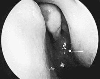
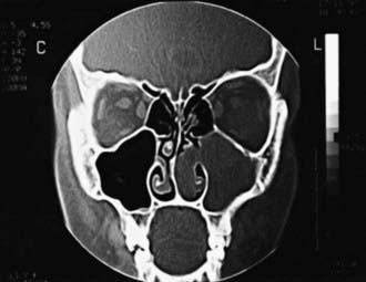
Examination of the external nose and rhinoscopy is performed. Ethmoidal polyps can be readily distinguished from the well-vascularized turbinate tissue, which is pink or red; antrochoanal polyps may have a more fleshy appearance (Fig. 370-1). Antrochoanal polyps may prolapse into the nasopharynx; flexible nasopharyngoscopy can assist in making this diagnosis. Prolonged presence of ethmoidal polyps in a child can widen the bridge of the nose and erode adjacent osseous structures. Tumors of the nose cause more local destruction and distortion of the anatomy. CT scan of the midface is key to diagnosis and planning for surgical treatment (Fig. 370-2).
Basik S, Karaman CZ, Akdilli A, et al. Surgical approaches to antrochoanal polyps in children. Int J Pediatr Otorhinolaryngol. 1998;46:197-205.
Magit AE. Tumors of the nose, paranasal sinuses, and nasopharynx. In: Bluestone CD, Stool SE, Kenna MA, editors. Pediatric otolaryngology. ed 3. Philadelphia: WB Saunders; 1996:893-904.
Orvidas LJ, Beatty CW, Weaver AL. Antrochoanal polyps in children. Am J Rhinol. 2001;15:321-325.
Scadding GK, Durham SR, Mirakian R, et al. British Society for Allergy and Clinical Immunology: BSACI guidelines for the management of rhinosinusitis and nasal polyposis. Clin Exp Allergy. 2008;38:260-275.
Stjärne P, Mösges R, Jorissen M, et al. A randomized controlled trial of mometasonc furoate nasal spray for the treatment of nasal polyposis. Arch Otolaryngol Head Neck Surg. 2006;132:179-185.
Thomas M, Yawn BP, Price D, et al. European Position Paper on Rhinosinusitis and Nasal Polyps Group. EPOS Primary Care Guidelines: European Position Paper on the Primary Care Diagnosis and Management of Rhinosinusitis and Nasal Polyps 2007—a summary. Prim Care Respir J. 2008;17:79-89.


