Mycoplasmas, Filamentous Bacteria, and Bacteroides
I Mycoplasma and Ureaplasma Species
1. The smallest free-living bacteria capable of passing through a 0.45-μm filter, which retains most other bacteria
2. Lack peptidoglycan-containing cell wall and thus are very pleomorphic and not visualized with Gram stain
• Slow-growing, obligate aerobe forming granular colonies on Eaton agar
• Serologic tests for antigen-specific immunoglobulin M (IgM) antibodies
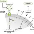
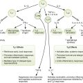
• P1 adhesin protein mediates adherence to respiratory epithelium.
• Damage to ciliated epithelium by lytic enzymes and hydrogen peroxide (H2O2) leads to decreased clearance of upper airways, facilitating the spread of bacteria to the lungs.
• Superantigen activity is related to systemic symptoms of M. pneumoniae infection.
3. Diseases caused by M. pneumonia
• Primary atypical (“walking”) pneumonia
a. Interstitial (no alveolar exudate) pneumonia that differs clinically from typical community acquired pneumonia caused by pneumococcus (Table 14-1)
TABLE 14-1
Mycoplasmal Versus Pneumococcal Pneumonia
| Characteristic | Mycoplasmal pneumonia | Pneumococcal pneumonia |
| Type of pneumonia | Atypical (interstitial) | Typical (alveolar) |
| Preceding pharyngitis | Common | Never |
| Onset | Gradual | Rapid with chills |
| Fever | Low grade | High |
| Cough | Nonproductive, paroxysmal | Productive |
| Sputum | Usually clear | Purulent |
| Pleuritic chest pain | Absent | Present |
| Leukocytosis | Absent | Present |
| Age of highest incidence | Young adults | Older adults |
| Complications | Otitis media, erythema multiforme, hemolytic anemia, myocarditis, pericarditis, bullous otitis media | Bacteremia, meningitis, otitis media |
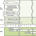
b. Initial malaise, low-grade fever, and headache followed after 2 to 4 days by nonproductive cough, rales, rhonchi, and myalgia, and, rarely, an IgM-mediated hemolytic anemia
a. Inflammation of the bronchi marked by nonproductive cough, fever, headache, sore throat, pharyngeal exudates, and cervical lymphadenopathy
• Organisms in the genera Nocardia and Actinomyces species form long branching filaments looking like fungi (Table 14-2).
TABLE 14-2
| Characteristic | Nocardia | Actinomyces |
| Morphology | Filamentous rods | Filamentous rods |
| Gram staining | Positive (weak) | Positive |
| Acid fast | Yes (weak) | No |
| Growth | Aerobic, slow growing | Anaerobic, slow growing |
| Part of normal human flora | No | Yes |
| Common disease manifestations | Lung disease, brain abscess, mycetoma and other skin infections; mostly in immunocompromised patients | Abscesses with draining sinus tracts usually after surgery or trauma; mycetoma |
• Nocardiae are ubiquitous soil organisms that cause exogenous infections primarily in immunocompromised individuals.
• Very slow growing, aerobic, and weakly gram positive
• Partially acid fast owing to the presence of mycolic acids in the cell wall (similar to Mycobacterium species)
a. Inhaled organisms (primarily Nocardia asteroides) colonize the oropharynx and then are aspirated into the lower airways.
b. Cough, dyspnea, and fever are usually present and pneumonia with cavitation may develop.
c. May disseminate to the central nervous system, forming brain abscesses, or to skin.
• They cause endogenous infections when a mucosal barrier is compromised.
• Actinomyces species are normal colonizers of the upper respiratory tract or the gastrointestinal tract.
• Slow growing, anaerobic, gram positive, and not acid fast
• “Sulfur” granules (1- to 5-mm yellow-orange masses of organisms resembling grains of sand) visible in clinical specimens
• Actinomyces israelii, which colonizes the oropharynx, is the most common cause of actinomycosis in humans.
• Cervicofacial disease is marked by tissue swelling, fibrosis, and scarring along draining sinus tracts at the angle of the jaw and neck.
• Abdominal and pelvic infections usually occur following surgery or trauma.
• Thoracic infection is associated with history of aspiration.
• Central nervous system infection (abscess and meningitis) usually results from spread from other foci.
• Mycetoma may represent an exogenous infection caused by soil-borne actinomycetes that enter wounds.
• B. fragilis is the most clinically significant of the numerous gram-negative, nonsporulating, anaerobic colonizers of the respiratory, gastrointestinal, and genitourinary tracts (Box 14-2).
1. Gram-negative, encapsulated, pleomorphic organisms that produce foul-smelling short-chain fatty acids
• Surgery and trauma promote endogenous spread of normal colonic flora to sterile sites, where they cause disease.