Multiple Sclerosis

Published on 03/03/2015 by admin

Filed under Neurology

Last modified 22/04/2025

Print this page

rate 1 star rate 2 star rate 3 star rate 4 star rate 5 star
Your rating: none, Average: 5 (1 votes)

This article have been viewed 4551 times

46 Multiple Sclerosis

Clinical Vignette

A 23-year-old woman suddenly noted pain behind her right eye; this discomfort was exacerbated whenever she moved her eyes horizontally or vertically. Within 2 days, she lost central vision in her right eye; she could not read with that eye alone; however, her left eye was normal. She was otherwise perfectly healthy.

Neurologic examination demonstrated her visual acuity as 20/200 OD and 20/30 OS; associated findings confined to her right eye included a slightly irregular, poorly reactive pupil, diminished color vision, and papilledema. A magnetic resonance image (MRI) of the brain demonstrated a few periventricular demyelinating lesions. Treatment was begun with daily intravenous methylprednisolone infusions for 10 days. Her vision returned to normal within a few weeks.

She was well for another 4 years until the sudden onset of an annoying swollen and numb feeling in her left leg and concomitant occurrence of an electric shock–like sensation that spread down her back whenever she bent her neck. Examination demonstrated brisk muscle stretch reflexes, left greater than right, a left Babinski sign, ipsilateral decreased position sense, and contralateral diminished temperature appreciation from her right foot to her high thoracic region. MRIs demonstrated demyelination within her left cervical-thoracic spinal cord junction, and concomitant increased T2 signal within the corpus callosal and periventricular cerebral white matter. Although she once again responded well to methylprednisolone, and despite beginning ongoing therapy with beta-interferon, this young woman continued to have occasional exacerbations with some degree of remission. Eventually, she developed a persistent gait ataxia; nevertheless, she has maintained a very successful professional career, is married, and has a 3-year-old child.

Comment: This is a classic example of a previously healthy young person, whose initial optic neuritis held a relatively poorer prognosis as her brain MRI at that time showed signs of “silent” central nervous system (CNS) demyelination. Today, with that initial combination of optic neuritis and MRI findings, a diagnosis of multiple sclerosis (MS) would be made and an “ABC” medication begun at the outset of her illness.

Multiple sclerosis is the most common central nervous system disorder to affect young and middle-aged adults. Because the disease process has such protean manifestations and a variable course, demyelinating disorders have a broad clinical spectrum, ranging from a single benign episode to one that is potentially fatal. Epidemiologic studies demonstrate that MS is more prevalent in northern latitudes, twice as common in women, and most often presents during the third and fourth decades. Caucasians are twice as likely as people of color to acquire MS in the United States and Canada. Interestingly, when MS develops among Asian populations, it predominantly affects the optic nerves and spinal cord with much less involvement of the brain in contrast to North American and European persons. Individuals born in a northern latitude, who are race- and age-matched, gain the innate diminished risk of equatorial region habitats if they move south before age 15 years. Other persons who also move from the north to the south, but do not do so until after age 15 years, maintain their innate higher risks. Although there are some yet to be precisely identified genetic predisposing factors for MS, these alone cannot account for the above-defined variabilities, as genetically comparable populations vary in MS prevalence depending on place of birth and age of migration.

Pathology

With classic MS, the primary process is one of demyelination leading to loss of myelin from central nervous system (CNS) axons. Myelin loss (a nonspecific term) occurs concurrently with other pathologic processes that also affect the axons, glial elements, or vasculature. CNS oligodendrocytes are responsible for the elaboration of brain myelin. This is a predominantly lipid-based (70%) structure, with the remainder being protein based. One part, myelin basic protein, is particularly immunologically susceptible and experimentally encephalitogenic.

Inspection of the gross brain of an MS subject does not demonstrate any sign of abnormalities that would indicate the presence of the myriad histologic changes that are to be found on microscopic evaluation. However, the optic nerves, optic chiasm, and spinal cord may be atrophic to the native eye. Sometimes, areas of patchy demyelination are seen on the basis pontis surface, the cerebellar peduncles, and the surface of the medulla and floor of the fourth ventricle.

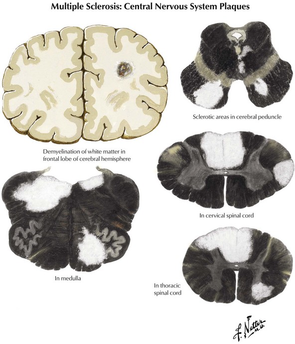
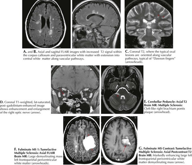
Coronal brain sections reveal changes similar to those noted on MRI, where variously sized MS plaques are apparent. Recently acquired lesions are pink and soft, whereas chronic MS lesions are gray, translucent, and firm (Fig. 46-1). It is often difficult to correlate the multiple lesions found at autopsy or by MRI throughout the neuraxis with a patient’s history. Sometimes classic MS plaques exist in patients who were never clinically suspected of harboring it.

Microscopic analysis demonstrates that many plaques have no relation to specific nerve tracts. Often, the plaques have a perivenular and paraventricular distribution. Myelin loss from a nerve fiber is distinct and best defined by toluidine blue stains. Macrophage accumulation is a frequent accompaniment. Active plaques contain myelin debris. Severe loss of oligodendrocytes within MS plaques is associated with the concomitant nonspecific finding of hypertrophic astrocytes. Signs of leptomeningeal inflammation, not unlike that found in acute disseminated MS, may be evident.

There is also a very significant component of axonal and neuronal damage in multiple sclerosis. This is particularly relevant to the long-term outcome and eventual disability. One can find evidence of axonal injury early on in the disease process. This is found in areas of obvious demyelination as well as areas of white and gray matter that appear normal to gross inspection. It is proposed that an antigen-specific destructive component related to both T cells and autoantibodies as well as the effects of activated macrophages and microglia lead to very significant axonal damage in the pathogenesis of MS. Mitochondrial function may also be impaired as various cellular substrates further contribute to this pathologic component.

Clinical Subtypes

The natural history of MS varies with the subtype of disease. Functional consequences may relate to some degree of axonal loss occurring after demyelination.

Benign MS

Clinical Vignette

A 52-year-old woman with migraine headaches since childhood, tobacco use throughout her adult years, and a history of transverse myelitis at age 14 years with complete recovery was subsequently noted to have a normal neurologic examination. She remained well until 65 years of age when she had an acute episode of left hemisensory deficit. An MRI scan of the brain revealed an acute lacunar infarct in the right thalamus on diffusion-weighted imaging, a chronic lacunar infarct in the left basal ganglia, and 10 bright lesions on T2 and FLAIR images, several of which were linear to ovoid in shape, configured perpendicularly to the ventricular walls, none enhancing with contrast. MRI of the cervical spine showed mild myelomalacia at C5 to C7 without other abnormality. She quit smoking and recovered with very little sensory impairment throughout her left side and lived without further neurologic symptoms until the age of 72 years, when she died of lung cancer.

Comment: This patient experienced a single bout of transverse myelitis as an adolescent, often a harbinger of more generalized MS. Medical technology at that time did not afford a simple way to explore her central nervous system for occult signs of multiple sclerosis. Clinical signs of more diffuse disease never developed. However, approximately 50 years later, when the patient required an MRI for a question of stroke, clinically inactive, more widespread MS became evident. In fact there was more than one explanation for the periventricular white matter abnormalities on MRI, namely, microvascular disease and migraine headaches. However, taken in the context of her prior episode of transverse myelitis, and the number and configuration of her white matter lesions, a diagnosis of relatively benign MS is likely. Certainly, whatever the idiosyncrasies of her immune system are that protected her, thus preventing further expression of more diffuse signs of clinical multiple sclerosis over her lifetime, once unlocked, they might provide the key for better appreciating the pathophysiology of MS and therefore direct more specific therapeutic research intervention in the future.

Relapsing–Remitting MS

Clinical Vignette

A 47-year-old woman sustained lower back and neck injury and concussion at 25 years of age in a motor vehicle accident. She underwent a C2-3 fusion and L4-5 diskectomy at that time and made a good recovery. She delivered two healthy children when she was ages 28 and 30 years. Five months after the birth of the first child, she began to experience numbness in the left leg and urinary urgency, with occasional leakage of urine. She was felt to have a recrudescence of her lumbar radicular symptoms and postpartum stress incontinence. Her primary care physician referred her for physical therapy, and her symptoms improved over the ensuing 2 months.

Just 1.5 years later, at age 32 years, she experienced increased difficulty lifting simple kitchen objects such as a milk carton and a frying pan and noted some numbness in her hands. She had increasing problems carrying her children, particularly placing them into their car seats. Her internist found Tinel signs at each wrist and thought she might have bilateral carpal tunnel syndrome; wrist splints were not helpful, and she remained relatively symptomatic. Over the years, she tried to remain active with her children, but increasingly found herself altering bicycle routes that they had once taken with ease as a family. She could not drink anything for 3 hours prior to going out on a bike ride to avoid urinary incontinence. Because she more commonly fatigued with any sustained walking, particularly during the warmer summer months, she curtailed her shopping.

Eventually she was referred to a neurologist after she developed a right foot drop, a subsequent series of falls, and then an exacerbation of her bladder symptoms. Examination demonstrated tandem ataxia, circumduction of her right leg, with diminished right arm swing, very brisk muscle stretch reflexes more on the right, sustained ankle clonus, bilateral Babinski signs, and markedly diminished vibration sense. Brain MRI revealed scattered bihemispheric periventricular and subcortical T2 and FLAIR hyperintensities without enhancement. MRI of the cervical and thoracic spine showed a stable C2-3 fusion, myelomalacia from T7 to T9 with patchy hyperintensity throughout this region, and an enhancing lesion at the conus. Cerebrospinal fluid examination demonstrated five oligoclonal bands without pleocytosis.

Comment: This is a relatively classic case of relapsing–remitting multiple sclerosis (RRMS) beginning subtly during the childbearing years. On occasions the onset may be more precipitous, almost mimicking a stroke; however, the clinical setting is with rare exceptions not quite as abrupt, and the findings do not replicate the damage one finds when a specific vessel is obstructed. Often, as in this instance, the symptoms are attributed to more benign entities such as various mononeuropathies, for example, the median nerve with her first visit to an internist, and to a peroneal lesion when the foot drop developed and she first visited an osteopath. Her limited exercise reserve, as exemplified by her diminished ability to walk or bike-ride any significant distance, could easily have been ascribed to fatigue, hysteria, or depression, something that commonly occurs during the first years of a demyelinating illness. This is particularly the case for a young woman who has recently taken on the new responsibility of caring for two babies. With time, the clinical setting clarifies itself.

Even the most skilled neurologist often had trouble confirming an MS diagnosis prior to the era of MRI. Today the brain MRI is sometimes normal or nonspecific early on, and it is not until one also obtains spinal imaging that the MS diagnosis is established. Lastly one needs to fastidiously maintain an objective approach to this type of patient, especially early on in the illness. It is important to be willing to reevaluate PRN and/or at a set interval. Corollary to this is that it is blatantly unfair to label the patient with new-onset symptoms as being histrionic when she has a normal neurologic examination and especially a normal brain MRI.

Secondary Progressive

Clinical Vignette

This now 50-year-old woman presented with a 29-year history of neurologic difficulties. She had reported having diplopia at age 21 years, making a good recovery. She was diagnosed with RRMS just 2 years later, after an episode of subacute right-sided weakness and cerebrospinal fluid (CSF) analysis demonstrating oligoclonal bands. She was treated with adrenocorticotropin hormone (ACTH). Although subsequently she required a right ankle–foot orthosis (AFO), she was able to return to an active life. In the early 1990s, she was prescribed subcutaneous interferon beta-1b that she tolerated poorly because of excessive injection-site reactions; she discontinued these within a few months. Subsequently she developed increasingly severe relapses every 1 to 2 years, each time undergoing treatment with intravenous (IV) methylprednisolone. She than began interferon beta-1a intramuscularly in the late 1990s when she visited a new neurologist because of increasing dependence on a cane and frequent bouts of urinary incontinence. Subsequently, mounting lower extremity weakness, spasticity, and fatigue evolved but she no longer experienced distinct flare-ups of her disease. It became increasingly difficult for her to keep up with the frequent travel that her work as an executive required.

Neurologic examination revealed an internuclear ophthalmoplegia (INO) and spastic paraparesis. Ultimately, the patient required an electric wheelchair to maintain mobility.

Comment: Typically, RRMS continues over many years, gradually leading to incremental decline in a range of neurologic functions resulting in varying degrees of neurologic disability. Eventually, the severity and discreteness of relapsing symptoms tend to wane, but dysfunction concomitantly becomes more insidious, resulting in secondary progressive MS (SSMS). Rarely, MS begins with a fulminant course followed by poor recovery, rapid progression, or both, ending in significant disability or death in a relatively short period after disease onset.

Primary Progressive

Clinical Vignette

A 45-year-old man who worked as a New York stockbroker had a gradual change in his commuting routine to and from work. For years, he would forego the subway for a brisk walk to the stock exchange after taking commuter rail from the suburbs. By the age of 40, he had universally taken to riding the train the entire distance. He attributed his “laziness” to aging and knee problems related to his high school basketball career. No longer could he keep himself out in front when bicycle-riding with his wife nor could he lift nearly the weight with his legs during his gym workouts that he had earlier on. To his embarrassment, he tripped on the stairs when walking with a coworker and knocked out his two front teeth.

Neurologic examination demonstrated a spastic gait with mild circumduction of the right leg, mild weakness of the iliopsoas bilaterally, brisk muscle stretch reflexes in the legs, bilateral Babinski signs, and loss of vibration perception at the toes, ankles, and knees.

Comment: In approximately 15% of patients with MS, the clinical course is more chronically progressive from the outset, in contrast to the more typical history of remissions and exacerbations of relapsing–remitting disease. This clinical setting defines another group of patients, those with primary progressive MS (PPMS). Most of these individuals have clinical evidence of significant spinal cord disease but a paucity of intracranial findings. Typically, progressive gait dysfunction and spasticity characterize their clinical course. Unlike RRMS, this subtype exhibits a male predominance, older age of onset, and poor response to disease-modifying strategies.

Therefore, the MS clinical spectrum can range from a relatively asymptomatic disorder, after an initial few benign episodes of CNS or optic nerve disorder, to an acute life-threatening illness that may mimic a brain tumor.

Differential Diagnosis

Because MS can affect any area of the CNS from the optic nerves to the distal spinal cord, patients present with extremely varied manifestations occurring over a wide age range from midchildhood into the early sixth decade. Whenever one is confronted with an acute neurologic process primarily affecting the CNS in a previously healthy young person, the physician must always consider if this could be the initial presentation of MS. There is a very broad differential diagnosis predicated on the patient’s initial symptomatology.

Ocular

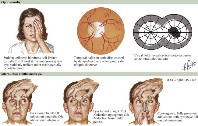
Blurred vision, likely a manifestation of acute optic neuritis, is a very common initial symptoms of MS. This may have great variability in degree from gross total unilateral blindness to a subtle change in visual acuity. Vision changes may also be related to a poorly expressed diplopia secondary to an INO, one of the most common neuro-ophthalmologic manifestations of MS (Fig. 46-2). In the first instance, the differential diagnosis includes any process affecting the retina or cornea, and in the second, the diplopia may be related to a pseudo-INO, mimicking MS. This is typical of myasthenia gravis. Sometimes a true INO secondary to a stroke may occur in older hypertensive patients.

At times a subtle demyelinating lesion may be clinically uncovered by exposure to heat. For example, during a summer heat wave, or even just taking a shower, a patient may note relatively short-lived symptomatology, sometimes visual blurring or just a change in gait; this is known as Uhthoff syndrome. The symptoms are usually fleeting. These routinely disappear when the patient returns to more normal ambient temperatures.

The prognostic significance of an acute episode of optic neuritis (ON) has been the subject of numerous investigations. Today MRI provides an important means to put the ON event into a long term perspective. A recent ON study, with a median 6 years’ follow-up, demonstrated that slightly less than 50% of these patients had converted to clinically definite MS (CDMS). Most importantly, both the presence and the number of MRI-defined spinal cord lesions, and to a lesser extent gadolinium-enhancing lesions, as well as the number of infratentorial lesions at baseline were significant independent predictors of an even higher disability manifested as CDMS.

When temporally associated with a myelopathy, this clinical presentation is referred to as Devic syndrome, or neuromyelitis optica (NMO). This is a humorally mediated autoimmune demyelinating disorder that is immunologically distinct from MS. It is characterized by an autoantibody that recognizes aquaporin 4 (AQP4), a water channel expressed on astrocyte podocytes. NMO may sometimes have a poorer prognosis than MS although on occasion it is more acutely responsive to immunosuppression or chemotherapy. This Devic syndrome does not typically benefit from first-line long-term MS disease modifiers (Chapter 47).

Inner Ear or Cerebellum

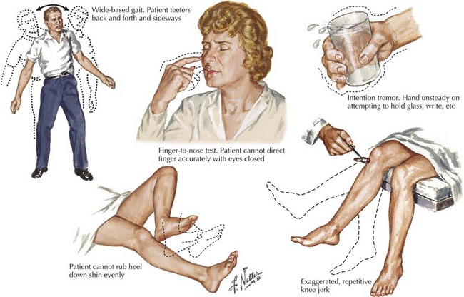
A vague feeling of unsteadiness, dizziness, and sometimes a whirling vertigo with nausea are frequent symptoms of MS. Precise definition of exactly what patients mean by “dizzy” is essential. Frequently, patients are in fact reporting a sense of dysequilibrium likely representing cerebellar dysfunction and less commonly reflecting vestibular pathways. Such patients typically demonstrate a broad-based gait, various signs of dysmetria, a tremor not seen in patients with a primary vestibular lesion, and often vertical nystagmus (Fig. 46-3).

The most common cause of true vertigo is either a primary vestibular neuronitis or the recurrent Ménière syndrome associated with hearing loss. These are both benign processes originating within the inner ear. However, one needs to always consider the possibility that the patient’s dizziness/vertigo has a primary CNS basis. One very important differential diagnostic clue to the latter is the finding of vertical nystagmus on the neurologic examination. This is not found when a peripheral mechanism for vertigo is present. The clinical demonstration of vertical nystagmus in a patient with vertigo requires an MRI to look for primary CNS disease.

Although spontaneous downbeat nystagmus is often the hallmark of another specific central nervous system lesion, namely an Arnold-Chiari malformation, this may also occur in MS patients reporting feelings of dizziness.

Myelopathies

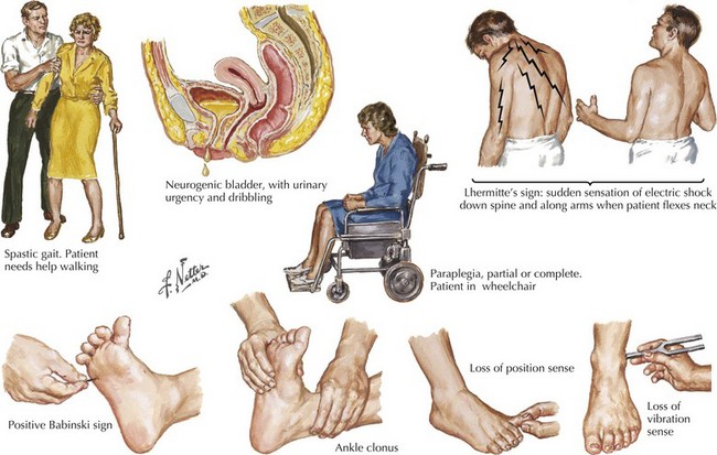
Sensations of tingling, tightness, pins and needles, and electric shocks are common MS symptoms. Relatively early in the disease course, some of these patients report an electric shock–like sensation that usually radiates down the back or arms, occurring spontaneously when the patient bends his or her neck. This is known as Lhermitte’s sign (Fig. 46-4). This most commonly occurs with MS patients but often the patient does not report this per se. Rather the skilled examiner is trained to ask about such as it is pathognomonic and indicative of spinal posterior column dysfunction. Lhermitte’s sign can be reported with almost any form of spinal cord disease, including spondylosis with significant cervical spinal stenosis, a space-occupying lesion, vitamin B12 (cobalamin) deficiency, or copper deficiency syndromes, especially in individuals who have had major gastrectomies or unusual diets, such as excess zinc that blocks copper absorption.

Other patients with MS frequently report a tight bandlike distribution of numbness. When circumferential or hemicircumferential, particularly in the midtrunk, the immediate concern is a possible transverse myelitis or primary spinal cord mass lesion. Sometimes patients describe a distribution suggestive of a polyneuropathy with bilateral symmetric numbness. On other occasions, numbness may occur acutely in the hand, compatible with mononeuropathy or stroke. Other myelopathies deserving consideration relate to tropical spastic paraparesis or AIDS.

Clinical Vignette

A 58-year-old school teacher with a history of depression and sciatica noted the presence of left-sided weakness that progressed gradually over a year. She found it difficult to maneuver her left leg into a pair of pants and occasionally experienced buckling of the right leg when dressing. Her left-handed grasp of a pen was increasingly compromised and her handwriting was changing. Advancing fatigue was making it increasingly difficult for her to get through her work day. She began to experience difficulty driving due to stiffness and shaking of the right leg, and, eventually, she could not exit her car without lifting her right leg with her hands. This was associated with a band-like constriction sensation around her thorax and intermittent urinary incontinence that she attributed to child-bearing and age.

Neurologic examination demonstrated diminished fine coordinated movements of her dominant left hand, bilateral hand clumsiness, and right greater than left leg weakness, with increased tone distally in the lower extremities, and bilateral Babinski signs. Her gait was wide-based and she had a right foot drop. Sensory examination was normal except for mild symmetrically decreased vibration in the distal lower extremities.

MRI scan of the brain with and without contrast showed scattered focal areas of T2 and FLAIR hyperintensity in the periventricular white matter, some oriented perpendicularly to the ventricles. There was a single lesion in the anterior corpus callosum and one area of faint enhancement in the left cerebral peduncle. Diffusion-weighted imaging was normal. Thoracic spine MRI showed an enhancing lesion in the cord at T9. Visual evoked responses showed some delay on the right, and CSF analysis revealed four oligoclonal bands and negative Lyme testing.

In retrospect, the patient recalled a transient visual disturbance in the right eye at age 19 years that was construed as migraine. When questioned, she also recalled an occasional Lhermitte sign occurring as a young woman.

A wider diagnostic net needs to be cast in older patients who are considered to possibly have had a cryptogenic stroke who do not have underlying risk factors for stroke. As so well illustrated in this vignette, a careful history is once again the essence of neurologic diagnosis, especially knowledge of what are the best questions to ask. Diffusion-weighted imaging and MRI perfusion studies can be very helpful in distinguishing subacute ischemic lesions from demyelinating lesions.

Clinical Vignette

A 30-year-old woman, previously healthy, began to experience episodes of déjà vu and derealization. In her work as a bank teller, colleagues noted that she had distinct periodic slowness in her work and an occasional latency in responsiveness when dealing with customers. A friend suggested that she was suffering from anxiety, and she consulted a therapist who witnessed one of her spells. Suspecting a seizure disorder, the patient was referred to a neurologist, who found her to be diffusely hyperreflexic with a left Babinski sign, mildly increased tone in the lower extremities, and a jaw jerk. EEG demonstrated left temporal lobe slowing with sharp waves. MRI scan of the brain revealed a focal enhancing mass lesion in the left temporal lobe with modest edema and three tiny, non-enhancing white matter lesions located subcortically as well as in the periventricular regions. A glioblastoma multiforme was suspected; however, stereotactic biopsy showed demyelination with axonal sparing, sparse perivascular lymphocytic infiltration, and numerous periodic acid Schiff-positive macrophages.

The patient received a short course of IV methylprednisolone, and phenytoin therapy was initiated. Her episodes resolved and repeat MRI in 3 months showed near complete resolution of the mass-like region.

Occasionally an acute focal cerebral lesion may be the presenting sign of MS leading the clinician to suspect that the patient has a glioblastoma multiforme, the most malignant of brain tumors. Fortunately, stereotactic biopsy occasionally demonstrates a rare monofocal form of MS, monofocal acute inflammatory demyelination (MAID) (Fig. 46-5). This can mimic a primary brain tumor or abscess. It is a rare and unique demyelinating disorder that may herald, or be superimposed on, more typical MS or may be seemingly unassociated with MS. MAID usually affects the cerebral hemispheres, and its clinical and radiologic features suggest a brain tumor. In contrast, an acute myelitis or optic neuritis is a more common example of a monofocal demyelinating process. Patients with the “cerebral form” of demyelinating disease have symptoms atypical for MS, including an acute hemiplegia, hemisensory complaints, and visual field deficits. Less common symptoms include headaches, seizures, aphasia, an alteration in level of consciousness, and cognitive or psychiatric manifestations.

Rarely, patients with a similar acute clinical presentation have multifocal CNS lesions, diagnosed as acute disseminated encephalomyelitis (ADEM) (Chapter 47). ADEM usually has widespread or multifocal MRI abnormalities.

Diagnostic Approach

Although there are no specific criteria for making the diagnosis of MS, MRI is the most helpful diagnostic adjunct. When typical MRI findings are found in the presence of a classic history and physical findings, there is little need to resort to other testing modalities in most clinical settings.

Magnetic Resonance Imaging

The diagnosis of MS has become very well defined with MRI; a positive study provides the physician with a solid diagnostic base leading in most instances to an accurate MS diagnosis in upwards of 95% of patients. New lesions typically have uniform gadolinium enhancement, whereas a ringlike enhancement is compatible with reactivation of prior lesions. Acute-phase plaques appear as rounded areas of high-signal intensity on FLAIR and T2 sequences. Gadolinium enhancement on T1 sequences is secondary to inflammatory damage to the blood–brain barrier. Earlier-onset, but nonreactivated, lesions do not enhance at all. T2-weighted MRI has the highest degree of sensitivity and is particularly useful for demonstrating disease dissemination; however, this lacks somewhat in specificity. A primary value of T1-weighted gadolinium-enhanced imaging is that it provides an increased specificity by differentiating enhancing from nonenhancing lesions.

Classic MRI findings are typified by multiple well-demarcated ovoid plaques whose long axis are situated perpendicularly along callososeptal interfaces and demonstrate a perivenular extension within the corpus callosum (Dawson fingers). Furthermore, plaques have a propensity for the periventricular and subcortical white matter, middle cerebellar peduncle, pons, or medulla (see Fig. 46-5). Within the spinal cord per se, white matter lesions can involve any part of the many afferent or efferent tracts, particularly the dorsal columns. At times the lesion can be so large that it mimics an intramedullary tumor as characterized by a large expansile hyperintense upper cervical cord plaque as seen in Figure 46-6. MRI has unequivocally replaced CSF analysis, the oldest methodology, as well as various forms of evoked neurophysiologic potentials, as the primary diagnostic methodology.

Increasingly powerful magnets and techniques, able to resolve variations from the norm, that sometimes mimic MS but may not be pathologic, sometimes complicate interpretation of abnormalities. “Unidentified bright objects (UBOs)” are the classic imitators seen in those who smoke, have hypertension, migraine headaches, or Lyme disease, or are present for indeterminate reasons. Similarly, Virchow-Robin spaces, dilated CSF-filled vascular areas, may give high T2 signal intensity. However, these can be distinguished from demyelinated plaques of MS because they are not present with proton density images.

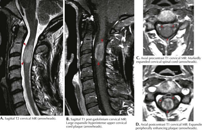
MRI of the spinal cord may be particularly useful in substantiating a diagnosis (see Fig. 46-6). In clinically suspicious cases with normal or equivocal brain MRI, spinal cord lesions can be compelling, even without symptoms referable to the spinal cord. Spinal cord lesions, however, are sometimes not as easily identified because of the constraints of motion artifact. The increased availability of 3-Tesla magnets in the coming years will add further image refinement, particularly with primary spinal forms of MS.

There is a most interesting subset of patients who eventually prove to have MS but are first evaluated with symptoms that are seemingly nonspecific and definitely not classic for MS. They undergo MRI scanning primarily to assuage patient fears of a structural abnormality, such as a tumor or perhaps MS. In a serendipitous fashion, the study then uncovers unexpected white-matter demyelinating lesions compatible with MS. For the physician, these MR findings do not seem to collate with the patient’s presenting symptoms. However, these observations need to be taken seriously. Such results are extending our definition of the initial clinical picture of multiple sclerosis and are making the neurologic clinician even more alert to the relative subtleties of this disorder early in its course.

Clinical Vignettes

A 27-year-old woman with a history of depression since early adolescence experienced painful spasm in her neck after her psychiatrist increased her dose of Zyprexa. An MRI of her cervical spine revealed an abnormality in the left dorsolateral spinal cord at C7. It was a nonenhancing lesion, bright on T2 and FLAIR image. Her torticollis resolved following reduction of her Zyprexa dosage. Brain MRI showed two lesions within the corpus callosum. Visual evoked response was significantly delayed on the right eye compatible with a lesion between the retina and the optic chiasm.

A 34-year-old landscaper was struck by the trunk of a tree that was felled by a coworker, failing to heed a warning to stand clear. He lost consciousness and was taken to a local emergency room. He had awakened and was noted to be fully alert and oriented. CT of the spine showed a T3 compression fracture. The day after his injury, he developed a severe headache. Magnetic resonance angiography of the head and neck revealed no dissection or any other vascular abnormality. Although brain MRI revealed no hemorrhages, there were 11 periventricular lesions seen on T2 and FLAIR images. Some of these were oriented perpendicularly to the ventricles, the largest of which, in the right frontal lobe, showed some cystic changes with low attenuation on T1 images and mild enhancement. CSF analysis demonstrated six oligoclonal bands and the presence of myelin basic protein. None of these findings could be related to his long history of ADH.

These two vignettes emphasize how MRI can serendipitously demonstrate findings of MS in the absence of active clinical symptoms, raising the question, “How early is too early to offer immunomodulatory therapy?” There is no consensus on the approach to such patients. How to identify patients with a potentially benign course versus those at risk for significant future disease with later disability remains unknown. MRI will be a keystone as this treatment algorithm is eventually defined. With the increasing evidence of the efficacy of the various ABC drugs in lessening long-term deficits, if these medications are begun when the diagnosis of MS is well defined, there is an increased tendency to begin active therapy once the diagnosis is ensured in patients such as these.

Cerebrospinal Fluid

Lumbar puncture for CSF is becoming superfluous when the diagnosis is established by history, examination, and unequivocal and very supportive MRI images. MRI is also very useful in excluding other diagnoses, particularly Lyme disease, sarcoidosis, lymphoma, or multi-infarcts secondary to a primary CNS vasculitis. However, when there is no definite MRI confirmation or collation of an MS diagnosis, a CSF examination can still be a very useful diagnostic procedure. When indicated, this procedure is typically performed by neurologists and neuroradiologists.

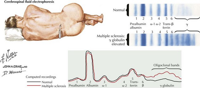
The typical CSF profile of a patient with MS is summarized in Figure 46-7. The white blood cell count is typically normal, with less than 6 mononuclear cells/mm3 being present; however, occasionally one may see MS patients with up to 50 lymphocytes per cubic millimeter. This usually does not exceed 25 cells/mm3. Lymphocyte counts greater than 100/mm3 must arouse suspicion of other inflammatory or even neoplastic processes. Given the expectation of subtle, if any, pleocytosis, it is paramount to strive for an atraumatic tap. The number of cells present may correlate with enhancing MRI lesions and thus with disease activity. However, there is no apparent correlation between cell count and T2 lesion burden. The CSF glucose level is normal, appropriate to the generally benign cell counts.

The CSF total protein is normal in 60% of MS cases; rarely is it greater than 70 mg/dL. Healthy individuals may have a CSF banding pattern similar to that found within a concomitant serum immunoglobulin electrophoresis. In contrast, if CSF bands outnumber serum bands, an abnormal CNS immune response is suggested. Whenever a few well-defined B-cell clones migrate to the CNS, these cells begin to produce distinct populations of immunoglobulins that separate into the bands seen on electrophoresis. CSF IgG is relatively increased when compared with albumin and with the IgG present in the serum of the same subject. This is related to changes in the permeability of the blood–brain barrier and IgG synthesis by plasma cells within the abnormal CNS. In healthy patients, there is no discrete CSF banding; instead, a diffuse single band represents a mixture of polyclonal immunoglobulins that do not segregate into distinct groups. An in-situ CNS hyperimmune response may be associated with oligoclonal band production. However, this is not specific for MS as it may occur with other CNS inflammatory or infectious diseases.

Myelin basic protein is a molecularly large component of myelin that can be detected in a degraded form by CSF immunoassay. Although its presence is thought to correlate with active demyelination, this is a relatively nonspecific finding. When detected, the level seems to be related to the amount and rate of myelin destruction. Thus, a relatively inactive instance of “smoldering” MS is not typically associated with the presence of myelin basic protein. In contrast, other pathologic processes that rapidly destroy large amounts of myelin (e.g., stroke) also produce high levels of CSF myelin basic protein.

Evoked Potentials

Neurophysiologic studies provide a means to objectively analyze the integrity of neuronal pathways in both the central and peripheral nervous systems. Prior to the availability of MRI, EPs were a useful modality to identify subclinical CNS disease. Testing is relatively easy to perform and requires minimal patient cooperation, particularly when testing the visual and, less commonly, auditory pathways. Today the primary value of EP testing occurs when a patient presents with a myelopathy and one is attempting to define the presence of multiple areas of clinical involvement in space and time to establish a diagnosis of MS. As some patients either forget or previously had a prior subclinical optic neuritis, visual evoked responses (VERs) may still be quite useful in this one instance. However, brainstem and somatosensory EPs are no longer of major diagnostic value when considering a diagnosis of MS.

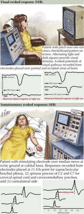
With VERs, a peripheral stimulus, typically a reversing high-contrast checkerboard pattern, provides a means to study the integrity of the visual system. The response latencies can yield objective data regarding the ability of the nervous system to transmit impulses efficiently from the optic nerve to the occipital cortex. If there is a unilateral absence or delay, one can conclude that there is slowing in conduction between the retina and the optic chiasm (Fig. 46-8). This is typical for a unilateral optic neuritis. It is particularly helpful as even if the patient totally regains his or her vision, the remyelination is not perfect; thus a marker is left for future reference. When bilateral changes are present with the potentials delayed, attenuated, or blocked because of a prior instance of CNS demyelination, one cannot precisely localize the lesion to the optic nerve as the lesion could be any place in the visual pathway.

If a number of years later a patient develops a myelopathy, VER testing can provide an opportunity to define the previous presence of some damage to these pathways. The combination of an abnormal VER response and a myelopathy is almost unique for a diagnosis of MS.

Multiple Sclerosis Diagnostic Criteria

Although MRI provides a superb diagnostic testing modality, it always needs to be accompanied by an appropriate clinical setting to make an unequivocal diagnosis of MS. Clinicians utilize a set of consensus criteria that continually evolve as new technological advances become available. Clinical history and examination were the mainstays of diagnosis in 1965 when Schumacher et al. issued the first widely used criteria for MS; these required that five of six of the conditions shown in Box 46-1 be met before making a clinically definite diagnosis of multiple sclerosis.

MS diagnostic criteria were revised in 1983 as evoked response testing and neuroimaging began to allow the identification of lesions that were not clinically evident and gave credence to laboratory data supportive of MS. The latter included CSF findings of specific increased number of oligoclonal bands in the CSF in comparison to the serum, elevated IgG levels, and increased IgG index. Most recently in 2001, the McDonald criteria became the standard almost universally applied in clinical research as well as to justify disease-modifying therapies specific for MS treatment.

These preserved the traditional requirement of multiple attacks of disease separated in time and space. Additionally, these new criteria provided for consideration of MRI and CSF findings when only one objective MRI lesion is found, only a single clinical attack has occurred, or when disease progression is insidious. The McDonald Diagnostic Criteria were revised in 2005 to incorporate the growing role of MR imaging for making a diagnosis of multiple sclerosis as well as to foster early treatment without sacrificing sensitivity and specificity (Box 46-2).

The ability to make a diagnosis of MS as early and accurately as possible is critical to patient care and to producing the most meaningful clinical research. Once the diagnosis of MS can be confirmed, the issue of when to initiate therapy becomes paramount.

Management And Therapy

Immunomodulatory Therapy

Medical management of MS was once limited to palliative care. Although symptom treatment remains integral to MS care, immunomodulatory therapies are changing treatment. Pharmacologic interventions modify the course of MS through the reduction of relapse rate and MRI lesion burden, both of which are likely to affect disease progression.

Five drugs are approved by the USFDA for treatment of RRMS: interferon beta-1b, intramuscular interferon beta-1a, glatiramer acetate, subcutaneous interferon beta-1a, and natalizumab infusion. Subcutaneous interferon beta-1b (8 million U every other day) became available early in the 1990s, followed by intramuscular interferon beta-1a (30 µg once weekly) later in that same decade. An alternative form of subcutaneous interferon beta-1a used in Europe became available in the United States in 2002 and is injected three times per week (22–44 µg), delivering a greater weekly dose of interferon beta-1a. Flulike symptoms, the most prominent adverse effect, occur in approximately 60% of those who use interferons, but these effects are mitigated over time and can be treated effectively with NSAIDs.

Glatiramer acetate (20 mg subcutaneously daily) became available in the United States in 1996 and is an unrelated compound of polypeptides that may have fewer potential adverse effects and perhaps more than one mechanism of action on the aberrant immune response. Some of its benefit may be realized later in the course of treatment than with the interferons.

In 2004, natalizumab was approved for use in the United States and the European Union. It was temporarily withdrawn by the manufacturer in February 2005 due to the occurrence of two cases of progressive multifocal leukoencephalopathy (PML) in MS patients treated with a combination of interferon beta 1-a and natalizumab. PML is a rare, progressive, and universally fatal central nervous system infection primarily occurring in immunodeficient individuals, particularly those with HIV-AIDS or very rarely those taking long-term immunosuppressive medications, particularly corticosteroids. PML is caused by the unmasked virulence of the fairly ubiquitous papillomavirus, JC virus.

In 2006, natalizumab returned to the market with more stringent guidelines for administration and solely as monotherapy for relapsing forms of MS. To date, however, additional cases of PML continue to accrue at a low incidence even with natalizumab monotherapy. Although the risk of evolving PML remains the greatest deterrent for many patients and clinicians, cases of liver toxicity and a questionable association with melanoma have since come to light. Thus far, there have been no controlled trials to adequately assess the efficacy of natalizumab for secondary progressive multiple sclerosis (SPMS).

Each of these agents reduces relapse rates and decreases the MRI burden of lesions within the central nervous system. Differences in trial design make effective drug-to-drug comparisons very difficult. To date, the reduction in relapse rate over 2 years hovers around 30–35% each for interferon therapies and for glatiramer acetate, whereas natalizumab was shown to reduce relapse rate over 1 year by as much as 68% when compared to placebo. Preexisting factors may influence the choice of therapy in treatment-naive patients. For example, a history of depression, suicidal ideation, or both, which may be adverse effects of the interferon drugs, may make glatiramer acetate the drug of choice in certain individuals. Some patients may be more compliant with one medication than another, depending upon individual tolerances for site reactions, flu-like symptoms, frequency of treatment, and impact upon lifestyle. Patients who cannot achieve compliance with intramuscular or subcutaneous injection may be able to tolerate monthly infusion with natalizumab.

Neutralizing Antibodies

Although the long-term safety of available first-line immunomodulating therapies is unclear, each type seems to be relatively well tolerated. However, the interferons, glatiramer acetate, and natalizumab are not naturally occurring human proteins and, as such, are prone to inducing antibodies in human recipients. The propensity for antibody production depends on the agent, dose, and route of administration. Substantial controversy exists as to the impact of these antibodies on long-term effectiveness of immunomodulatory medications.

Drug-induced antibodies simply bind to the molecules of the active agent and may have no significant function. Neutralizing antibodies are a subset of these binding antibodies that might interfere with drug activity by a steric hindrance of drug binding to its receptor or via remote effects on drug activity through binding at distant sites.

Some agents are more “foreign” than others and have a higher likelihood of inducing antibodies. Glycosylation renders substances more soluble in suspension; without it, aggregates that may be immunogenic are likely to form. This may account for the higher incidence of neutralizing antibodies with the use of interferon beta-1b (30–35%) compared with interferon beta-1a (15%). The latter is glycosylated, has no amino acid substitutions, and looks almost exactly like naturally occurring IFN-β. Higher doses of the drug, more frequent exposures, and delivery via subcutaneous injection may affect antibody induction. Natalizumab therapy is associated with antibody production that persists for greater than 6 months in 6% of patients. Natalizumab antibodies present beyond 6 months of monthly treatments appear to neutralize the drug and negate its benefits completely; therefore, antibody testing for natalizumab has become routine under a carefully administered monitoring program (TOUCH program) required for patients who are treated with natalizumab and their prescribing physicians. True hypersensitivity reactions to natalizumab have occurred in 2–9% of patients administered the drug, and these patients are more inclined than those without reactions to evolve persistent antibodies.

Treatment of More Severe Multiple Sclerosis

The protocol for treating recalcitrant MS is not formulaic. Only two agents are approved by the USFDA for rapidly progressive MS: mitoxantrone and interferon beta-1b. Mitoxantrone (12 mg/m2 IV given every 3 months to a cumulative maximum dose of 144 mg) is a chemotherapeutic agent used to treat other immune-mediated disorders. Usually considered a treatment of last resort after multiple treatment failures, its categorization is due to its potential toxicities, the most worrisome of which is cardiac toxicity.

Two trials offered some evidence that interferon beta-1b favorably impacts secondary progressive disease with respect to various outcome measures (progression of disability scores, relapse rate reduction, MRI activity), not all of which were significant in both studies. High-dose interferon beta-1a has also been investigated in secondary progressive disease; two studies demonstrated efficacy compared with placebo. Although heterogeneity of study populations and outcome measures make these studies difficult to compare, they offer some rationale for choosing a new therapy in patients who have advanced from relapsing–remitting disease to a more aggressive RRMS or secondary progressive disease, regardless of previous treatments. Multi-interferon therapy and interferon plus glatiramer acetate are being investigated. Despite multiple investigations focused on other treatment strategies, their efficacies have remained largely anecdotal.

Azathioprine, Cyclosporine, and Cyclophosphamide

The equivocal benefits of azathioprine (2–3 mg/kg daily) in MS are realized in decreasing relapse rates and MRI lesion burden. Cyclosporine inhibits the production of lymphokines and seems to foster the expansion of certain populations of suppressor T cells. One cyclosporine and azathioprine comparison showed no differences between the two agents with respect to relapse frequency or expanded disability status scale, but therapy complications were more than twice as likely in the cyclosporine group as in the azathioprine group. Other studies of cyclosporine have been marred by high attrition in the treatment groups due to toxicities (renal failure, hypertension) that likely occur before any benefit is achieved. These observations have taken cyclosporine out of the usual arsenal of immunosuppressive therapy for the treatment of MS in many centers.

Cyclophosphamide is another immunosuppressive agent widely used to treat various neoplastic and autoimmune disorders. One study suggested some response based on expanded disability status scale scores to induction with booster infusions in patients younger than age 40 years. Toxicities include hemorrhagic cystitis, leukopenia, myocarditis, pulmonary interstitial fibrosis, malignancy, interstitial pulmonary fibrosis, and infertility. Mitoxantrone tends to be better tolerated, with fewer adverse effects, and has better evidence of efficacy in secondary progressive MS.

Methotrexate. Low-dose weekly oral methotrexate is not approved for use in MS but is used as an adjunct therapy in off-label use based on a limited number of studies in patients with MS and its efficacy in rheumatoid arthritis, another autoimmune disease. Methotrexate is a competitive inhibitor of dihydrofolate reductase and interferes with the production of reduced cofactors required for the synthesis of DNA and RNA. It seems to have both immunosuppressive and antiinflammatory effects and immunoregulatory action. Toxicities include pulmonary and liver fibrosis, cirrhosis, and bone marrow suppression.

Clinical Vignette

A 31-year-old ICU nurse had no explanation for her complaints of fatigue. She had long worked 36 hours per week in three 12-hour shifts with additional shifts in overtime, more often than not. She was not married, had no children, and had always made exercise a regular part of her routine. She was in the habit of running with her dog in the mornings and, while she used to find this invigorating and an energy booster for the coming day, she began to feel that she was really pushing herself to keep up with her usual activities on most days. By late morning, she would feel exhaustion setting in, even though she made it more of a point than ever to get plenty of sleep in her down time and began taking vitamin supplements.

She found herself cutting back on social activities, and her friends and colleagues saw the change and wondered whether she was depressed. Overall she had no history of mood disorder or any other perceived stressors, and had been happy with her life until she began to have significant physical and mental fatigue. From the neurologic perspective, she had no overt limb weakness, subjective sensory impairment, imbalance, visual difficulties, or pain. Similarly, there was nothing to suggest any underlying systemic disorder. However, the fatigue was taking over her life and she decided to seek further medical evaluation.

Her primary care physician found her thyroid function and B12 levels normal; an ANA was positive with a speckled pattern. Consideration of an occult depression diagnosis led to a trial of an SSRI. However, she felt no better. Soon thereafter, she began to experience a vibrating sensation in her feet and a feeling of tightness in her upper thorax, as if she was wearing a bra that was too tight. She became convinced that either she was becoming a hypochondriac or her doctor was “missing something.” When she voiced this concern, her physician made referrals to a rheumatologist, an endocrinologist, and a neurologist. The endocrinologist felt that she had mild vitamin D deficiency and recommended some additional supplementation to her multivitamin, but was otherwise unconcerned. The rheumatologist felt there was no evidence for systemic lupus erythematosus or any connective tissue disorder, apart from possible fibromyalgia.

Her neurologist elicited a history of urinary frequency with relatively little urine volume with each void. On examination, she found diminished vibratory perception in the patient’s lower extremities up to the knees and diffuse hyperreflexia. A postvoid residual of 180 mL was measured by ultrasound. MRI revealed multifocal areas of chronic demyelination in the brain, cervical, and thoracic spinal cord.

Comment: This clinical scenario is not all that uncommon and emphasizes the importance of a careful neurologic evaluation in previously well-functioning young to middle-aged persons who begin to develop limitations in their abilities to perform their daily activities. One must be careful to not invoke the common diagnosis of chronic fatigue syndrome in such individuals until careful and often ongoing neurologic evaluations precisely exclude a diagnosis of MS.

Related MS Management Problems

Fatigue

Fatigue is often one of the most debilitating symptoms, particularly early on in MS. The primary “lassitude” of MS is an overwhelming sense of physical and mental exhaustion that has no identifiable cause but significantly interferes with normal activity. The majority of patients with MS rank it as their most disabling symptom affecting daily living. This eclipses bowel and bladder dysfunction, weakness, and balance problems. Although fatigue can be secondary to deconditioning, the advanced symptoms of MS that limit physical activity are not a prerequisite for fatigue. It has been associated with every stage of the disease, regardless of clinical subtype, and is often present in an apparently well-compensated individual who otherwise has little objective disability.

Persistent but ineffective neuronal firing of short-circuited neural pathways that do not affect movement in weak limbs, transmit cohesive visual impulses, or recruit the reflexes necessary to maintain balance may be a pathophysiologic correlate of primary MS fatigue. There is no specific link evident to provide objective measures of disease activity such as commonly used MS functional scales or MRI findings.

Additionally, some evidence exists of an association among MS-related fatigue, depression, and cognitive dysfunction. That this is true in patients without significant deficits and in patients who have not received a diagnosis suggests that the mental fatigue is not simply an epiphenomenon of a reactive depression. The fatigue of MS is likely a direct consequence of demyelination, inflammation, and axonal injury along shared neuronal networks that affect attention, depression, and cognitive dysfunction. Hypometabolism in the frontal cortex and basal ganglia resulting from this type of damage has been implicated in primary MS fatigue.

Sufficient rest is critical to managing fatigue in MS. Additionally some pharmacologic therapy is a useful adjunct to behavioral and lifestyle strategies. Despite the incomplete understanding of the pathophysiology of MS-related fatigue, clearly valuable pharmacotherapies mitigate a profoundly low energy level through modification of altered neurochemistry that results from damage to the CNS.

Wakefulness involves various arousal systems with complex circuits that project between the reticular activating system, limbic system, and frontal cortex. From the brainstem, monoamine-mediated pathways (dopamine, norepinephrine, serotonin, acetylcholine) ascend through the reticular activating system. Several useful agents operate along this pathway. The stimulant medications (methylphenidate, amphetamine salts, dextroamphetamine [Dexedrine], pemoline) affect this mesocorticolimbic system by enhancing global CNS activation but may produce undesirable side effects such as dependence through stimulation of central “reward mechanisms” (governed by the nucleus accumbens in the striatum), insomnia, appetite suppression, and peripheral autonomic effects, including tachycardia, dysrhythmia, and hypertension. Amantadine enhances dopamine release and seems to have some benefit in improving central attention and processing speed. Modafinil, used to treat narcolepsy, is also helpful in treating MS fatigue by increasing activity in the frontal cortex via activation of histaminergic neurons arising from the hypothalamus.

Bladder Dysfunction

Bladder symptoms, one of the most vexing problems commonly experienced, significantly influence quality of life in 50–80% of patients with MS. The degree of impairment usually parallels the degree of other neurologic deficits. Bladder hygiene is compromised by increased spasticity, loss of strength, and diminished mobility. Although bladder dysfunction rarely leads to life-threatening infection or secondary renal failure, the resultant recurrent or chronic urinary tract infections can potentiate other MS symptoms and are a common trigger for significant exacerbations.

On rare instances, an acute pyelonephritis may develop with a high fever; this may lead to a temporary exacerbation of the underlying neurologic deficits. In a way, this mimics the Uhthoff phenomena. This emphasizes the need to look for evidence of an underlying infection in anyone with MS who has an acute exacerbation; appropriate antibiotics will not only clear the infection but also lead to the resolution of the MS exacerbation.

Control of urination depends on the complex coordination of reflexive and volitional activities that rely on the integrity of complex pathways that traverse the subcortical white matter, spinal cord tracts, and reflex spinal cord centers. Lateral and posterior cervical spinal tracts are among the most common sites of demyelination in MS and are critical for all aspects of coordinating voluntary voiding.

Bladder dysfunction in MS is managed both pharmacologically and with adaptations in toileting. The most common pharmacologic agents diminish bladder contraction. Typically this is an antispasmodic anticholinergic, oxybutynin chloride. Dosage is usually initiated at 5 mg, the maximum being 20 mg daily. These are generally more effective than attempts to improve bladder emptying with cholinergic agents. Alpha-adrenergic agents increase bladder outlet resistance and sometimes help to treat urinary incontinence. Emptying the hyporeflexic or areflexic bladder is best accomplished by clean, intermittent self-catheterization.

Additional Resources

Comi G, Filippi M, Barkhof L, et al. Effect of early interferon treatment on conversion to definite multiple sclerosis: a randomized trial. Lancet. 2001;357:1576-1582.

European Study Group on Interferon beta-1b in Secondary Progressive MS. Placebo-controlled multicentre randomized trial of interferon beta-1b in treatment of secondary progressive multiple sclerosis. Lancet. 1998;352:1491-1497.

Fletcher SG, Castro-Borrero W, Remington G, et al. Sexual dysfunction in patients with multiple sclerosis: a multidisciplinary approach to evaluation and management. Nat Clin Pract Urol. 2009;6:96-107. An important review of particular importance to any MS patient with spinal cord dysfunction

Goodin DS, Cohen BA, O’Connor P, et al. Assessment: the use of natalizumab (Tysabri) for the treatment of multiple sclerosis (an evidence-based review). Neurology. 2008;71:766-773.

Guo S, Bozkaya D, Ward A, et al. Treating relapsing multiple sclerosis with subcutaneous versus intramuscular interferon-beta-1a: modelling the clinical and economic implications. Pharmacoeconomics. 2009;27(1):39-53. This study provides a useful cost analysis comparing effectiveness of two of the most common preparations of interferon beta-a

Hartung HP. New cases of progressive multifocal leukoencephalopathy after treatment with natalizumab. Lancet Neurol. 2009;8:28-31.

Kappos L, Pohlman CH, Freedman MS, et al. Treatment with interferon beta 1-b delays conversion to clinically definite and McDonald MS in patients with clinically isolated syndromes. Neurology. 2006;67(7):1242-1249.

Kumar N, McEvoy K, Ahlskog JE. Myelopathy due to copper deficiency following gastrointestinal surgery. Arch Neurol. 2003;60:1782-1785.

Lassmann H. Axonal and neuronal pathology in multiple sclerosis: What have we learnt from animal models. Exp Neurol. 2009 Oct 17. [Epub ahead of print]. A review of the increasingly recognized importance of axonal damage in the early pathophysiology of MS

Lovblad KO, Anzalone N, Dorfler A, et al. MR imaging in multiple sclerosis: review and recommendations for current practice. AJNR Am J Neuroradiol. 2009 Dec 17. Epub ahead of print

Pohlman CH, O’Connor PW, et al. A randomized, placebo-controlled trial of natalizumab for relapsing remitting multiple sclerosis. N Engl J Med. 2006;354(9):899-910.

Pohlman CH, Reingold SC, et al. Diagnostic criteria for multiple sclerosis: 2005 revisions to the “McDonald criteria. Ann Neurol. 2005;58(6):840-846.

Swanton JK, Fernando KT, Dalton CM, et al. Early MRI in optic neuritis: The risk for disability. Neurology. 2009;72:542-550. This is an important prospective analysis of the risk of development of clinically active MS among a cohort of newly diagnosed optic neuritis patients