Moraxella catarrhalis

Published on 22/03/2015 by admin

Filed under Pediatrics

Last modified 22/04/2025

Print this page

rate 1 star rate 2 star rate 3 star rate 4 star rate 5 star
Your rating: none, Average: 0 (0 votes)

This article have been viewed 1236 times

Chapter 188 Moraxella catarrhalis

Moraxella catarrhalis, an unencapsulated gram-negative diplococcus, is a human-specific pathogen that colonizes the respiratory tract beginning in infancy. Colonization and infection with M. catarrhalis are increasing in countries in which pneumococcal conjugate vaccines are used widely. The most important clinical manifestation of M. catarrhalis infection in children is otitis media.

Epidemiology

The ecological niche of M. catarrhalis is the human respiratory tract. The bacterium has not been recovered from animals or environmental sources. Age is the most important determinant of the prevalence of upper respiratory tract colonization. Common throughout infancy, nasopharyngeal colonization is a dynamic process with active turnover due to acquisition and clearance of strains of M. catarrhalis. Some geographic variation in rates of colonization is observed. On the basis of monthly or bimonthly cultures, colonization during the 1st year of life may range from 33% to 100%. Several factors likely account for this variability among studies, including living conditions, daycare attendance, hygiene, environmental factors (e.g., household smoking), and genetics of the population. The prevalence of colonization steadily decreases with age. Understanding nasopharyngeal colonization patterns is important, because the pathogenesis of otitis media involves migration of the bacterium from the nasopharynx to the middle ear via the eustachian tube.

The widespread use of pneumococcal polysaccharide vaccines in some countries has resulted in alteration of patterns of nasopharyngeal colonization in the population. A relative increase in colonization by non-vaccine pneumococcal serotypes, nontypable H. influenzae, and M. catarrhalis has occurred. These changes in colonization patterns may account for the increased rates of otitis media due to nontypable H. influenzae and M. catarrhalis. Similar shifts in etiology are being observed in children with sinusitis as well.

Clinical Manifestations
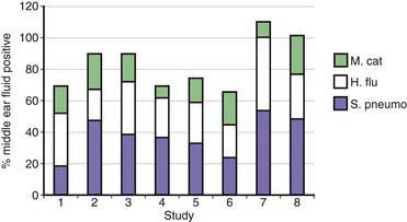
M. catarrhalis causes predominantly mucosal infections in children. The mechanism of infection is migration of the infecting strains from the nasopharynx to the middle ear in the case of otitis media or to the sinuses in the case of sinusitis. The inciting event for both otitis media and sinusitis is often a preceding viral infection.

Acute Otitis Media

Approximately 80% of children have one or more episodes of otitis media by age 3 yr. Otitis media is the most common reason for which children receive antibiotics. On the basis of culture of middle ear fluid obtained by tympanocentesis, the predominant causes of acute otitis media are S. pneumoniae, H. influenzae, and M. catarrhalis (Fig. 188-1). Overall, M. catarrhalis causes 15-20% of cases of otitis media. The distribution of the causative agents of otitis media is changing as a result of widespread administration of pneumococcal conjugate vaccines, with a relative increase in H. influenzae and M. catarrhalis.

Acute otitis media due to M. catarrhalis is clinically milder than otitis media due to H. influenzae or S. pneumoniae, with less fever and lower prevalence of a red, bulging tympanic membrane. However, substantial overlap in symptoms is seen so that it is not possible to predict etiology in an individual child on the basis of clinical features. Tympanocentesis is required to make an etiologic diagnosis but is not performed routinely, and thus, treatment of otitis media is generally empirical.

Diagnosis

The clinical diagnosis of otitis media is made by demonstration of fluid in the middle ear by pneumatic otoscopy. A tympanocentesis is required to establish an etiologic diagnosis, but this procedure is not performed routinely. Thus, the choice of antibiotic for otitis media is empirical and generally based on guidelines. Management of bacterial sinusitis is also empirical, because determining the etiology of sinusitis requires a sinus puncture, also a procedure that is not performed routinely.

The key to making a microbiologic diagnosis is distinguishing M. catarrhalis from commensal Neisseria that are part of the normal upper respiratory tract flora. Indeed, the difficulty in distinguishing colonies of M. catarrhalis from Neisseria species explains in part why M. catarrhalis has been overlooked in the past as a respiratory tract pathogen. M. catarrhalis produces round, opaque colonies that can be slid across the agar surface without disruption, the “hockey puck sign.” In addition, after 48 hr, M. catarrhalis colonies tend to be larger than Neisseria and take on a pink color. A variety of biochemical tests distinguish M. catarrhalis from Neisseria species, and commercially available kits based on these tests are available.

Sensitive tests that employ PCR to detect respiratory tract bacterial pathogens in human respiratory tract secretions are in development. The application of such assays when they become available is likely to contribute new information about the epidemiology and disease patterns of M. catarrhalis.

Bibliography

Deshpande LM, Sader HS, Fritsche TR, Jones RN. Contemporary prevalence of BRO beta-lactamases in Moraxella catarrhalis: report from the SENTRY antimicrobial surveillance program (North America, 1997 to 2004). J Clin Microbiol. 2006;44:3775-3777.

Hall-Stoodley L, Hu FZ, Gieseke A, et al. Direct detection of bacterial biofilms on the middle-ear mucosa of children with chronic otitis media. JAMA. 2006;296:202-211.

Heiniger N, Spaniol V, Troller R, et al. A reservoir of Moraxella catarrhalis in human pharyngeal lymphoid tissue. J Infect Dis. 2007;196:1080-1087.

Murphy TF, Paramswaran GI. Moraxella catarrhalis, a human respiratory tract pathogen. Clin Infect Dis. 2009;49:124-131.

Revai K, McCormick DP, Patel J, et al. Effect of pneumococcal conjugate vaccine on nasopharyngeal bacterial colonization during acute otitis media. Pediatrics. 2006;117:1823-1829.

Ruckdeschel EA, Kirkham C, Lesse AJ, et al. Mining the Moraxella catarrhalis genome: identification of potential vaccine antigens expressed during human infection. Infect Immun. 2008;76:1599-1607.

Slevogt H, Seybold J, Tiwari KN, et al. Moraxella catarrhalis is internalized in respiratory epithelial cells by a trigger-like mechanism and initiates a TLR2- and partly NOD1-dependent inflammatory immune response. Cell Microbiol. 2007;9:694-707.

Tan TT, Riesbeck K. Current progress of adhesins as vaccine candidates for Moraxella catarrhalis. Expert Rev Vaccines. 2007;6:949-956.

Verhaegh SJ, Streefland A, Dewnarain JK, et al. Age-related genotypic and phenotypic differences in Moraxella catarrhalis isolates from children and adults presenting with respiratory disease in 2001–2002. Microbiology. 2008;154:1178-1184.

Wirth T, Morelli G, Kusecek B, et al. The rise and spread of a new pathogen: seroresistant Moraxella catarrhalis. Genome Res. 2007;17:1647-1656.