Molecular Basis for Treating Cutaneous Melanoma

Published on 09/04/2015 by admin

Filed under Hematology, Oncology and Palliative Medicine

Last modified 09/04/2015

Print this page

rate 1 star rate 2 star rate 3 star rate 4 star rate 5 star
Your rating: none, Average: 0 (0 votes)

This article have been viewed 2881 times

Figure 42-1 Development of melanoma Although melanomas can develop from melanocytes at the epidermal-dermal junction directly or in a preexisting nevus, an orderly progression from melanocyte to neoplasm (benign nevus) to dysplasia to noninvasive (in situ) cancer to overt invasive malignancy can also be seen in clinical material. This figure shows the histopathologic features of a compound nevus, dysplastic nevus, melanoma in situ, and invasive melanoma.
Melanoma typically appears on the sun-exposed areas of the skin, and the amount of sunlight exposure over life is a risk factor. Melanomas arising in sun-damaged skin differ in their molecular pathway expression compared to melanomas from non–sun-damaged skin. 17
Cutaneous melanomas appear in four major distinct clinical and histologic variants: nodular, superficial spreading, lentigo maligna, and acral lentiginous melanomas. Although molecular pathways may differ between these variants, the risk for metastasis remains linked to the depth of invasion into the skin. Superficial spreading melanoma (SSM) is the most common form of the disease and is typically present for years on the skin. It has the hallmark of the classic ABCDE characteristics popularly known as asymmetry, irregular borders, variegated color, larger diameter (greater than 6 mm), and evolving over time. 18 SSM has radial growth and nodular or vertical growth. Nodular melanoma has a vertical growth phase without any evidence of horizontal growth. Nodular melanomas usually appear rapidly and invade the dermis quickly. Acral lentiginous melanoma is seen on the palms, soles, and nail beds and is the most frequent melanoma in dark-skinned people. Lentigo maligna melanoma is the invasive form of lentigo maligna (LM), which is a melanoma in situ arising in sun-damaged skin. LMs are typically noted on the skin for many years.

Melanoma Therapy and Molecular Targets

Historically, melanoma has been one of the most unresponsive cancers to traditional chemotherapy approaches. Dacarbazine, an agent that methylates the 7-position of guanine on DNA, crosslinks DNA strands, leading to inhibition of DNA, RNA, and protein synthesis. Treatment with this agent demonstrates an 8% objective response rate in metastatic melanoma. 19 Studies in the 1980s suggested that multiagent chemotherapy could enhance the activity of dacarbazine in patients with metastatic melanoma, but a subsequent randomized study failed to show improvement in overall survival with multiagent chemotherapy compared to dacarbazine alone. 19 The first modern-era immune therapies for melanoma, interferon alpha and interleukin-2, introduced in the 1980s, showed tantalizing results and are still considered part of the therapeutic armamentarium despite recent advances. 20,21
As molecular pathways related to melanoma carcinogenesis and progression have been discovered, novel targets and agents specific for these targets have also been identified. In addition, the molecular pathways mediating immune response to melanoma may also provide novel strategies for treatment. The most relevant new approaches for melanoma in 2012 target BRAF, c-Kit, and CTLA4. 2224 We review these approaches first and explore other pathways that will likely be relevant for the future discovery of additional therapeutic agents.

Mapk-Braf and Intersection of Other Pathways
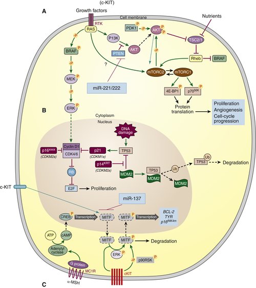
The mitogen-activated protein kinase pathway (MAPK) is an oncogenic pathway that mediates growth and progression (Figure 42-2 ). Growth factor receptor tyrosine kinases (RTK) associated with RAS initiate this pathway, among others, leading to activation of BRAF, MEK, and ERK in the cytoplasm. Phosphorylated ERK migrates to the nucleus and causes cell proliferation. The three members of the RAS proto-oncogene family include HRAS, NRAS, and KRAS, all with GTPase activity. NRAS is the most frequently mutated gene of this family and is seen in 25% to 35% of melanomas. 25,26 The most frequent mutation in NRAS is arginine (R) substituted for a glutamine (Q) at position 61 (Q61R). Downstream from NRAS is RAF. There are three human isoforms of RAF (ARAF, BRAF, and CRAF), which have different cellular locations and different activation sequences. BRAF has constitutive phosphorylation of the N terminus, which is different than for ARAF and CRAF and thus can be activated directly by RAS. 27 The most common RAF mutation is in BRAF, which has been identified in as many as 66% of melanomas. 28 BRAF mutation is also seen in a high percentage of benign nevi, suggesting that mutation of BRAF is seen early in the transformation of melanocytes at the basal layer of the dermal-epidermal junction into tumor (benign and malignant). 28,29 The highest frequency mutation of BRAF occurs at the 600 position with a substitution of a glutamic acid (E) for valine (V) (BRAF[V600E]). This mutation constitutively maintains activation of a downstream event on MEK and ERK, a process referred to as addiction to an oncogenic pathway.
Cancers addicted to an oncogenic pathway are susceptible to inhibition by blockade of that pathway. RAS activation through a number of RTKs, including epidermal (EGFR), platelet-derived (PDGFR), and vascular endothelial (VEGFR) growth factor receptors, mediate not only tumor growth but also tumor-associated angiogenesis. Sorafenib (Bay 43-9006) is a biaryl urea developed as a RAF inhibitor and was the first of its kind to enter clinical trials. It had demonstrable activity against RAF targets, including the mutant BRAF (BRAF[V600E], BRAF[V600K], and BRAF[V600M]). 30 In a Phase II melanoma study, 19% of 37 patients had stable disease, but there was no relationship of response to BRAF(V600E) status. 31 Subsequent studies with combined chemotherapy and sorafenib failed to demonstrate significant activity. 32,33
Using a structure-guided approach to drug discovery, PLX4720 (vemurafenib) was developed as a selective BRAF(V600E) inhibitor. 34,35 PLX4720 binds to the ATP binding site on active BRAF but not BRAF in its inactive conformation. Inhibitors that bind in and around the region occupied by the adenine ring of ATP are referred to as type I inhibitors, whereas sorafenib is a type II inhibitor that instead occupies a hydrophobic site directly adjacent to the ATP pocket. 35 The first Phase I study of PLX4720 or vemurafenib demonstrated significant activity in metastatic melanoma patients, and subsequent randomized studies confirmed the activity, which led to FDA approval for its use in the treatment of metastatic melanoma. 22,36
In the Phase III study, 675 eligible patients with metastatic melanoma stage IIIC, M1a, M1b, and M1c were randomized in a 1:1 ratio to receive oral vemurafenib (960 mg twice daily) or dacarbazine (1000 mg/m2 IV every 3 weeks). At the time of analysis, the hazard ratio for death was 0.37 (95% confidence interval 0.26 to 0.55; P < .0001) and for progression-free survival 0.26 (95% confidence interval 0.20 to 0.33; P < .0001) favoring the vemurafenib treatment. Two complete responses and 104 partial responses were reported in the 219 evaluable subjects who received vemurafenib. Although the toxicity profile for vemurafenib was mild, with cutaneous toxicity being most significant, dose interruption and modification were required in 38% of subjects. The paradoxical activation of CRAF leads to the development of squamous cell cancers and keratoacanthomas. 37 Subsequent studies have demonstrated a median duration of response to BRAF blockade of approximately 7 months. 38 Other BRAF-targeted molecules are under development. 39
Although targeting mutant BRAF has had significant impact on the therapeutic paradigm, development of resistance continues to be a barrier for prolonged responses in the majority of patients. Development of resistance to BRAF-targeted agents is complex and multifaceted but appears to be driven by reactivation of the MAPK pathway. One mechanism that has been identified is a 61-kDa splice variant of BRAF(V600E) that lacks the RAS-binding domain at exon 4-8 (p61BRAF[V600E]). 40 p61BRAF(V600E) shows enhanced dimerization and is resistant to known RAF inhibitors. Increased expression of serine/threonine kinases on BRAF, CRAF, or COT1 may also be involved. 4042 The activating mutations Q61K/R on N-RAS and C212S on MEK1 have also been implicated in BRAF inhibitor resistance by phosphorylation of ERK. 43,44 The development of multiple pathways of resistance to BRAF inhibition has also been demonstrated from cloning melanoma BRAF mutated resistant cell lines, suggesting that overcoming clinical resistance will be a formidable barrier going forward. 45
image
Figure 42-2 (A, B) RTK-RAS activation of MAPK and AKT pathways. Arrows represent activation pathways and T (⊥) represents inhibition. In the AKT pathway, PTEN (phosphatase and tensin homologue) dephosphorylates PIP2 (phosphatidylinositol bisphosphate) and acts in an inhibitory manner. miRNA 221/222 can modulate this pathway by inhibiting PTEN. Activation of AKT is associated with a conformational change and translocates close to the cell membrane where it is phosphorylated by PDK1 (3-phosphoinositide-dependent kinase 1) and then can inhibit TSC2 (tuberous sclerosis protein 2) and activate mTORC2 (mammalian target of rapamycin C2). The MAPK pathway is triggered through RAS activation, ultimately leading to phosphorylated ERK translocating to the nucleus, leading in turn to cellular proliferation through cyclin D1 complex. DNA damage activates TP53 (tumor protein 53), which in turn activates CDKN1a (cyclin-dependent kinase inhibitor 1a), suppresses cyclin D1-CDK4/6 complex, and inhibits transcription via RB (retinoblastoma). MDM2 (mouse double minute-2) inhibits TP53 by binding it and making available for degradation through the ubiquitin pathway. p14(ARF) sequesters MDM2 and thus inhibits TP53 degradation. (C) α-MSH (melanocyte-stimulating hormone) binds to MC1R (melanocortin-1 receptor), stimulating a cascade that transcribes MITF (microphthalmia transcription factor) gene. miR-137 can modulate the MITF pathway by exerting its effects on transcription of MITF. (Adapted with permission from Ibrahim N, Haluska FG. Molecular pathogenesis of cutaneous melanocytic neoplasms. Annu Rev Pathol Mech Dis. 2009;4:551-579.)
Another mechanism of resistance is signaling through an alternative pathway, PI3K/AKT. 46 PI3K/AKT can be activated through persistence of tyrosine kinase activity of platelet-derived growth factor receptor or insulin growth factor 1 receptor, RAS signaling, RAS independent signaling, or loss of PTEN activity, as well as other mechanisms. PI3K/AKT and MAPK pathways are co-activated in many melanomas. 47 Resistance to mutant BRAF(V600) inhibitors develops through co-option of the PI3K/AKT pathway and RAF isoform switching. 48 These mechanisms provide the foundation for combining BRAF blockade with inhibition downstream in the MAPK pathway at MEK and inhibition of the PI3K/AKT pathway at AKT or mTOR. Another interconnection between the MAPK pathway and mTOR pathway is through phosphor-ERK (pERK) activation of RSK and TORC1, and inhibition of AMPK. AMP-activated protein kinase is activated during metabolic stress and inhibits protein and fatty acid synthesis.
The AMPK-TORC pathway can be explored using metformin, a drug that activates AMPK and is used to treat type 2 diabetes. Metformin inhibits the growth of NRAS-mutant melanoma but not BRAF-mutant melanoma cells in culture. 49 BRAF-mutant melanoma drives activation of RSK and TORC1, thus overcoming the effects of metformin on AMPK and inhibition of TORC1. Metformin, through activation of AMPK, increases degradation of dual-specificity protein phosphatase (DUSP6), thereby increasing pERK, then VEGF-A, and stimulating BRAF-mutant melanoma growth in a mouse xenograft model. Blockade of VEGF-A with an antibody (bevacizumab) and combination with metformin inhibits tumor growth. These laboratory observations may have significant implications in the choice of agents to control type 2 diabetes in melanoma patients on BRAF targeted therapy and have further implications for multiagent targeted therapy.
Notch protein, so named because it controls notch formation in the wings of fruit flies, has also been implicated in melanoma formation. 50 Notch signaling has been shown to suppress both MAPK and PI3K/AKT pathways indirectly. 51 Inhibition of one of these two pathways reverses the effects of Notch on melanoma progression. 52 A better understanding of the interaction between these pathways may also provide novel strategies for therapy.
Translocation of pERK to the nucleus leads to cell proliferation through CyclinD1. P16 or cyclin-dependent kinase inhibitor 2a (CDKN2A), located at chromosome 9p21, has been associated with familial melanoma syndromes. 53 Alterations in p16 have been seen in benign and dysplastic nevi, suggesting a role in early melanoma development and progression. 54 Thus, independent activation of proliferative pathways in melanoma may also occur significantly downstream of MAPK and theoretically lead to autonomous growth resistant to BRAF targeted therapy. Activating mutations in cyclin-dependent kinase 4 (CDK4) have also been shown to predispose to melanoma and represent yet another checkpoint that may be involved with developing resistance to BRAF targeted therapy. 55
Melanocortin receptors are G-protein–coupled receptors and comprise a family of five different receptors. When melanocyte-stimulating hormone (α-MSH) binds to MC1R, cyclic adenosine monophosphate is generated and leads to transcription of the microphthalmia transcription factor (MITF), leading to pigment production. Low levels of MITF in melanoma cells signal proliferation and survival. 56 MC1R polymorphisms have been associated with increased risk for melanoma and nonmelanoma skin cancer. Phospho-ERK phosphorylates MITF, and pMITF is degraded through the ubiquitin pathway. Constitutive activation of pERK through mutated BRAF subsequently leads to low expression of MITF and enhances melanoma cell proliferation and survival, most likely through interaction with CDKN2a and BCL-2. 57 This pathway as a role in development of BRAF targeted therapy is not yet well elucidated, but has at least theoretical implications. MITF is also controlled by c-Kit, which is discussed later.
Heat shock protein 90 (HSP90) cooperates with its co-chaperone Cdc37 in supporting a variety of protein kinases involved with cancer progression, including BRAF. HSP90 inhibitors have had little single-agent activity in clinical trials. HSP activity across the spectrum of resistant pathways involved with BRAF inhibitors suggests a role for the combination of these agents. In cell culture experiments as well as in mouse xenograft models, HSP90 inhibitor XL888 was demonstrated to reverse vemurafenib resistance and was associated with degradation of other secondary receptor tyrosine kinases and their downstream constituents. 58

c-Kit

c-Kit mutations are rare in melanoma and are seen most commonly in tumors derived from mucosal and acral areas or in melanomas associated with sun-damaged skin. c-Kit is involved with the melanocyte pigmentary pathway through activation of MITF. As noted previously, imatinib is a protein tyrosine kinase inhibitor that blocks protein phosphorylation by the fusion protein BCR-abl in chronic myelogenous leukemia and blocks downstream c-KIT signaling in gastrointestinal stromal tumors. Thus, there is a rationale for using imatinib in the subset of patients whose melanoma overexpresses or has mutations in c-Kit. In a Phase II study of imatinib in 43 patients with metastatic melanoma and aberrations in c-KIT, 23% had objective partial responses, and the median progression-free survival was 3.5 months. 23 Of interest is the association of benefit with mutations in exon 11 or exon 13 of c-Kit, with 9 of the 10 responding patients having these mutations. Sunitinib, another agent that inhibits mutant c-Kit, has shown similar results, with response seen in melanomas expressing mutant c-Kit and much less so in tumors with overexpression of c-Kit. 59 Resistance to c-Kit targeted therapy has been associated with the development of NRAS mutations. 59

Epigenetic Pathways: MicroRNA

MicroRNAs (miRNAs) are single-stranded noncoding nucleotide sequences about 22 bases long. MiRNAs are generated through a double-stranded precursor that undergoes cleavage by Drosha (RNase type III endonuclease) to an approximately 70-nucleotide unit. 60 Drosha and the associated dsRNA binding protein DGCR8 complex are transported to the nucleus and cleaved by Dicer (RNase type III endonuclease) to the approximately 22-nucleotide double-stranded miRNA. One strand of the miRNA binds to the 3′-untranslated region of messenger (m) RNA, thereby blocking translation and causing cleavage and destruction of the mRNA. 61,62
MiRNAs are involved with regulation of a number of melanoma-related growth and proliferation pathways and are thus potential targets for therapy. Although the gain or loss of many miRNAs is shared across tumor types, a number of miRNAs are more specifically associated with melanoma. 63,64 MiR-137, located on chromosome region 1p22, and miR-182 (7q31-34) are putative negative regulators of MITF. 65,66 MITF, in turn, regulates the transcription of miR-221 and miR-222. 67 MiR-221/222 are located on chromosome X and function to inhibit expression of c-Kit receptor. Suppression of miR-221/222 with anti-mRNAs in melanoma cell lines resulted in decreasing melanoma cell proliferation and migration. 67
MiRNAs can be obtained from archival tissues such as blood and serum, allowing them to be considered as biomarkers and potential targets for new therapeutics. 68,69 For example, high expression of miR-15b was found to correlate with poor survival in melanoma patients. 70 The soybean isoflavone genistein inhibits human uveal melanoma cell growth in culture and in a murine model and is associated with alteration of miR-27a, again suggesting that targeting of miRs may have therapeutic importance. 71 MiR-193b is downregulated in human melanoma cell lines. 72 When miR-193b was transfected back into these cells, proliferation was suppressed because of miR-193b directly downregulating cyclin D1.

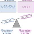
Paratumoral Pathways

The tumor microenvironment is composed of vascular, stromal, and immune cells that have an intimate spatial relationship with the cancer cell and influence cancer development and progression. As we gain more understanding of these interactions, these paratumoral pathways become targets for therapy.
It is well recognized that fibroblasts are associated with many tumors and that these cells can play a contributing role in cancer progression. Tumor-infiltrating fibroblasts (TIF) are typically spindle-like and express α-smooth muscle actin, resembling myofibroblasts. These cells can express transforming growth factor-β (TGFβ), VEGF, and provide extracellular matrix, all of which support tumor growth. In a 3D coculture model, fibroblasts migrate to and infiltrate human melanoma spheroids within 7 days through melanoma-derived motility factors. 73 These fibroblasts are active and produce extracellular matrix. Following targeted therapy, for example, with an EGFR inhibitor for lung cancer, a flare phenomenon has been observed with rapid progression of cancer. Similar observations have also been noted in vemurafenib-treated melanoma patients. TIFs have been proposed as one component contributing to this phenomenon. The role of these TIFs in producing a niche for melanoma stem survival is unexplored to date, but may provide a mechanism for establishment of a resistant phenotype.
Neoangiogenesis is another paratumoral event required for the establishment and progression of cancer. The pioneering work in this area was led by the late Judah Folkman. Following anecdotal reports of alpha interferon causing regression of benign hemangiomas in infants, a study of 20 subjects demonstrated the significant activity of interferon as an anti-angiogenesis therapy. 74 The role of anti-angiogenesis therapy is now well established in a number of tumor types, although its role in melanoma is not yet definitively demonstrated. The BEAM trial in melanoma evaluated the role of adding bevacizumab (blocking anti-VEGF antibody) to carboplatin and paclitaxel in a randomized Phase II study. 75 Outcomes for the 214 treated patients demonstrate a trend toward improvement in response and median progression-free survival (PFS) (16.4% response and 4.2 months PFS for chemotherapy vs. 25.5% response and 5.6 months for chemotherapy + bevacizumab). A trial of sorafenib with either temsirolimus (an mTOR inhibitor) or tipifarnib (a farnesyl transferase inhibitor required for RAS activation) also failed to show a difference between the arms or improved response or PFS compared to historical controls. 76 Although angiogenesis is an important component of melanoma and VEGF has been implicated, studies to date have failed to show that modification of this pathway results in significant clinical benefit.
Regulation of the immune system is another area of great expectation in melanoma. IFN alfa-2b and peg-IFN alfa-2b are the only agents thus far that have shown clinical benefit in the surgical adjuvant setting of melanoma patients with high risk for recurrence. 20,77 High-dose IL-2 has an established role in patients with good performance status who have metastatic melanoma, with a small percentage of patients (approximately 5%) reaching complete and durable remissions. 21,78 The understanding of immune regulatory checkpoints has provided a deeper understanding of immune response in cancer and therapeutic targets. Cytotoxic T lymphocyte antigen 4 (CTLA-4), a member of the immunoglobulin superfamily, has been identified as a co-stimulatory molecule involved with negative control. 79,80 Anti-CTLA-4 blocking antibodies have been developed that led to clinical trials assessing their role in the treatment of melanoma. 81,82
Preliminary data suggested benefit of CTLA-4 blockade, and subsequent randomized trials confirmed the activity of ipilimumab in the treatment of patients with metastatic melanoma. 24,83,84 The first study randomized patients between ipilimumab at 3 mg/kg combined with gp100 vaccine, ipilimumab alone, or gp100 vaccine alone. 24 , Six hundred seventy-six previously treated patients with metastatic melanoma who expressed HLA-A∗201 were assigned to ipilimumab and gp100 peptide vaccine, ipilimumab alone or gp100 vaccine alone in a 3:1:1 ratio. 24 Ipilimumab was given at 3 mg/kg every 3 weeks for up to four cycles. The ipilimumab arms had a median survival of 10 to 10.1 months compared to 6.4 months for the arm given gp100 vaccine alone. There was a 2.1% study-related mortality rate. Three patients (0.6%) on the ipilimumab arms entered a complete remission (CR). The second randomized study evaluated ipilimumab 10 mg/kg every 3 weeks with dacarbazine versus dacarbazine alone with placebo. 84 Ipilimumab or placebo was continued every 12 weeks for stable or responding patients. Five hundred two previously untreated patients were randomized in a 1:1 ratio. Median overall survival showed an improvement in the ipilimumab-dacarbazine arm from 9.1 to 11.2 months. Survival was improved in the combination arm at 1 year (47.3% vs. 36.3%), 2 years (28.5% vs. 17.9%), and 3 years (20.8% vs. 12.2%). Four patients (0.4%) had a CR in the combination arm compared to two in the dacarbazine-placebo arm, and no treatment-related deaths were reported.
It has been generally believed that immune therapy is not effective for the treatment of brain metastasis. Never- theless, 72 patients with melanoma brain metastases were treated with ipilimumab at 10 mg/kg every 3 weeks for 24 weeks with responding patients eligible for maintenance in a Phase II study. 85 In asymptomatic patients, 12 of 51 patients obtained control of their brain metastases and 8 had objective responses. In this cohort of patients, the median overall survival was 7 months with 26% of patients alive at 24 months. This study provides the first evidence of immune therapy benefit for melanoma brain metastasis.
Engagement by antigen of the T-cell receptor (TCR)-CD3 complex provides the first signal to the T cell but is not sufficient for activation. The necessary second signal is provided by CD28 binding to its ligand B7 (1 and 2 or CD80 and CD86, respectively) on antigen-presenting cells such as dendritic cells. 86 The formation of TCR-CD3 complex and CD28 in combination with co-receptors CD4 or CD8 forms the immunological synapse. 87 Once engaged, the co-receptors that are associated with the protein tyrosine kinase LCK, phosphorylates the immune receptor tyrosine-based activation motifs (ITAMS) in the TCRζ chain initiating a cascade. This cascade includes ζ-chain–associated protein kinase (ZAP), SYK (spleen tyrosine kinase), LAT (linker for activation of T cells), and SLP76 (SRC homology 2 (CH2)-domain-containing leukocyte protein of 76 Da), which phosphorylates phospholipase Cγ1, activating the TEC family of kinases. This increases intracellular Ca2+, which leads to activation of transcription factors. CTLA-4 is expressed on activated or memory T cells and has a 50- to 100-fold higher binding avidity to B7 than CD28, most likely because CTLA-4 dimer binds two bivalent B7 molecules, whereas CD28 binds to a single B7 domain. 88,89 Engagement of CTLA-4 leads to termination of the T-cell response. A number of different models have been proposed to explain the mechanism by which CTLA-4 controls regulation. Both CD28 and CTLA-4 bind PI3K at a motif that closely resembles a similar motif on growth factor receptors. CTLA-4 does not have binding domains for LCK or growth-factor receptor 2 (GRB2) whereas CD28 does. This may be one explanation of CTLA-4 activation.
It has long been the practice for oncologists to combine multiple approaches to treat patients with cancer. Thus, the use of cytoreductive surgery followed by radiation and drug therapy has become commonplace. Combining drug therapy using non–cross-resistant multiagent combinations with different mechanisms of action has also become a standard approach for many cancers. This concept is supported by mathematical modeling and observations of clinical benefit. 9093 There is good rationale for combining targeted and immune therapies in patients with melanoma, based on the recent success. The magnitude and speed of response seen with targeted therapy may affect immune pathways, including reducing tumor-associated immune suppression and enhancing antigen availability to dendritic cells for processing and activation. 94
The expanding list of agents available for regulating immune and signaling pathways provides tools for exploring this complex cross talk. A mutant BRAF melanoma cell line treated with a BRAF target blocker upregulates melanoma differentiation antigens and can be better recognized and killed by melanoma-specific cytotoxic T cells. 95 Although MEK inhibition also upregulates melanoma differentiation antigens in this model, blocking MEK also inhibits T-cell killing whereas BRAF blockade does not. An increase in tumor-infiltrating T cells (TILs) has been observed in tumors from patients treated with vemurafenib. 96 We have observed a similar TIL infiltration in a mouse model of BRAF mutant melanoma treated with BRAF blockade (Turk MJ, personal communication). Targeted agents can have both a negative and positive interaction with immune regulation. 97 In addition to the BRAF(V600E) targeted agents, other melanoma-relevant agents that can be considered for combination with immune therapy include imatinib, which blocks c-kit and can also decrease indoleamine dioxygenase (IDO), a negative regulator of immune function, as well as promote dendritic and natural killer cell communication; bevacizumab, a VEGF-neutralizing antibody, which increases DC maturation; bortezomib, a proteasome inhibitor, which can sensitize tumor to immune-mediated killing; PI3K-AKT inhibitors that also enhance tumor cell sensitivity to immune-mediated killing; and HSP90 inhibitors, which can decrease immune suppression and increase NK cell targets on the tumor. 97 As discussed earlier, understanding the molecular pathways of immune cells in the context of cancer will also lead to new strategies of targeted therapy directed at enhancing immune activation and inhibiting regulation. 98,99

Conclusions

Recent updates of ongoing clinical trials provided with BRAF inhibitors and ipilimumab demonstrate the promise of these new therapeutic tools as well as their limitations. High response rates to vemurafenib and dabrafenib are met with the development of early clinical resistance. 100,101 Although both progression-free survival and overall survival are now extended in patients with metastatic melanoma with BRAF(V600E) mutation, the median overall survival in the BRIM3 trial of 13.2 months is short and demonstrates the need for strategies to convert the initial response to long-term survivorship in a higher percentage of patients. 101,102 The challenge may be met with the identification of resistant pathways and the development of additional agents targeted to these pathways, such as the combination of MEK and BRAF inhibitors. The pipeline in the pharmaceutical industry is full of agents that target MEK, ERK, and others. MiRNA inhibitors are also being developed. Our knowledge of how these signals intersect with the molecular pathway and how miRNAs are regulated should provide additional therapeutic strategies that will need to be tested in the clinical setting.
Another approach is to use pathway inhibitors early in the setting of high-risk stage II and III disease. The questions of which pathway, which agent, and for what duration of therapy can only be answered through the experience gained in clinical trials. The early development of heterogeneity of molecular profiles within cancer 103 may ultimately allow for aggressive and resistant clones to threaten patients’ lives and prove to be a barrier that will require combination of therapeutic strategies.
The low objective response rate noted with anti-CTLA4 antibody is counterbalanced by the observation of long complete remissions seen in a small percentage of patients no longer needing additional therapy. This improvement of the tail of the curve is an important concept and should not be underestimated as we seek to enhance the long-term goal of cure of metastatic melanoma. 104 Our understanding of the complex inflammatory and regulatory networks of immune pathways has led to the development of new therapeutic tools that are being and will need to be explored through clinical trials with correlative biological studies. Our recognition of the molecular pathways activated in immune cells from patients with cancer 98,99 will provide new strategies to regulate those pathways.
There is a long history of recognizing that the inflammatory responses in cancers treated with chemotherapy contribute to benefit. We have now identified inflammatory responses in tumors treated with the new targeted therapies, providing an additional rationale for combining these therapeutic strategies.
We have begun to unravel the molecular mysteries of melanoma carcinogenesis, progression, and metastasis, as well as the molecular pathways regulating the host response to this disease. Through judicious and thoughtful manipulation of these pathways, melanoma will likely be tamed to a chronic illness that does not cause death or can be cured outright.
References

1. Rudkin C.T. , Hungerford D.A. , Nowell P.C. DNA contents of chromosome Ph1 and chromosome 21 in human chronic granulocytic leukemia . Science . 1964 ; 144 : 1229 1231 .

2. Rowley J.D. Letter: A new consistent chromosomal abnormality in chronic myelogenous leukaemia identified by quinacrine fluorescence and Giemsa staining . Nature . 1973 ; 243 : 290 293 .

3. Rosenberg N. , Baltimore D. A quantitative assay for transformation of bone marrow cells by Abelson murine leukemia virus . J Exp Med . 1976 ; 143 : 1453 1463 .

4. Witte O.N. , Rosenberg N. , Paskind M. et al. Identification of an Abelson murine leukemia virus-encoded protein present in transformed fibroblast and lymphoid cells . Proc Natl Acad Sci U S A . 1978 ; 75 : 2488 2492 .

5. de Klein A. , van Kessel A.G. , Grosveld G. et al. A cellular oncogene is translocated to the Philadelphia chromosome in chronic myelocytic leukaemia . Nature . 1982 ; 300 : 765 767 .

6. Daley G.Q. , McLaughlin J. , Witte O.N. et al. The CML-specific P210 bcr/abl protein, unlike v-abl, does not transform NIH/3T3 fibroblasts . Science . 1987 ; 237 : 532 535 .

7. Druker B.J. , Tamura S. , Buchdunger E. et al. Effects of a selective inhibitor of the Abl tyrosine kinase on the growth of Bcr-Abl positive cells . Nat Med . 1996 ; 2 : 561 566 .

8. Jemal A. , Saraiya M. , Patel P. et al. Recent trends in cutaneous melanoma incidence and death rates in the United States, 1992-2006 . J Am Acad Dermatol . 2011 ; 65 S17-S25, e1-3 .

9. Merrill R.M. Risk-adjusted melanoma skin cancer incidence rates in Whites (United States) . Melanoma Res . 2011 ; 21 : 535 540 .

10. Soong S.J. , Ding S. , Coit D. et al. Predicting survival outcome of localized melanoma: an electronic prediction tool based on the AJCC Melanoma Database . Ann Surg Oncol . 2010 ; 17 :

Buy Membership for Hematology, Oncology and Palliative Medicine Category to continue reading. Learn more here