Metastatic Melanoma

Published on 19/07/2015 by admin

Filed under Radiology

Last modified 22/04/2025

Print this page

rate 1 star rate 2 star rate 3 star rate 4 star rate 5 star
Your rating: none, Average: 0 (0 votes)

This article have been viewed 2281 times

 Most common sites of metastases: Skin, lymph nodes (75%), lung (70%), liver (58%), CNS (54%), GI tract (40%)

image Most common sites in abdomen: Liver and small bowel
• Melanoma metastases are often T1 hyperintense on MR due to melanin content
• Lymph nodes

image 1st nodes to be involved are usually regional lymph nodes with contiguous spread through lymphatic chains
image Metastatic nodes may enlarge or change in morphology (↑ enhancement, loss of fatty hilum)
• Liver

image Most common site of visceral organ involvement
image May be hypervascular on arterial phase and usually hypodense on venous phase
• Gastrointestinal tract

image Small bowel most common site (75% of cases)
image May present as lead point of small bowel intussusception
image Soft tumor that does not usually cause obstruction
• Gallbladder

image Melanoma is most common metastasis to gallbladder
• Kidney

image Can involve kidney, bladder, or collecting systems
image Unique predisposition for perirenal space
image Consider melanoma with isolated mass in perirenal space

TOP DIFFERENTIAL DIAGNOSES

• Leukemia and lymphoma
• Metastases from other primary tumors
• Primary GI malignancies
• Kaposi sarcoma

CLINICAL ISSUES

• Risk of metastasis correlates with depth of primary tumor into dermis
image
(Left) Axial CECT in a patient with known metastatic melanoma demonstrates mass-like wall thickening and aneurysmal dilatation image of 2 segments of colon, in keeping with bowel metastases.

image
(Right) Coronal volume-rendered CECT in the same patient demonstrates 3 different metastases image
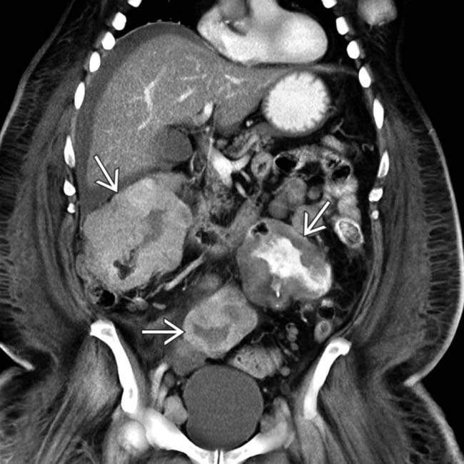
, with several others scattered throughout the small and large bowel (not shown). Lymphoma and GI stromal tumors can also cause similar aneurysmal dilatation.
image
(Left) Axial CECT in a patient with melanoma demonstrates a nodular hypervascular metastasis image 
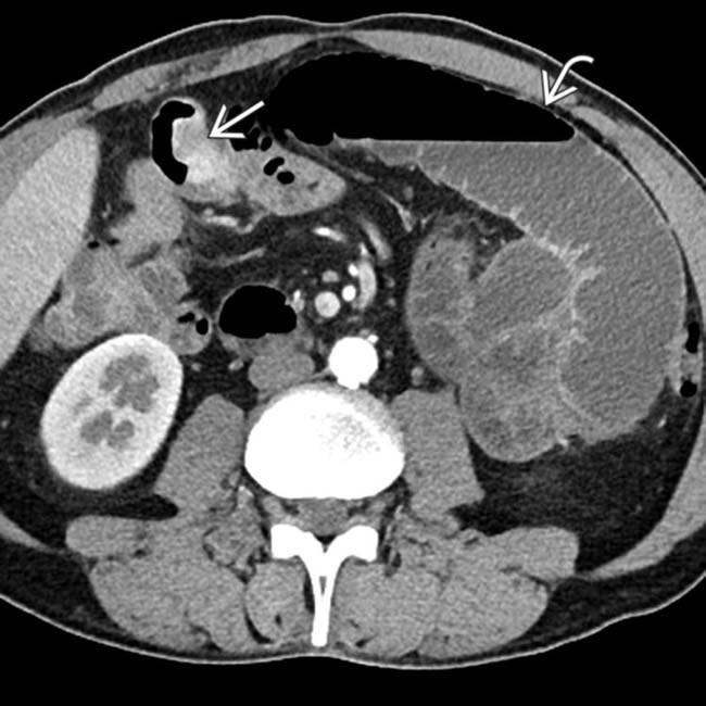
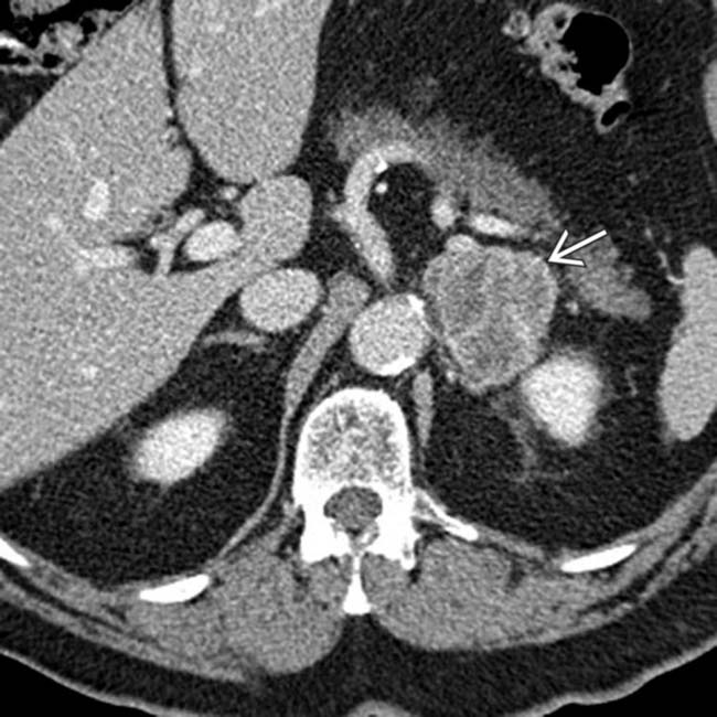
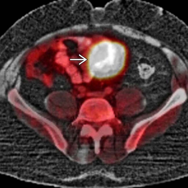
in the small bowel causing proximal bowel obstruction image.

image
(Right) Axial CECT in the same patient demonstrates multiple other sites of nodular enhancing soft tissue image 
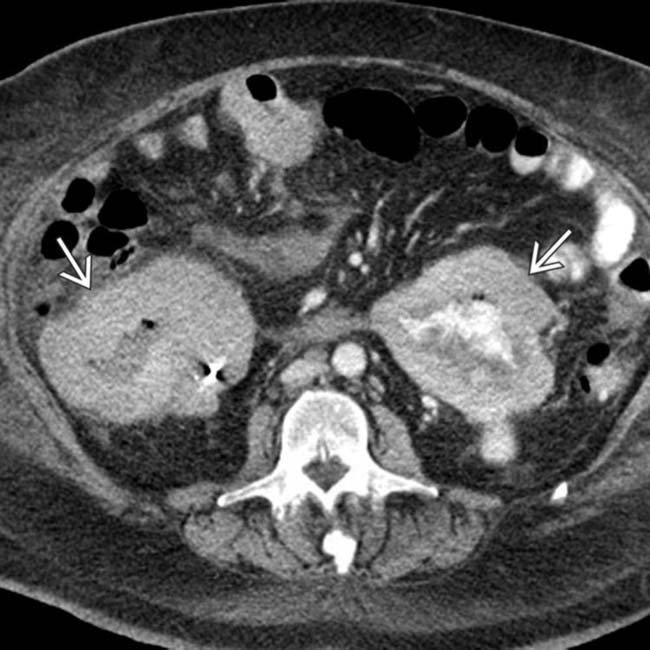
in the small bowel. Multifocal metastases to the bowel are not uncommon in melanoma.

TERMINOLOGY

Definitions

• Spectrum of metastatic lesions originating from known or occult malignant melanoma

IMAGING

General Features

• Best diagnostic clue

image Multiple “bull’s-eye” lesions of variable size in GI tract of patient with history of melanoma
• Location

image Unique predisposition for metastatic disease to unusual locations (gallbladder, small bowel, spleen, subcutaneous soft tissues, etc.) 

– Can metastasize to nearly any location and may have an isolated metastasis in atypical location
– Distant metastases depend on site of primary tumor

image Lower extremity melanomas often spread to pelvis
image Ocular melanomas frequently spread to liver
image Most common sites of metastases: Skin, lymph nodes (75%), lung (70%), liver (58%), CNS (54%), GI tract (40%)
image Most common sites in abdomen: Liver and small bowel
• Morphology

image Typically multiple, in any site of body
image Well-circumscribed, spherical or oval
image Nodule, plaque, polypoid mass
image “Bull’s-eye” or “target” lesion (central ulceration)

Imaging Recommendations

• Best imaging tool

image PET/CT (from vertex through feet) with diagnostic CECT for total body screening

– Sensitivity and specificity are ↑ by simultaneous interpretation of diagnostic quality CT
– Melanoma may not be FDG avid or may be misinterpreted as normal bowel or kidney on PET
• Protocol advice

image Multiphase CECT: Melanoma may be hypervascular and metastases may not be visualized on monophasic CECT

Radiographic Findings

• Radiography

image Rarely, calcification may be seen in hepatic lesions

CT Findings

• Lymph nodes

image First nodes to be involved are usually regional lymph nodes with contiguous spread through lymphatic chains

– Careful assessment necessary of lymph node stations adjacent to primary tumor
– Abdominal nodal involvement in 30% of cases
image Metastatic nodes may enlarge or change in morphology (↑ enhancement, loss of fatty hilum, irregular margins)

– Involved lymph nodes may enlarge and bleed
– Rarely necrotic with peripheral enhancement
• Liver

image Most common site of visceral organ involvement

– Particularly common with ocular melanoma, and can occur years after initial diagnosis
image Single or multiple lesions of variable size ± calcification

– May be hypervascular on arterial phase
– Most (86%) lesions hypodense on portal venous phase
image Rim enhancement in lesions with central necrosis
image Subcapsular hematoma may result from spontaneous bleeding of hepatic metastases
• Gastrointestinal tract

image Can involve any portion of GI tract, but small bowel is most common site (75% of cases)
image Can present with a single or multiple lesions, often with central necrosis or ulceration
image May lead to aneurysmal dilation of bowel lumen
image Predilection for antimesenteric border of small bowel
image May present as lead point of small bowel intussusception
image Esophagus

– Very rare lesion
– Bulky soft-tissue mass with esophageal dilatation upstream
– More common in distal 1/2
image Stomach

– Sessile or pedunculated intraluminal soft-tissue masses ± “target” appearance
image Duodenum

– Multiple sessile intramural or intraluminal masses, ± “target” appearance, ± fold thickening
image Small bowel

– Single or multiple intramural or intraluminal soft-tissue masses; ± extraluminal component
– Central necrosis (“target” appearance)
image Colon

– Single or multiple “target” lesions ± wall thickening, may simulate diverticulitis

image Due to eccentric wall thickening and infiltration of pericolonic fat
image Soft tumor that does not usually cause obstruction
• Mesenteric involvement

image Mesentery and omentum are often both involved
image Single or multiple nodules of variable size with peritoneal/omental stranding, nodularity, and ascites
image Melanoma should be considered with large solitary peritoneal soft-tissue mass without known primary
• Gallbladder

image Melanoma is most common metastasis to gallbladder

– Seen in 15% of patients (according to 1 series)
image Small, flat, subepithelial nodule gradually progresses to discrete polypoid mass
• Pancreas

image Peripancreatic nodes may simulate pancreatic mass
image Single or multiple enhancing nodules on CECT
image May be hypervascular on arterial phase and mimic neuroendocrine tumor
• Spleen

image Melanoma is common source of metastasis to spleen
image Seen in 30% of cases according to 1 report
image Multiple ill-defined low-attenuation lesions or well-defined “cystic” lesions (usually with solid component) 

– Single or multiple
– Homogeneous hypodense lesion may mimic cyst, but usually shows enhancement and solid component on CECT and US
• Kidney

image 3rd most common metastasis to kidney (after lung/breast cancer)

– Seen in 35% of cases
image Unique predisposition for perirenal space 

– Large isolated mass in perirenal space should prompt consideration of melanoma
– Can involve kidney, bladder, or collecting systems
image Single or multiple lesions of variable size which can appear solid or “cystic” (never simple cystic appearance)
• Adrenal

image Seen in 11% of cases
image Usually unilateral (but can be bilateral)
image Indistinguishable from other metastases on imaging

– Usually hypodense, lobulated, and heterogeneous
image May destroy underlying adrenal gland and result in adrenal insufficiency (Addison syndrome)

MR Findings

• T1WI

image Hyperintense to liver (due to melanin content)

– Almost unique to melanoma metastases
– Amelanotic melanoma may be T1 hypointense
– T1WI hyperintensity may occur in other metastases with fat or hemorrhage
• T2WI

image Hyperintense to liver: Similar to other metastases
• T1WI C+

image May be hyperenhancing on arterial phase imaging
image Most are maximally conspicuous on portal venous phase

Ultrasonographic Findings

• Liver metastases

image Single or multiple hypoechoic lesions with variable size
image Displacement of adjacent vessels (no invasion)
image Subcapsular hematoma may be seen
• Gallbladder metastases

image Single or multiple masses with broad-based attachment to wall; classically thought to be hyperechoic, but hypoechoic more common
image Increased vascularity on color Doppler

Fluoroscopic Findings

• Upper GI

image Single or multiple well-defined filling defects in wall of stomach or gut

– Bull’s-eye or “target” lesion (central umbilication): Mass with barium collection in center
– “Spoke-wheel” pattern: Superficial fissures radiating towards central ulceration
image Amorphus contrast collection (cavitated mass)
image May present as submucosal nodules, polypoid mass, or pedunculated intraluminal nodule
image Usually results in luminal narrowing, but may cause aneurysmal dilation, indistinguishable from lymphoma
image Esophagus

– Indistinguishable from primary carcinoma by esophagram
– Bulky, smooth, polypoid filling defect
– Extrinsic compression on esophagus by enlarged mediastinal lymph nodes
image Stomach

– Infrequent site of involvement
– Usually associated with small-bowel lesions
– Early lesions are submucosal nodules
image Duodenum

– Most common metastatic tumor of duodenum
– Multiple flat filling defect ± central umbilication (“bull’s-eye” lesion)
– Duodenal fold thickening
image Small bowel

– Most common site in GI tract
– Single or multiple intraluminal filling defect(s)
– Predilection for antimesenteric border
– Central ulceration (“bull’s – eye” lesion)

Nuclear Medicine Findings

• PET/CT

image ↑ FDG avidity in most sites of melanoma
image Higher accuracy than CT or PET alone

– Sensitivity of 98% and specificity of 94%
– PET often shows metastases to atypical sites that may be difficult to detect on CT
– Used for surveillance of recurrence
image Detection of lymph node metastases

– Limited role in stage I or II disease; unlikely to detect microscopic nodal disease in normal-sized lymph nodes below size threshold of PET/CT
– May play a role in detecting lymph node metastases in stage III or IV (particularly if surgery is considered for limited metastatic disease)
• Lymphoscintigraphy

image Injection around primary tumor site in skin with Tc-99m-labeled nanocolloid
image Allows mapping of pattern of nodal drainage sites that can be sampled at time of initial surgical resection

– Negative sentinel node biopsy implies stage I or II disease
– Positive sentinel node biopsy suggests stage III disease

DIFFERENTIAL DIAGNOSIS

Leukemia and Lymphoma

• May have widespread lymphadenopathy
• Bowel involvement may be identical to melanoma: “Bull’s-eye” lesions and aneurysmal dilation of lumen

Metastases From Other Primary Tumors

• Melanoma may mimic other hypervascular metastases: Hepatocellular carcinoma, neuroendocrine tumor, carcinoid, renal cell carcinoma
• History of primary cancer

Intestinal Intramural Benign Tumor

• Usually single and large

Primary GI Malignancies (Small Bowel, Gastric, or Colon Adenocarcinoma)

• May appear as annular constricting lesion, but aneurysmal dilatation uncommon
• May obstruct bowel (uncommon with melanoma)

Kaposi Sarcoma

• Disseminated lymphadenopathy with multifocal hepatic or intestinal nodules (often with “target” lesions)
• More common in stomach and duodenum
• May appear identical to melanoma, but Kaposi most common in AIDS patients with mucocutaneous lesions

PATHOLOGY

General Features

• Etiology

image Originates from melanocytes (derived from neural crest cells during embryonic life)
image Most melanomas arise from benign skin nevi 

– Can also arise in eyes or other mucosal surfaces
– Primary tumor not identified in ∼ 3% of cases which initially present with metastatic disease
image Exposure to ultraviolet radiation is most important carcinogenic factor
image Mechanism of metastases

– Direct spread
– Lymphatic spread (1st to regional nodes)
– Hematogenous spread (throughout body)
• Genetics

image Multiple gene mutations, including  CDKN2A/p16, which accounts for ∼ 40% of familial melanomas

Staging, Grading, & Classification

• Clinical staging

image I: Localized melanoma, no metastasis (78%)
image II: Regional lymph node involvement (18%)
image III: Distant metastasis (4%)
• Breslow staging

image Thin: Depth < 0.75 mm (46% lead to metastases)
image Intermediate: Depth 0.76-3.99 mm (50% lead to metastases)
image Thick: Depth > 4 mm (84% lead to metastases)
• AJCC TNM classification

image T1: ≤ 1 mm deep; T2: 1-2 mm deep; T3: 2-4 mm deep; T4: > 4 mm deep
image N0: No node; N1: 1 node involved; N2: 2-3 nodes involved; N3: ≥ 4 nodes involved
image M0: No metastases; M1: Distant metastasis
image Stage I: T1 or T2a with N0M0; Stage II: T2b-T4b with N0M0; Stage III: N1-N3; Stage IV: Distant metastatic disease (M1)

Gross Pathologic & Surgical Features

• Well-circumscribed, round or oval
• Soft tumor (very little stroma)
• Central ulceration if tumor outgrows blood supply

Microscopic Features

• Enlarged cells containing large pleomorphic hyperchromic nuclei with prominent nucleoli
• Numerous mitotic figures
• Positive S100 and homatropine methylbromide (HMB-45) stains

CLINICAL ISSUES

Presentation

• Most common signs/symptoms

image Asymptomatic or vague abdominal pain
image Nausea, vomiting, anorexia with GI involvement

– Especially with intussusception or obstruction
• Other signs/symptoms

Demographics

• Age

image Risk increases with age
image 3rd most common cancer in young adults 

– 15-39 years old
• Gender

image Slightly more common in men, who have higher mortality
image Incidence still rising in men
• Epidemiology

image Melanoma constitutes 1% of all cancers in adults
image Lifetime risk in USA: 1 in 75
image Highest prevalence reported in Australia and Israel

– Probably due to immigration of fair-skinned Caucasians to sunny climate
image Incidence of melanoma has ↑ in past several decades

– Incidence increased 2.5% per year (1990 and 1995)
– Secondary to better screening and ↑ UV exposure

Natural History & Prognosis

• Rate of metastasis correlates with depth of primary tumor into dermis

image 46% in thin (< 0.75 mm), 50% in intermediate (0.75-3.99 mm), and 84% in thick (> 4 mm) tumors
• 5-year survival rate correlates with stage of tumor

image Clinical staging: Stage I (80%), stage II (45%), and stage III (15%)
image TNM staging: Stage I (> 90%), stage II (45-77%), stage III (27-70%), and stage IV (< 20%)
• Complications include gut perforation and intussusception 

image Obstruction is rare as tumor is soft (like lymphoma)

Treatment

• Resect local primary tumor and sentinel nodes for staging (and for cure if not metastatic)

image May resect symptomatic metastases to bowel
image Resection of “limited” nodal or other metastases may offer survival benefit
• Combination chemotherapy (dacarbazine, cisplatin, vinblastine, carmustine, tamoxifen)
• Growing role for immunotherapy: High-dose interleukin-2 (IL-2), anti- CTLA-4 monoclonal antibodies (ipilimumab), or anti- PD1 monoclonal antibodies (pembrolizumab)
• Interferon α-2b (IFN α-2b) as adjuvant immunotherapy for patients with node-positive melanoma after resection or negative nodes with high risk of recurrence

image Not typically utilized for melanoma with distant metastases

DIAGNOSTIC CHECKLIST

Consider

• Melanoma and lymphoma have overlapping imaging features but may be distinguished by presentation/biopsy

Image Interpretation Pearls

• PET/CT detects metastases missed by other imaging modalities

image Should be performed before extensive surgery
• Check atypical sites for melanoma metastases, including gallbladder, muscles, soft tissues, and bowel
• Ocular melanoma has high incidence of liver metastases

image
(Left) Axial CECT demonstrates an avidly enhancing, lobulated, heterogeneous mass image in the left adrenal gland in a patient with melanoma treated 10 years ago. Biopsy of the mass found this to be metastatic melanoma.
image
(Right) Axial CECT demonstrates several discrete lobulated, heterogeneous perirenal masses image 
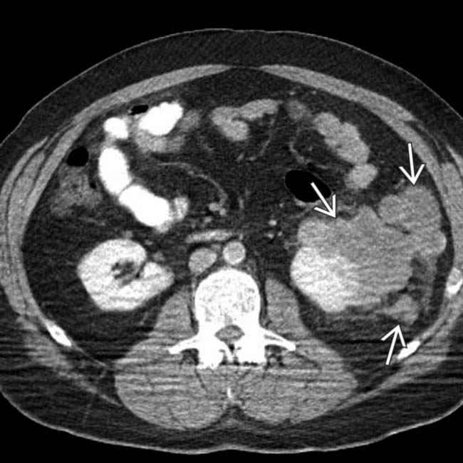
in a patient with melanoma. Metastases from melanoma have a unique predisposition for involving the perirenal space.
image
(Left) Coronal CECT demonstrates an enhancing mass image 
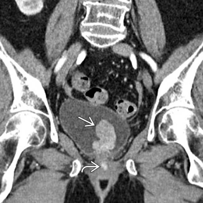
in the bladder. Notice the additional enhancing lesion image in the urethra. Both lesions were found to represent metastatic melanoma. The bladder lesion is indistinguishable from a primary bladder tumor without a clinical history.

image
(Right) Axial CECT shows an enhancing mass image 
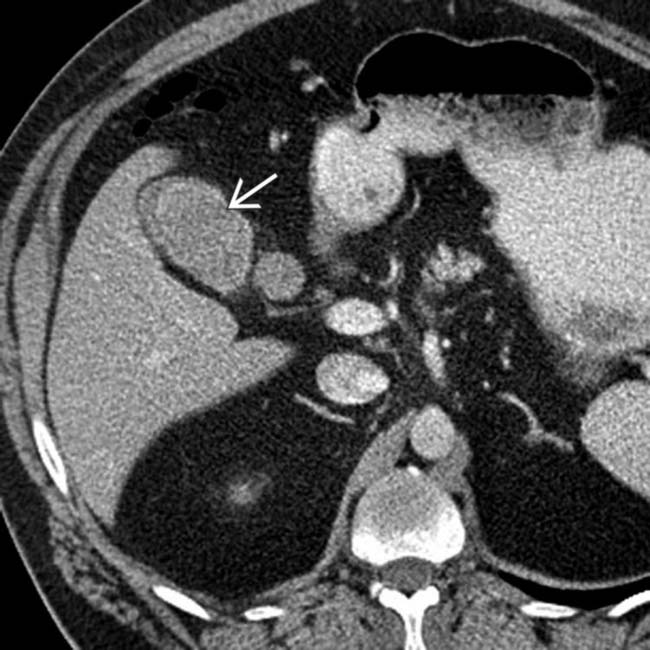
in the gallbladder. The patient had a history of melanoma and the lesion had been slowly growing over time. Melanoma is the most common cause of metastases to the gallbladder.
image
(Left) Axial T1WI MR shows multiple metastases image 
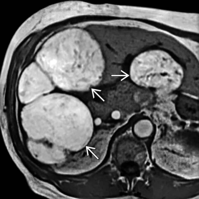
that have the peculiar feature of being hyperintense on T1WI, which is attributed to the melanin in these lesions. In some instances, metastases with fat or hemorrhage can also appear hyperintense on T1WI.

image
(Right) Upper GI series shows classic bull’s-eye lesions, consisting of small, intramural masses with central ulceration image. Lymphoma and Kaposi sarcoma can also result in bull’s-eye lesions.
image
Axial CECT shows massive, extensive lymph node metastases image 
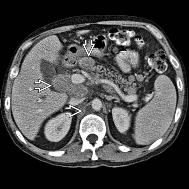
throughout the retroperitoneum, mesentery, and retrocrural spaces. There was also metastasis to the wall of the proximal small bowel (not shown).

image
Axial CECT shows a mesenteric mass image 
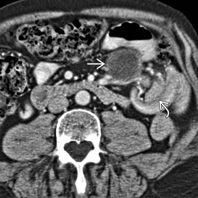
and a classic intussusception image. The lead point of the intussusception is a metastasis in the wall of the intussusceptum.
image
Axial CECT shows enhancing soft-tissue nodules image 
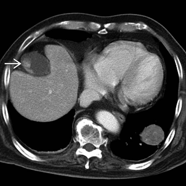
in the wall of the gallbladder. The gallbladder lesions progressed over time and represent metastases from the patient’s known melanoma.
image
CT shows a hypodense splenic metastasis image 
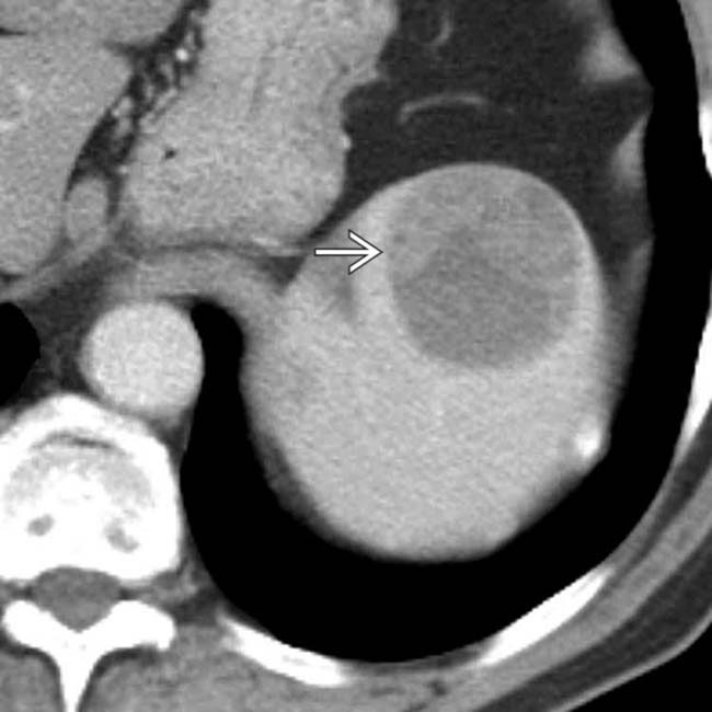
from melanoma. While the lesion is partially cystic, note that there is clearly a solid soft-tissue component differentiating this lesion from a cyst.
image
Axial CECT in an asymptomatic patient demonstrates a large, hypodense mass image 
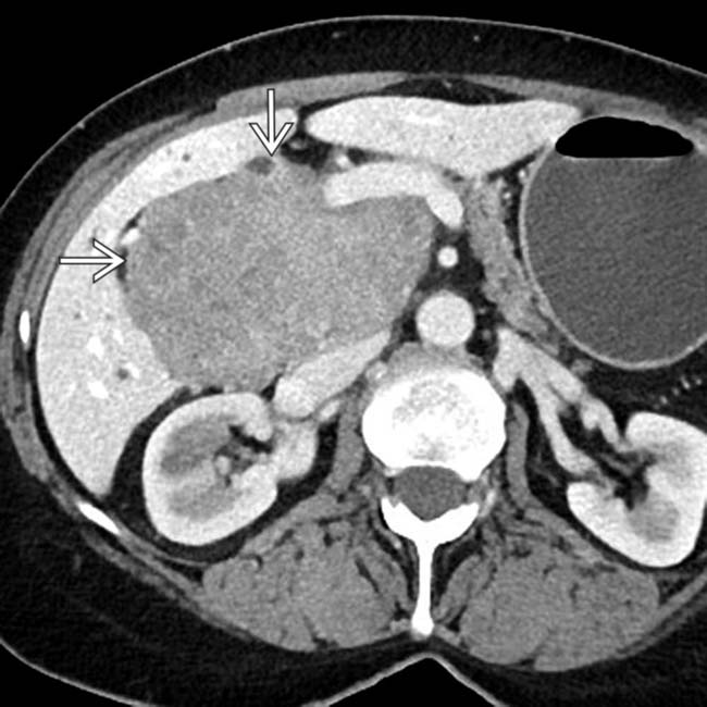
centered in the porta hepatis.
image
Coronal CECT in the same patient demonstrates the large mass image
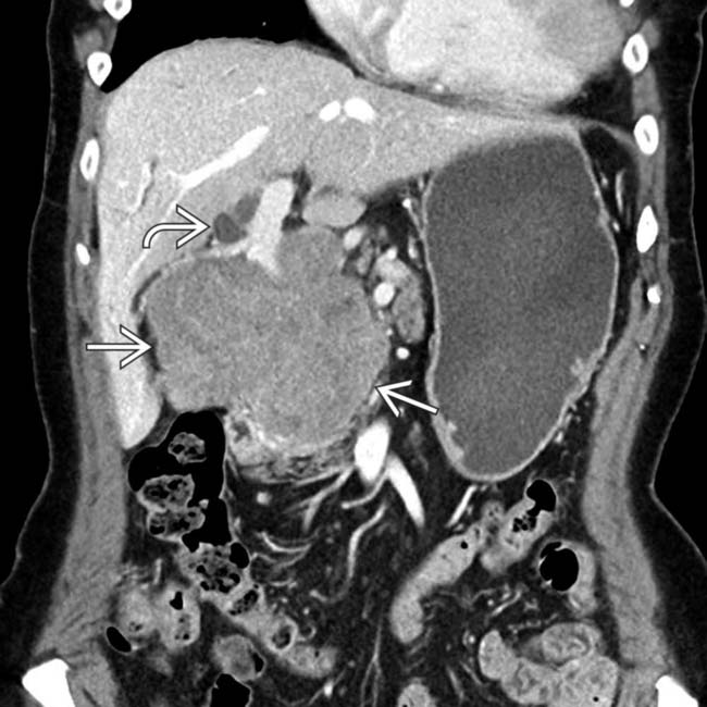
, which causes mild biliary dilatation image due to mass effect. The mass was biopsied and found to represent a large nodal metastasis from melanoma. The primary site of tumor was never found, something which occurs in roughly 3% of cases.
image
Axial CECT demonstrates extensive carcinomatosis, omental caking image
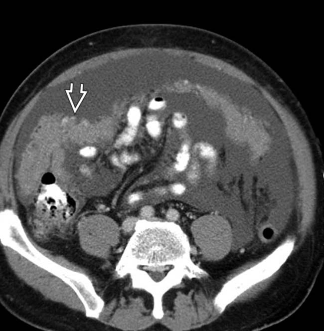
, and ascites from metastatic melanoma. There are multiple manifestations of widespread metastases from malignant melanoma.
image
Axial CECT in a patient with known melanoma demonstrates a polyploid mass image 
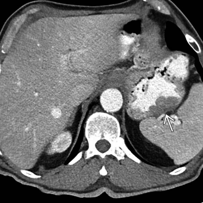
incidentally discovered in the proximal stomach. Note that the mass is subtly hypodense to the adjacent normal gastric wall.
image
Axial NECT shows subtle hypodense metastases image 
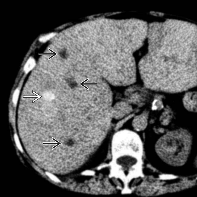
and others that are hyperdense image. The latter may be due to calcification, large amounts of melanin, or hemorrhage.
image
Axial CECT shows massive bilateral adrenal masses image 
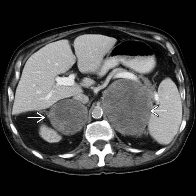
due to malignant melanoma metastases. These destroyed the adrenal parenchyma and resulted in adrenal insufficiency.
image
Axial CECT shows a classic intussusception image 
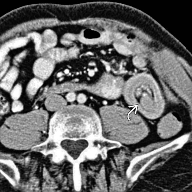
in a patient with a history of cutaneous melanoma.
image
Axial CECT in the same patient shows that the lead point of the intussusception is a metastasis image 
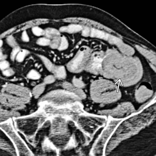
in the wall of the small intestine.
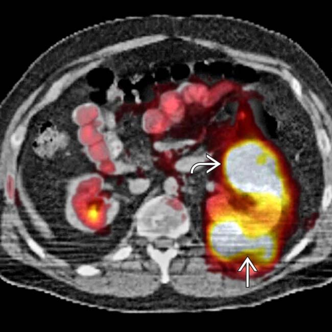
image
Axial PET/CT shows FDG-avid metastases in the kidney image and perirenal space image.
image
Axial PET/CT in the same patient shows an additional lesion in the bowel image. Despite the large size of the metastasis, there was no bowel obstruction.
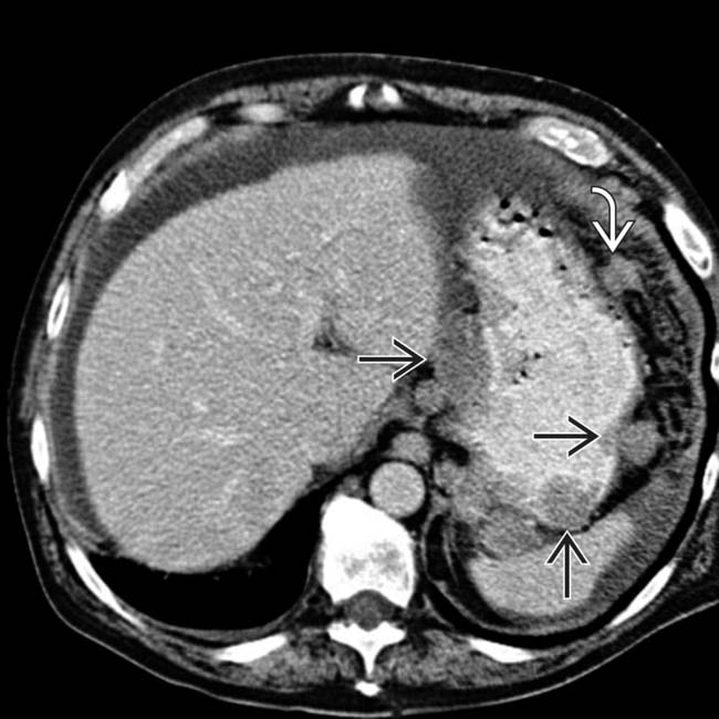
image
Axial CECT shows classic widespread metastases from melanoma to the gastric wall image and peritoneum image, resulting in ascites.
image
Axial CECT in the same patient shows additional metastases to the omentum as large and small nodules image
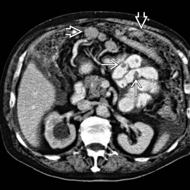
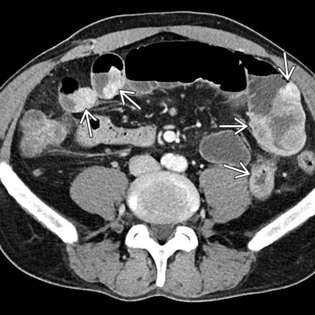
. Note the nodal and small-bowel metastases image. The right ureter was obstructed due to a ureteral or retroperitoneal metastasis.
image
Axial CECT in the same patient shows additional metastases to the peritoneum image, abdominal wall image, lymph nodes, and perirenal space image. “Unusual” sites of metastases are typical in patients with melanoma.

SELECTED REFERENCES

1. Baker, JJ, et al. Routine restaging PET/CT and detection of initial recurrence in sentinel lymph node positive stage III melanoma. Am J Surg. 2014; 207(4):549–554.

Choi, WS, et al. CT findings of gallbladder metastases: emphasis on differences according to primary tumors. Korean J Radiol. 2014; 15(3):334–345.

Trout, AT, et al. Melanoma metastases in the abdomen and pelvis: Frequency and patterns of spread. World J Radiol. 2013; 5(2):25–32.

Sofue, K, et al. MR imaging of hepatic metastasis in patients with malignant melanoma: evaluation of suspected lesions screened at contrast-enhanced CT. Eur J Radiol. 2012; 81(4):714–718.

Barretta, ML, et al. Gallbladder metastasis: spectrum of imaging findings. Abdom Imaging. 2011; 36(6):729–734.

Patnana, M, et al. Multimethod imaging, staging, and spectrum of manifestations of metastatic melanoma. Clin Radiol. 2011; 66(3):224–236.

Intenzo, CM, et al. Lymphoscintigraphy in cutaneous melanoma: an updated total body atlas of sentinel node mapping. Radiographics. 2009; 29(4):1125–1135.

Martel, JP, et al. Melanoma of the gallbladder. Radiographics. 2009; 29(1):291–296.

Pleiss, C, et al. Role of FDG-PET in the assessment of survival prognosis in melanoma. Cancer Biother Radiopharm. 2007; 22(6):740–747.

Strobel, K, et al. High-risk melanoma: accuracy of FDG PET/CT with added CT morphologic information for detection of metastases. Radiology. 2007; 244(2):566–574.

King, DM. Imaging of metastatic melanoma. Cancer Imaging. 2006; 6:204–208.

Vucetiimage, B, et al. The role of preoperative lymphoscintigraphy in surgery planning for sentinel lymph node biopsy in malignant melanoma. Wien Klin Wochenschr. 2006; 118(9–10):286–293.

Petro, A, et al. Current melanoma staging. Clin Dermatol. 2004; 22(3):223–227.

Hubalewska, A, et al. Use of Tc99m-nanocolloid for sentinel nodes identification in cervical cancer. Nucl Med Rev Cent East Eur. 2003; 6(2):127–130.

Sumner, WE, 3rd., et al. Implications of lymphatic drainage to unusual sentinel lymph node sites in patients with primary cutaneous melanoma. Cancer. 2002; 95(2):354–360.

Blake, SP, et al. Liver metastases from melanoma: detection with multiphasic contrast-enhanced CT. Radiology. 1999; 213(1):92–96.

Kamel, IR, et al. Imaging of abdominal manifestations of melanoma. Crit Rev Diagn Imaging. 1998; 39(6):447–486.

Fishman, EK, et al. CT of malignant melanoma in the chest, abdomen, and musculoskeletal system. Radiographics. 1990; 10(4):603–620.

Stutte, H, et al. Ultrasonographic diagnosis of melanoma metastases in liver, gallbladder, and spleen. J Ultrasound Med. 1989; 8(10):541–547.

Daunt, N, et al. Metastatic melanoma in the biliary tree. Br J Radiol. 1982; 55(659):873–874.