Mesenteric Adenitis and Enteritis

Published on 19/07/2015 by admin

Filed under Radiology

Last modified 22/04/2025

Print this page

rate 1 star rate 2 star rate 3 star rate 4 star rate 5 star
Your rating: none, Average: 0 (0 votes)

This article have been viewed 3293 times

 Mesenteric adenopathy is often much more evident on coronal-reformatted CT

• Ileal ± cecal wall thickening, sometimes with regional ileus

image Mucosal hyperenhancement, submucosal edema
• Normal-appearing appendix

TOP DIFFERENTIAL DIAGNOSES

• Appendicitis
• Crohn disease

image Early Crohn disease may be impossible to distinguish
image Time course and likelihood of recurrence are different
• Cecal or appendiceal carcinoma

image Affects older adults, not children

PATHOLOGY

• Reactive lymph node enlargement secondary to enteric pathogens
• Viral (most common)
• Bacterial (especially Yersinia and Campylobacter species)

CLINICAL ISSUES

• Commonly seen in children and young adults < 25 years old

image 8-12% of young patients with acute RLQ pain have mesenteric adenitis
• Pain, fever, nausea, vomiting

image Leukocytosis
• Self-limited, usually resolves without treatment
image
(Left) Axial CT in a 25-year-old woman presenting with fever and RLQ tenderness shows wall thickening and mucosal hyperenhancement of the terminal ileum and cecum image.

image
(Right) Another CT section in the same patient shows a normal appendix image
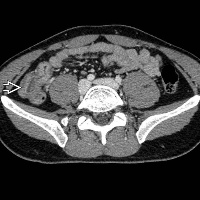
, excluding appendicitis as the diagnosis.
image
(Left) Coronal-reformatted image in the same patient shows a cluster of mildly enlarged ileocolic mesenteric nodes image
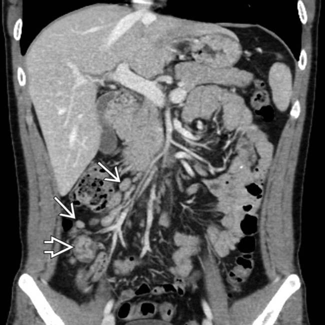
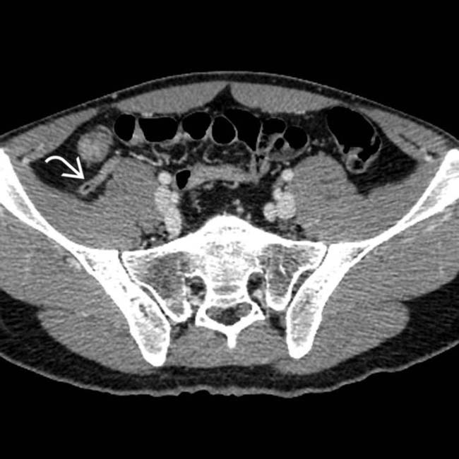
, along with the thick-walled, inflamed terminal ileum image.

image
(Right) Another coronal CECT section shows enlarged nodes and engorged vessels in the ileocolic mesentery image
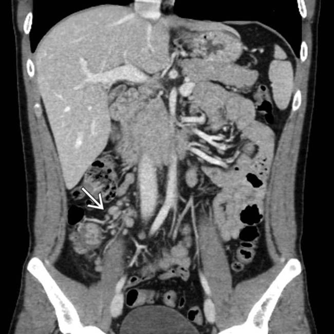
, along with the thick-walled terminal ileum. These are classic imaging and CT features of mesenteric adenitis and enteritis, and the patient made an uneventful recovery without specific therapy.

TERMINOLOGY

Definitions

• Benign inflammation of lymph nodes in ileal mesentery, often with terminal ileitis

IMAGING

General Features

• Best diagnostic clue

image Cluster of slightly prominent (≥ 5 mm) mesenteric lymph nodes in right lower quadrant (RLQ)
image Ileal/ileocolic wall thickening
image Normal-appearing appendix
• Size

image Nodes ≥ 5 mm in short axis
image Rarely exceed 10 mm
image Clustered (≥ 3 nodes)

Imaging Recommendations

• Best imaging tool

image CECT with coronal reformations

Radiographic Findings

• Regional ileus, bowel wall thickening may be seen

Ultrasonographic Findings

• Nodal tenderness in response to transducer pressure
• Round and hypoechoic nodes measuring ≥ 5 mm
• Terminal ileal wall thickening may be seen
• Nonvisualization of inflamed appendix

CT Findings

• Cluster of mildly enlarged ileocolic mesenteric lymph nodes (≥ 5 mm)

image Mesenteric adenopathy is often much more evident on coronal-reformatted CT
• Bowel wall thickening of terminal ileum ± cecum

image Mucosal hyperenhancement and submucosal edema

– More evident with water as oral contrast agent
image ± engorged ileocolic vessels
• Normal appendix

DIFFERENTIAL DIAGNOSIS

Appendicitis

• May have similar cluster of nodes
• Thick-walled appendix with mural and mucosal enhancement

image Periappendiceal inflammation
• Total diameter of appendix > 7 mm
• Appendicolith often seen on CT (33-50%)

Crohn Disease

• Segmental areas of ileo-colonic ulceration
• Discontinuous and asymmetric bowel wall thickening (> 1 cm)
• More mesenteric hyperemia (comb sign)
• History of prior similar episodes of pain in most cases

Cecal or Appendiceal Carcinoma

• Also associated with RLQ clustered nodes
• Look for soft tissue mass and omental metastases
• Affects older adults, not children

PATHOLOGY

General Features

• Etiology

image Viral (most common)
image Bacterial: Yersinia enterocolitica, Yersinia pseudotuberculosis, Helicobacter pylori, Campylobacter jejuni, Salmonella, Shigella
image Mycobacterial
• Associated abnormalities

image Ileitis, ileocolitis
• Pathophysiology

image Reactive lymph node enlargement secondary to enteric pathogens

CLINICAL ISSUES

Presentation

• Most common signs/symptoms

image Pain, fever, nausea, vomiting
image Leukocytosis
• Other signs/symptoms

image Diarrhea, RLQ tenderness

Demographics

• Age

image Commonly seen in children and young adults < 25 years old
image Concurrent enteric disease commonly seen in children, especially those < 5 years old
• Epidemiology

image 8-12% of young patients presenting with acute RLQ pain have mesenteric adenitis

Natural History & Prognosis

• Self-limited, usually resolves without treatment
• Concurrent enterocolitis may be severe (rarely)

Treatment

• Conservative, nonoperative management

DIAGNOSTIC CHECKLIST

Consider

• Mesenteric adenitis is diagnosis of exclusion

SELECTED REFERENCES

1. Patlas, MN, et al. Cross-sectional imaging of nontraumatic peritoneal and mesenteric emergencies. Can Assoc Radiol J. 2013; 64(2):148–153.

2. Toorenvliet, B, et al. Clinical differentiation between acute appendicitis and acute mesenteric lymphadenitis in children. Eur J Pediatr Surg. 2011; 21(2):120–123.

3. Lee, MW, et al. Sonography of acute right lower quadrant pain: importance of increased intraabdominal fat echo. AJR Am J Roentgenol. 2009; 192(1):174–179.

4. Pickhardt, PJ, et al. Unusual nonneoplastic peritoneal and subperitoneal conditions: CT findings. Radiographics. 2005; 25(3):719–730.

5. Rao, PM, et al. CT diagnosis of mesenteric adenitis. Radiology. 1997; 202(1):145–149.

6. Puylaert, JB. Mesenteric adenitis and acute terminal ileitis: US evaluation using graded compression. Radiology. 1986; 161(3):691–695.

Frisch, M, et al. Appendicitis, mesenteric lymphadenitis, and subsequent risk of ulcerative colitis: cohort studies in Sweden and Denmark. BMJ. 2009 Mar 9; 338:b716. [doi: 10. PubMed Central PMCID: PMC2659291, 1136].