Meningitis, Encephalitis, and Other Infections of the Central Nervous System

Published on 08/02/2015 by admin

Filed under Basic Science

Last modified 22/04/2025

Print this page

rate 1 star rate 2 star rate 3 star rate 4 star rate 5 star
Your rating: none, Average: 0 (0 votes)

This article have been viewed 1937 times

Meningitis, Encephalitis, and Other Infections of the Central Nervous System

Objectives

1. Describe the anatomy of the central nervous system (CNS), and list the anatomic structures that compose it.

2. Define meninges; name the three separate layers and describe their function.

3. Define the cerebrospinal fluid, and list the functions of the cerebrospinal fluid (CSF).

4. Describe the routes of infection for the central nervous system.

5. Explain the host defense mechanisms that protect the central nervous system from infection.

6. Define meningitis, and describe the two major types of meningitis including the etiologic agents.

7. When discussing the etiology of acute meningitis, explain the host predisposing factors for the neonate and identify the most commonly associated bacterial pathogens.

8. Discuss how the advent of the Hib vaccine in the United States has helped to prevent pediatric cases of meningitis.

9. Compare acute and chronic meningitis; outline the distinguishing symptoms and CSF findings for each, including cell counts and chemistry laboratory results.

10. Explain the disease processes for encephalitis and meningoencephalitis.

11. Discuss two ways that meningoencephalitis infections, brain abscesses, or other CNS infections are caused by parasites; identify the associated infecting organisms and the population of patients at increased risk for developing these conditions.

12. Explain the collection, transport, and specimen storage requirements for CSF; include specimen processing and the appropriate distribution of specimen throughout the laboratory.

13. List the culture media used to identify the causative agent of meningitis in bacterial, mycobacterial, and fungal infections; what incubation conditions are required for each type of organism?

General Considerations

Anatomy

Diagnosis of an infection involving the central nervous system (CNS) is of critical importance. Most clinicians consider infection in the CNS to be a medical emergency. An understanding of the basic anatomy and physiology of the CNS is helpful for the microbiologist to ensure appropriate specimen processing and interpretation of laboratory results.

Coverings and Spaces of the CNS

The central nervous system consists of the brain and the spinal cord. Because of the vital and essential role of the CNS in the body’s regulatory processes, the brain and spinal cord have two protective coverings: an outer covering consisting of bone and an inner covering of membranes called the meninges. The outer bone covering encases the brain (i.e., cranial bones or skull) and spinal cord (i.e., the vertebrae). The meninges is a collective term for the three distinct membrane layers surrounding the brain and spinal column:

The pia mater and the arachnoid membrane are collectively called the leptomeninges. The portion of the arachnoid that covers the top of the brain contains arachnoid villi, which are special structures that absorb the spinal fluid and allow it to pass into the blood.

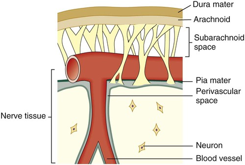
Between and around the meninges are spaces that include the epidural, subdural, and subarachnoid spaces. The relative location of the meninges and spaces to one another in the brain are depicted in Figure 71-1. The location and nature of the meninges and spaces are summarized in Table 71-1.

TABLE 71-1

Inner Coverings (Meninges) of the Brain, Spinal Cord, and Surrounding Spaces

Anatomic Structure Relative Location Key Features
Epidural space Outside the dura mater yet inside the skull Cushion of fat and connective tissues
Dura mater Outermost membrane Membrane that adheres to the skull; white fibrous tissue
Subdural space Between the dura mater and the arachnoid membrane Cushion of lubricating serous fluid
Arachnoid membrane Between the dura mater and pia mater Delicate, cobweb-like membrane covering the brain and spinal cord
Subarachnoid space Beneath the arachnoid membrane Contains a significant amount of CSF in an adult (~125-150 mL)
Pia mater Beneath the subarachnoid space Adheres to the outer surface of the brain and spinal cord; contains blood vessels

Cerebrospinal Fluid

Cerebrospinal fluid (CSF) surrounds the brain and spinal cord and has several functions. The CSF provides cushioning and buoyancy for the bulk of the brain, reducing the effective weight of the brain by a factor of 30. CSF carries essential metabolites into the neural tissue and cleanses the tissues of wastes as it circulates around the brain, ventricles, and spinal cord. Every 3 to 4 hours, the entire volume of CSF is exchanged. In addition to these functions, CSF provides a means by which the brain monitors changes in the internal environment.

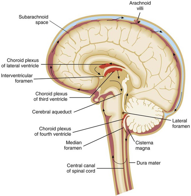
CSF is found in the subarachnoid space (see Table 71-1) and within cavities and canals of the brain and spinal cord. There are four large, fluid-filled spaces within the brain referred to as ventricles. Specialized secretory cells, called the choroid plexus, produce CSF. The choroid plexus is located centrally within the brain in the third and fourth ventricles. Approximately 23 mL of CSF are contained within these ventricles in an adult. The fluid travels around the outside areas of the brain within the subarachnoid space, driven primarily by the pressure produced initially at the choroid plexus (Figure 71-2). By virtue of its circulation, chemical and cellular changes in the CSF may provide valuable information about infections within the subarachnoid space.

Routes of Infection

One of the most important defense mechanisms of the CNS is the blood-brain barrier. The blood-brain barrier functions to maintain homeostasis in the brain through restricting the flow of chemical constituents from the blood to the CNS. In order for the CNS to become infected with bacteria, parasite, or virus, the blood-brain barrier must be penetrated.

Organisms may gain access to the CNS through several primary routes:

• Hematogenous spread: followed by entry into the subarachnoid space through the choroid plexus or through other blood vessels of the brain. This is the most common route of infection for the CNS.

• Direct spread from an infected site: the extension of an infection close to or contiguous with the CNS can occasionally occur; examples of such infections include otitis media (infection of middle ear), sinusitis, and mastoiditis.

• Anatomic defects in CNS structures: anatomic defects as a result of surgery, trauma, or congenital abnormalities can allow microorganisms easy and ready access to the CNS.

• Travel along nerves leading to the brain (direct intraneural): the least common route of CNS infection caused by organisms such as rabies virus, which travels along peripheral sensory nerves, and herpes simplex virus.

Diseases of the Central Nervous System

Meningitis

Infection within the subarachnoid space or throughout the leptomeninges is called meningitis. Based on the host’s response to the invading microorganism, meningitis is divided into two major categories: purulent and aseptic meningitis.

Purulent Meningitis.

A patient with purulent meningitis typically has a marked, acute inflammatory exudative cerebral spinal fluid containing large numbers of polymorphonuclear cells (PMNs). Frequently, the underlying CNS tissue, in particular the ventricles, may be involved. If the ventricles become involved, this process is referred to as ventriculitis. Bacterial organisms are usually the cause of these infections.

Pathogenesis.

The outcome of a host-microbe interaction depends on the characteristics of both the host and the microorganism. As previously indicated, an important host defense mechanism within the CNS is the blood-brain barrier; this barrier involves the choroid plexus, arachnoid membrane, and the cerebral microvascular endothelium. The unique structural properties of the vascular endothelium, such as the continuous intercellular tight junctions, provide a barrier minimizing the passage of infectious agents into the CSF. The normal function of the vascular endothelium includes regulating the transport of nutrients in and out of the CSF, including low-molecular-weight plasma proteins, glucose, and electrolytes.

The host’s age and other underlying factors contribute to whether an individual is predisposed to the development of infectious meningitis. Neonates have the highest infection rate for meningitis, because of the immature neonatal immune system, the increased permeability of the blood-brain barrier in newborns, and the presence of colonizing bacteria in the female vaginal tract that can pass to the infant during childbirth. The most common bacterial pathogens responsible for meningitis in newborns are group B streptococci, Escherichia coli, and Listeria monocytogenes. Prior to the advent of the Hib vaccine in the United States in 1985, Haemophilus influenza type b (Hib) was a common cause of meningitis in children 4 months to 5 years of age. Because of the incorporation of Hib into childhood immunization programs, childhood Hib disease has dramatically declined.

Among young adults, Neisseria meningitidis is typically the agent that is associated with meningitis. N. meningitidis has been identified in epidemics among young adults in crowded conditions (e.g., military recruits and college dormitory mates). There are two meningococcal vaccines (vaccines for N. meningitidis) available in the United States. The meningococcal polysaccharide vaccine (MPSV4) is used for individuals older than 55 years of age, and the meningococcal conjugate vaccine (MCV4) is used for adolescents. Streptococcus pneumoniae is frequently the cause of meningitis in young children and the elderly; often this meningitis develops from bacteremia or from infection of the sinuses or middle ear. There are two pneumococcal vaccines (vaccines for S. pneumoniae) that are recommended currently in the United States. The pneumococcal conjugate vaccine (PCV13) protects against infection from 13 different serotypes of S. pneumoniae and is used for vaccination of children and adults. The second vaccine, pneumococcal polysaccharide vaccine (PPSV), provides protection from 23 serotypes of S. pneumoniae, including those associated with serious life-threatening infections. This vaccine is recommended for adults 65 years of age and older or anyone over the age of 2 who has long-term health problems or is immunocompromised.

Because the respiratory tract is the primary portal of entry for many etiologic agents of meningitis, factors that predispose adults to meningitis are often the same factors that increase the likelihood for the development of pneumonia or other respiratory tract colonization or infection. Alcoholism, splenectomy, diabetes mellitus, prosthetic devices, and immunosuppression contribute to increased risk. Finally, patients with prosthetic devices, particularly CNS and ventriculoperitoneal shunts, are at increased risk for developing meningitis.

For organisms to reach the CNS (primarily by the blood-borne route), host defense mechanisms must be overcome. Most cases of meningitis are a result of bacteria that share a similar pathogenesis. The successful meningeal pathogen must first sequentially colonize and cross host mucosal epithelium, then enter and thrive within the bloodstream. The most common causes of meningitis possess the ability to evade host defenses at each of these levels. For example, clinical isolates of Streptococcus pneumoniae and N. meningitidis secrete IgA proteases capable of destroying the host’s secretory IgA, thereby facilitating bacterial attachment to the epithelium. In addition, all of the most common etiologic agents of bacterial meningitis possess an antiphagocytic capsule that allows the organisms to evade destruction by the host immune system.

Organisms appear to enter the CNS by interacting and subsequently breaking down the blood-brain barrier at the level of microvascular endothelium. One of the least understood processes in the pathogenesis of meningitis is how organisms cross this barrier into the subarachnoid space. Nevertheless, there appear to be specific bacterial surface components, such as pili, polysaccharide capsules, and lipoteichoic acids, that facilitate adhesion of the organisms to the microvascular endothelial cells and subsequent penetration into the CSF. Organisms can enter (1) through loss of capillary integrity by disrupting tight junctions of the blood-brain barrier, (2) through transport within circulating phagocytic cells, or (3) by crossing the endothelial cell lining within endothelial cell vacuoles. After gaining access, the organism multiplies within the CSF, a site initially free of antimicrobial antibodies or phagocytic cells.

Acute.

Symptoms of acute meningitis include fever, stiff neck, headache, nausea and vomiting, neurologic abnormalities, and change in mental status.

In acute bacterial meningitis, the CSF usually contains large numbers of inflammatory cells (>1000/mm3), primarily polymorphonuclear cells (PMNs). The CSF shows a decreased glucose level relative to the serum glucose level and an increase in protein concentration. In a healthy individual, the normal CSF glucose level is 0.6 of the serum glucose level and ranges from 45 to 100 mg/dL; the CSF protein range in an adult is 15 to 50 mg/dL; newborn CSF protein ranges run as high as 170 mg/dL with an average of 90 mg/dL.

The sequelae of acute bacterial meningitis in children are frequent and serious. Seizures can occur in 20% to 30% of patients, and other neurologic changes are common. Acute sequelae include cerebral edema, hydrocephalus, cerebral herniation, and focal neurologic changes. Permanent deafness can occur in 10% of children who recover from bacterial meningitis. Other subtle physiologic and psychological sequelae may also follow an episode of acute bacterial meningitis.

Chronic.

Chronic meningitis can often occur in patients who are immunocompromised, although this is not always the case. Patients experience an insidious onset of disease, with some or all of the following symptoms: fever, headache, stiff neck, nausea and vomiting, lethargy, confusion, and mental deterioration. Symptoms may persist for a month or longer before treatment is sought. The CSF usually manifests an abnormal number of white blood cells (usually lymphocytic), elevated protein, and decrease in glucose content (Table 71-2). The pathogenesis of chronic meningitis is similar to that of acute disease.

TABLE 71-2

Guidelines for Interpretation of Results Following Hematologic and Chemical Analysis of Cerebrospinal Fluid (CSF) from Children and Adults (Excluding Neonates)

Clinical Setting Leukocytes/mm3 Predominant Cell Type Protein Glucose*
Normal 0-5 None 15-50 mg/dL 45-100 mg/dL
Viral infection 2-2000 (mean of 80) Mononuclear Slightly elevated (50-100 mg/dL) or normal Normal
Purulent infection 5-20,000 (mean of 800) PMN Elevated (>100 mg/dL) Low (<45 mg/dL), but may be normal early in the course of disease
Tuberculosis and fungi 5-2000 (mean of 100) Mononuclear Elevated (>50 mg/dL) Normal or often low (>45 mg/dL)

image

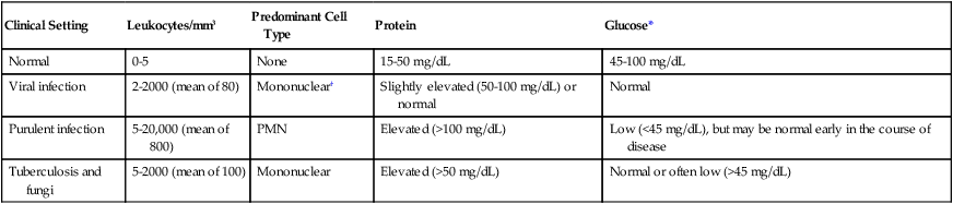
*Must consider CSF glucose level in relation to blood glucose level. Normally, the CSF glucose serum ratio is 0.6, or 50% to 70% of the blood glucose normal value.

About 20% to 75% of cases may have PMN leukocytosis early in the course of infection.

Epidemiology/Etiologic Agents-Acute Meningitis.

The etiology of acute meningitis depends on the age of the patient. Most cases in the United States occur in children younger than 5 years of age. Before 1985, H. influenzae type b H. influenzae type b was the most common infectious agent in children between 1 month and 6 years of age within the United States. Ninety-five percent of all cases were due to H. influenzae type b, Neisseria meningitidis, and Streptococcus pneumoniae. In 1985, the first Hib vaccine, a polysaccharide vaccine, was licensed for use in children 18 months of age or older but was not efficacious in children younger than 18 months. However, the widespread use of conjugate vaccine, Hib polysaccharide-protein conjugate, in children as young as 2 months of age has significantly affected the incidence of invasive H. influenzae type b disease; the total number of annual cases of H. influenzae disease in the United States have been reduced by 55% and the number of cases of H. influenzae meningitis by 94%. However, the risks for meningococcal and pneumococcal diseases resulting from agents other than H. influenzae have remained level. Children older than 6 years of age are less likely to develop meningitis, but the risk for meningitis infection increases when the child reaches early adulthood. As previously mentioned, neonates have the highest incidence of acute meningitis, with a concomitant increased mortality rate (as high as 20%). Organisms causing disease in the newborn are different from those that affect other age groups; many of them are acquired by the newborn during passage through the mother’s vaginal vault. Neonates are likely to be infected with, in order of incidence, group B streptococci, Escherichia coli, other gram-negative bacilli, and Listeria monocytogenes; occasionally other organisms may be involved. For example, Elizabethkingia meningoseptica has been associated with nursery outbreaks of meningitis. This organism is a normal inhabitant of water in the environment and is presumably acquired as a nosocomial infection.

Important causes of meningitis in the adult, in addition to the meningococcus in young adults, include pneumococci, Listeria monocytogenes, and, less commonly, Staphylococcus aureus and various gram-negative bacilli. Meningitis caused by the latter organisms results from hematogenous seeding from various sources, including urinary tract infections. The percentage of adults with nosocomial bacterial meningitis at large urban hospitals has been increasing. The various etiologic agents of chronic meningitis are listed in Box 71-1.

Aseptic Meningitis.

Aseptic meningitis is usually viral and characterized by an increase of lymphocytes and other mononuclear cells (pleocytosis) in the CSF; bacterial and fungal cultures are negative. (This is in contrast to bacterial meningitis, which is characterized by purulence and the polymorphonuclear [PMN] cell response in the CSF.) Aseptic meningitis is usually self-limiting with symptoms that may include fever, headache, stiff neck, nausea, and vomiting.

In addition to the increase of lymphocytes and other mononuclear cells in the CSF, the glucose level remains normal, whereas the protein CSF level may remain normal or be slightly elevated. Aseptic meningitis can also be a symptom for syphilis and some other spirochete diseases (e.g., leptospirosis and Lyme borreliosis). Stiff neck and CSF pleocytosis may also be associated with other disease processes, such as malignancy.

Encephalitis/Meningoencephalitis

Encephalitis is an acute inflammation of the brain parenchyma and is usually caused by direct viral invasion. Concomitant meningitis occurring with encephalitis is known as meningoencephalitis, and the cellular infiltrate present in the CSF is typically lymphocytic rather than polymorphonuclear cells.

The host response to these CNS infections can differ somewhat from those associated with purulent or aseptic meningitis. Early in the course of viral encephalitis, or when considerable tissue damage occurs as a part of encephalitis, the nature of the inflammatory cells found in the CSF may be no different from that associated with bacterial meningitis; cell counts, however, are typically much lower.

Viral.

Viral encephalitis, which cannot always be distinguished clinically from meningitis, is common in the warmer months. The primary agents are enteroviruses (coxsackie viruses A and B, echoviruses), mumps virus, herpes simplex virus, and arboviruses (West Nile virus, togavirus, bunyavirus, equine encephalitis, St. Louis encephalitis, and other encephalitis viruses). Other viruses—such as measles, cytomegalovirus, lymphocytic choriomeningitis, Epstein-Barr virus, hepatitis, varicella-zoster virus, rabies virus, myxoviruses, and paramyxoviruses—are less commonly encountered. Any preceding viral illness and exposure history are important considerations in establishing a cause by clinical means. Since 1999, with the first debut of West Nile in the United States, the West Nile virus has been an important consideration in the diagnosis of viral encephalitis. The Centers for Disease Control and Prevention (CDC) reports that the incidence of West Nile infection peaked in 2003 with 9862 cases of West Nile infection; 2860 were reported cases of meningitis and encephalitis, resulting in 264 deaths. Since then the rates of infection have dropped: human cases reported to the CDC in 2010 were significantly lower with 1021 total reported cases of West Nile; 629 were neuroinvasive cases resulting in 57 deaths; a state-by-state breakdown of the disease incidence is outlined in Table 71-3. In 2012, a deadly resurgence of West Nile virus occurred, including neuroinvasive and non-neuroinvasive, for a total of 4531 cases through mid-October, according to the CDC.

TABLE 71-3

Final 2010 West Nile Virus Human Infections in the United States*

State Neuroinvasive Disease Cases Non-neuroinvasive Disease Cases Total Cases Deaths Presumptive Viremic Donors*
Alabama 1 2 3 0 0
Arizona 107 60 167 15 31
Arkansas 6 1 7 1 0
California 72 39 111 6 24
Colorado 26 55 81 4 1
Connecticut 7 4 11 0 5
District of Columbia 3 3 6 0 0
Florida 9 3 12 2 1
Georgia 4 9 13 0 1
Idaho 0 1 1 0 0
Illinois 45 16 61 4 5
Indiana 6 7 13 1 0
Iowa 5 4 9 2 1
Kansas 4 15 19 0 1
Kentucky 2 1 3 1 7
Louisiana 20 7 27 0 7
Maryland 17 6 23 2 0
Massachusetts 6 1 7 0 1
Michigan 25 4 29 3 2
Minnesota 4 4 8 0 1
Mississippi 3 5 8 0 2
Missouri 3 0 3 0 0
Nebraska 10 29 39 2 10
Nevada 0 2 2 0 0
New Hampshire 1 0 1 0 0
New Jersey 15 15 30 2 0
New Mexico 21 4 25 1 6
New York 89 39 128 4 16
North Dakota 2 7 9 0 0
Ohio 4 1 5 0 0
Oklahoma 1 0 1 0 1
Pennsylvania 19 9 28 0 0
South Carolina 1 0 1 0 0
South Dakota 4 16 20 0 0
Tennessee 2 2 4 0 1
Texas 77 12 89 6 14
Utah 1 1 2 0 3
Virginia 4 1 5 1 2
Washington 1 1 2 0 0
Wisconsin 0 2 2 0 1
Wyoming 2 4 6 0 0
Totals 629 392 1021 57 144

image

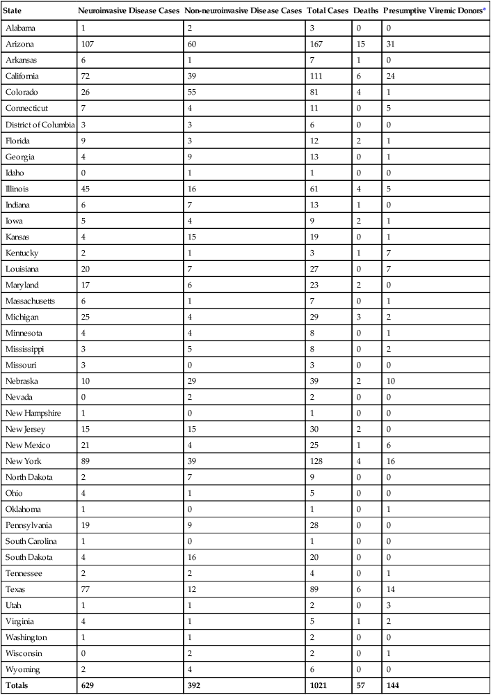
Neuroinvasive disease refers to severe cases of disease that affect a person’s nervous system. These include encephalitis, meningitis, and acute flaccid paralysis that is an inflammation of the spinal cord that can cause a sudden onset of weakness in the limbs or breathing muscles.

*Human Cases Reported to CDC.

Neuroinvasive infection with West Nile presents with symptoms of headache, fever, and a change in consciousness along with altered mental status. The examination of the CSF shows an increase in leukocytes with a marked increase in lymphocytes. Chemistries demonstrate an elevated protein count and normal glucose levels. Definitive diagnosis requires testing for the presence of the IgM antibody to West Nile in the serum or CSF, and because IgM does not cross the blood-brain barrier, presence of IgM antibody to West Nile in the CSF is a strong indicator for CNS infection. Polymerase chain reaction (PCR) can also be used to test for West Nile infection, but because West Nile infections have a transient and low viremia, results must be interpreted with caution. A negative result does not necessarily rule out West Nile infection.

Involvement of the nervous system in patients who are infected with the human immunodeficiency virus (HIV) is common. HIV is a neurotropic (attracted to nerve cells) virus capable of entering the CNS by macrophage transport and the cause of various neurologic syndromes. As HIV-infected individuals become progressively more immunosuppressed, the CNS becomes a target for opportunistic pathogens, such as cytomegalovirus, BK virus, and JC (John Cunningham) virus, which can produce meningitis or encephalitis. BK virus is named after the initials of the first renal transplant patient where the virus was identified in association with clinical disease.

Parasitic

Parasites can cause meningoencephalitis, brain abscess (see the following discussion), or other CNS infection via two routes. A rare but devastating meningoencephalitis is caused by the free-living amebae, Naegleria fowleri and Acanthamoeba spp., which invade the brain via direct extension from the nasal mucosa. These organisms are acquired during swimming or diving in natural, stagnating freshwater ponds and lakes.

Other parasites reach the brain via hematogenous spread. Toxoplasmosis, caused by an intracellular parasite that destroys brain parenchyma, is a common CNS affliction in HIV-infected patients with acquired immunodeficiency syndrome (AIDS). Entamoeba histolytica and Strongyloides stercoralis have been identified in brain tissue, and the larval form of Taenia solium (the pork tapeworm), called a cysticercus, can travel to the brain via the bloodstream and encyst within the brain tissue. Amebic brain infection and cysticercosis cause changes in the CSF similar to meningitis.

Brain Abscess

Brain abscesses (localized collections of pus in a cavity formed by the breakdown of tissue) may occasionally cause changes in the CSF and clinical symptoms similar to meningitis. Brain abscesses result from contiguous infection of the sinuses, middle ear, or mastoids (25%-50%), hematogeneously (15%-30%), or through direct inoculation as a result of trauma or surgery (8%-19%). Brain abscesses may rupture into the subarachnoid space, producing severe meningitis with a high mortality rate. If anaerobic organisms or viridans streptococci are recovered from CSF cultures, the diagnosis of brain abscess should be considered; however, CSF culture is typically negative in brain abscess. Patients who are immunosuppressed or who have diabetes with ketoacidosis may present with a rapid progressive fungal infection (phycomycosis) of the nasal sinuses or palatal region capable of traveling directly to the brain. The complex polymicrobial infections isolated from brain abscesses are far too extensive to list.

Laboratory Diagnosis of Central Nervous System Infections

Meningitis

Except in unusual circumstances, a lumbar puncture (spinal tap) is one of the first steps in the diagnosis of a patient with suspected CNS infection, in particular, meningitis. Refer to Table 5-1 to review the procedure for collecting, transporting, and processing specimens obtained from the central nervous system.
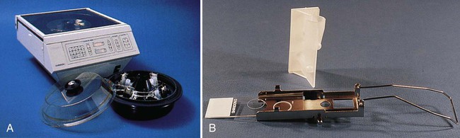
Specimen Collection and Transport

CSF is collected by aseptically inserting a needle into the subarachnoid space (lumbar puncture), at the lumbar spine region between L3, L4, or L5. Three or four tubes of CSF should be collected into sterile collection tubes that contain no additives. The tubes are numbered sequentially in the order in which they were collected along with the patient’s name. When processing the CSF collection tubes in the laboratory, tube 1 is used for chemistry studies, glucose and protein count, as well as immunology studies, as these tests are least affected by the presence of blood cells or bacteria introduced as a result of the spinal tap procedure; tube 2 is used for culture, allowing a larger proportion of the total fluid to be concentrated, which can facilitate the detection of infectious agents present in low numbers; tubes 3 and 4 are used for cell count and differential, as these tubes are least likely to contain cells introduced by the collection procedure. If a small capillary blood vessel is inadvertently broken during the spinal tap, blood cells picked up from this source will usually be absent from the last tube collected; comparison of counts between tubes 1 and 3 (4) is occasionally needed if a traumatic tap is suspected as well as to differentiate a traumatic bloody tap from a true subarachnoid hemorrhage. In a traumatic tap, the red blood cells will be unevenly distributed among the three tubes, with the heaviest concentration of red blood cells being in tube 1 and diminishing amounts in tubes 2 and 3. In an intracranial hemorrhage, the red blood cells will be evenly distributed among the three tubes. The volume of CSF that can be collected is based on the volume available in the patient (adult versus neonate) and the opening pressure of the CSF when the needle first punctures the subarachnoid space. An elevated pressure requires the CSF fluid to be withdrawn more slowly, which may prevent the collection of a larger volume. The volume of CSF is critical for detecting certain microorganisms, such as mycobacteria and fungi. A minimum of 5 to 10 mL is recommended for detecting these agents by centrifugation and subsequent culture. When the laboratory receives an inadequate volume of CSF, the physician should be consulted regarding the order of priority for laboratory tests. Processing too little specimen lowers the sensitivity of laboratory tests, which may lead to false-negative results. This is potentially more harmful to patient care than performing an additional lumbar puncture to obtain the necessary or required amount of sample.

CSF should be hand-delivered immediately to the laboratory. Certain agents, such as Streptococcus pneumoniae, may not be detectable after an hour or longer. Specimens for microbiology studies should never be refrigerated; if not rapidly processed, CSF should be incubated (35° C) or left at room temperature. One exception to this rule involves CSF for viral studies. These specimens may be refrigerated for as long as 23 hours after collection or frozen at −70° C if a longer delay is anticipated until they are processed and inoculated into culture media. CSF for viral studies should never be frozen at temperatures above −70° C. If not processed immediately, CSF specimen for hematology studies can be refrigerated, whereas the CSF for chemistry and serology can be frozen (−20° C).

Information gathered from specimen analysis should be promptly relayed to the clinician who can directly affect therapeutic outcome. Such specimens should be processed immediately upon receipt in the laboratory (STAT) and results reported to the physician as soon as possible.

Initial Processing

Initial processing of CSF for bacterial, fungal, or parasitic studies includes centrifugation of all specimens with a volume greater than 1 mL for at least 15 minutes at 1500× g. Specimens in which cryptococci or mycobacteria are suspected require special handling. (Discussions of techniques for culturing CSF for mycobacteria and fungi are found in Chapters 43 and 59, respectively.) If fewer than 1 mL of CSF is available, the specimens should be gram stained and plated directly to blood and chocolate agar plates. The supernatant is removed to a sterile tube, leaving approximately 0.5 mL of fluid. The remaining fluid is used to suspend the sediment for visual examination or culture. Mixing of the sediment after the supernatant has been removed is critical. Forcefully aspirating the sediment up and down into a sterile pipette several times will adequately disperse the organisms that remained adherent to the bottom of the tube after centrifugation. Laboratories that use a sterile pipette to remove portions of the sediment from underneath the supernatant will miss a significant number of positive specimens. The supernatant can be used to test for the presence of antigens, rapid diagnostic test (vertical flow immunochromatography), for N. meningitidis, or for chemistry evaluations (e.g., protein, glucose, lactate, C-reactive protein). As a safeguard, keep the supernatant even if it has no immediate use.

CSF Laboratory Results

As previously mentioned, CSF is also removed for analysis of cells, protein, and glucose. Ideally, the glucose content of the peripheral blood is determined simultaneously for comparison to CSF levels. General guidelines for the interpretation of results are shown in Table 71-2.

Because the results of hematologic and chemical tests directly relate to the probability of infection, communication between the physician and the microbiology laboratory is essential. Among 555 cerebrospinal fluid samples from patients older than 4 months of age tested at the University of California–Los Angeles, only 2 showed normal cell count and protein in the presence of bacterial meningitis. Thus, the diagnosis of acute bacterial meningitis can be excluded in patients with normal fluid parameters in almost all cases, precluding further expensive and labor-intensive microbiologic processing beyond a standard smear and culture (which must be included in all cases). Similar criteria have been used to exclude performance of smear and culture for tuberculosis, as well as syphilis serology, on CSF specimens.

Visual Detection of Etiologic Agents

Following centrifugation, the resulting CSF sediment may be visually examined for the presence of cells and organisms.

Stained Smear of Sediment.

Gram stain must be performed on all CSF sediments. False-positive smears have resulted from inadvertent use of contaminated slides. Therefore, use of alcohol-dipped and flamed or autoclaved slides is recommended. After thoroughly mixing the sediment, a heaped drop is placed on the surface of a sterile or alcohol-cleaned slide. The sediment should never be spread out on the slide surface, because this increases the difficulty of finding small numbers of microorganisms. The drop of sediment is allowed to air dry, is heat or methanol fixed, and is stained by either Gram (Figure 71-3) or acridine orange. The acridine orange fluorochrome stain may allow faster examination of the slide under high-power magnification (400×) and thus a more thorough examination. The brightly fluorescing bacteria will be easily visible. All suspicious smears can be stained using the Gram stain (directly over the acridine orange) to confirm the presence and morphology of organisms.

Using a cytospin centrifuge to prepare slides for staining has also been found to be an excellent alternative procedure. This method for preparing smears for staining concentrates cellular material and bacterial cells up to a 1000-fold. By centrifugation, a small amount of CSF (or other body fluid) is concentrated onto a circular area of a microscopic slide (Figure 71-4), fixed, stained, and then examined.

The presence or absence of bacteria, inflammatory cells, and erythrocytes should be reported following examination. Based on demographic and clinical patient data and Gram stain morphology, the etiology of the majority of bacterial meningitis cases can be presumptively determined within the first 30 minutes following receipt of the specimen.

Wet Preparation.

Amoebas are best observed by examining thoroughly mixed sediment as a wet preparation under phase-contrast microscopy. If a phase-contrast microscope is not available, observing under light microscopy with the condenser closed slightly can be used as an alternative. Amoebas are identifiable by their typical slow, methodical movement in one direction via pseudopodia. (The organisms may require a little time under the warm light of the microscope before they begin to move.) Organisms must be distinguished from motile macrophages, which occasionally occur in CSF. Following a suspicious wet preparation, a trichrome stain can assist in the differentiation of amoebas from somatic cells. The pathogenic amoebas can be cultured on a lawn of Klebsiella pneumoniae or Escherichia coli (see Chapter 47).

India Ink Stain.

The large polysaccharide capsule of Cryptococcus neoformans allows these organisms to be visualized by the India ink stain. However, latex agglutination testing for capsular antigen is more sensitive and extremely specific. Antigen testing is recommended over the use of an India ink stain. Furthermore, strains of C. neoformans that infect patients with AIDS may not possess detectable capsules making culture essential. To perform the India ink preparation, a drop of CSF sediment is mixed with one-third volume of India ink (Pelikan Drawing Ink, Block, Gunther, and Wagner; available at art supply stores). The India ink can be protected against contamination by adding 0.05 mL thimerosal (Merthiolate, Sigma Chemical Co., St. Louis, Missouri) to the stain. After mixing the CSF and ink to make a smooth suspension, a coverslip is applied to the drop and the preparation is examined under high-power magnification (400×) for characteristic encapsulated yeast cells, which can be confirmed by examination under oil immersion. The inexperienced microbiologist must be careful not to confuse white blood cells with yeast. The presence of encapsulated buds, smaller than the mother cell, is diagnostic.

Direct Detection of Etiologic Agents

Antigen.

Commercial reagents and kits are available for the rapid detection of antigen in the CSF; a review of the methodologies used will be discussed in the following sections; for more detailed specifics, please refer back to Chapter 9.

Bacteria.

Rapid antigen detection from CSF has been largely accomplished by the techniques of latex agglutination (see Chapter 9). All commercial agglutination systems use the principle of an antibody-coated particle capable of binding to specific antigen, resulting in macroscopically visible agglutination. The soluble capsular polysaccharide found in the common etiologic agents of meningitis, including the group B streptococcal polysaccharide, are well suited to serve as bridging antigens. The agglutination assays may contain either a polyclonal or monoclonal antibody or an antigen from an infectious agent.

In general, the commercial systems have been developed for use with CSF, urine, or serum, although results with serum have not been as diagnostically useful as those with CSF. Soluble antigens from Streptococcus agalactiae and Haemophilus influenzae may concentrate in the urine. Urine, however, seems to produce a higher incidence of nonspecific reactions than either serum or CSF. The manufacturers’ directions must be followed for performance of antigen detection test systems for different specimen types. Although some of the systems require pretreatment of samples (usually heating for 5 minutes), not all manufacturers recommend such a step. The reagents, however, may yield false positive or cross-reactions unless the specimen is pretreated. Interference by rheumatoid factor and other substances, more often present in body fluids other than CSF, has also been reported. The method of Smith and colleagues has been shown to effectively reduce a substantial portion of nonspecific and false-positive reactions, at least for tests performed with latex particle reagents. This pretreatment, called rapid extraction of antigen procedure (REAP; see Procedure 71-1 on the Evolve site), is recommended for laboratories that use commercial body fluid antigen detection kits. Certain commercial systems have an extraction procedure included in the protocol.

Based on the findings of several studies, only a limited number of clinically useful situations warrant bacterial antigen testing (BAT). Examples include CSF specimens from previously treated patients and Gram stain–negative CSF specimens with abnormal parameters (elevated protein, decreased glucose, or an abnormal white blood cell count). The assays are not substitutes for properly performed smears and cultures. Some of the assays demonstrate a decreased sensitivity and specificity. In light of these limitations, practice guidelines for the diagnosis and management of bacterial meningitis do not recommend routine use of BAT.

Cryptococcus neoformans.

Reagents for the detection of the polysaccharide capsular antigen of Cryptococcus neoformans are available commercially. CSF specimens that yield positive results for cryptococcal antigen should be tested with a second latex agglutination test for rheumatoid factor. The commercial test systems incorporate rheumatoid factor testing in the protocol. A positive rheumatoid factor test renders the cryptococcal latex test unable to interpret, and the results should be reported as such, unless the rheumatoid factor antibodies have been inactivated. Both latex agglutination assays (numerous commercial manufacturers) and enzyme immunoassays are available for the detection of cryptococcus antigen. Undiluted specimens containing large amounts of capsular antigen may yield a false-negative reaction caused by a prozone phenomenon. Patients with AIDS may have an antigen titer in excess of 100,000 requiring many dilutions to reach an end point. Serial dilution protocols are useful for monitoring a patient’s response to treatment, as well as for initial diagnosis.

Culture

The majority of cases of bacterial meningitis is usually caused by a single organism and requires a limited number of culture media.

Bacteria and Fungi.

Routine bacteriologic media should include a chocolate agar plate, 5% sheep blood agar plate, and an enrichment broth, usually thioglycolate without indicator. The chocolate agar plate is needed to recover fastidious organisms, most notably H. influenzae and isolates of N. meningitidis, which are unable to grow on blood agar plates; the use of the blood agar plate aids in the recognition of S. pneumoniae. After vortexing the sediment and preparing smears, several drops of the sediment should be inoculated to each medium. Plates should be incubated at 37° C in 5% to 10% carbon dioxide (CO2) for at least 72 hours. If a CO2 incubator is not available, a candle jar can be used. The broth should be incubated in air at 37° C for at least 5-10 days. The broth cap must be loose to allow free exchange of air. If organisms morphologically resembling anaerobic bacteria are seen on the Gram stain or if a brain abscess is suspected, an anaerobic blood agar plate may also be inoculated. These media will support the growth of almost all bacterial pathogens and several fungi.

The symptoms of chronic meningitis that prompt a physician to request fungal cultures are the same as those for tuberculous meningitis. Cultures for mycobacteria are addressed in Chapter 43. For CSF fungal cultures, two drops of the well-mixed sediment should be inoculated onto Sabouraud dextrose agar or other non-blood-containing medium and brain-heart infusion with 5% sheep blood. Fungal media should be incubated in air at 30° C for 4 weeks. If possible, two sets of media should be inoculated, with one set incubated at 30° C and the other at 35° C.

Parasites and Viruses.

Conditions for the culture of free-living amoebae and viral agents are discussed in Chapters 47 and 65, respectively. The physician must notify the laboratory to culture these agents.

Brain Abscess/Biopsies

Specimen Collection, Transport and Processing.

Whenever possible, biopsy specimens or aspirates from brain abscesses should be submitted to the laboratory under anaerobic conditions. Several devices are commercially available to transport biopsy specimens under anaerobic conditions. Swabs are not considered an optimum specimen, but if used to collect abscess material they should be sent in a transport device that maintains an anaerobic environment.

Biopsy specimens should be homogenized in sterile saline before plating and smear preparation. This processing should be kept to a minimum to reduce oxygenation.

Abscess and biopsy specimens submitted for culture should be inoculated onto 5% sheep blood and chocolate agar plates. Plates should be incubated in 5% to 10% CO2 for 72 hours at 35° C. In addition, an anaerobic agar plate and broth with an anaerobic indicator, vitamin K, and hemin should be inoculated and incubated in an anaerobic environment at 35° C. Anaerobic culture plates are incubated for a minimum of 72 hours but are examined after 48 hours of incubation. Anaerobic broths should be incubated for a minimum of 5 days. If a fungal etiology is suspected, fungal media, such as brain-heart infusion with blood and antibiotics or inhibitory mold agar, should be inoculated.

Chapter Review

1. Which of the following are specialized structures of the meninges that function to absorb the spinal fluid and allow it to pass into the blood?

2. What bacteria are responsible for outbreaks of meningitis among neonates in hospital nurseries?

3. All of the following are pathogenic sources capable of causing brain abscesses except:

4. When processing CSF specimens for laboratory diagnosis, the specimen appears red in some of the tubes, a sign of red blood cells and bleeding; to determine whether the blood is due to a bloody tap or a subarachnoid hemorrhage, cell counts are done on which of the following tubes:

5. Refer to the previous question. Which tube is used for chemistry and immunologic studies?

6. Culture for the determination of the etiologic agent causing meningitis is set up from which tube of CSF?

7. Which of the following organisms is a parasite that grows intracellularly, destroys brain parenchyma, and is a common CNS affliction in HIV-infected patients with AIDS?

8. Which of the following causes of pediatric meningitis was significantly reduced as the result of an effective vaccination program?

9. When a physician suspects Cryptococcus neoformans as the etiologic agent of a CNS infection, what is the best way to test for it?

10. The cerebrospinal fluid that surrounds the brain and spinal fluid functions to:

11. True or False

_____ In cushioning and providing buoyancy for the bulk of the brain, the effective weight of the brain is reduced by a factor of 30.

_____ The three layers of protective membranes, which surround the brain and spinal column, are called the meninges.

_____ The elderly population of patients have the highest prevalence of meningitis.

_____ Encephalitis is an inflammation of the brain parenchyma and is normally caused by bacteria.

_____ The first Hib vaccine was not efficacious in children younger than 18 months of age.

_____ The normal CSF glucose serum ratio is 0.6, or 50% to 70% of the blood glucose normal value.

_____ A component of syphilis infections is aseptic meningitis.

_____ CSF cultures from patients suffering from brain abscesses are typically positive for anaerobes or viridians streptococci.

_____ CSF is found in the subdural spaces of the brain.

_____ The entire volume of CSF is exchanged every 5 to 6 hours.

_____ When collecting CSF for culture studies, it is imperative to collect the correct volume of CSF.

_____ When transporting CSF to the laboratory for bacterial studies, the CSF must be refrigerated and kept at a temperature of 2° to 8°C.

_____ If a physician orders viral studies on a CSF and the transport to the laboratory will be longer than 2 to 3 hours after collection, the CSF specimen must be frozen at −20°C.

_____ The most sensitive method for detecting encephalitis-causing viruses in the CSF is PCR.

12. Matching: Match the term with the appropriate definition.

image

13. Short Answer