Ménétrier Disease

Published on 19/07/2015 by admin

Filed under Radiology

Last modified 22/04/2025

Print this page

rate 1 star rate 2 star rate 3 star rate 4 star rate 5 star
Your rating: none, Average: 3 (1 votes)

This article have been viewed 3868 times

 Engorged gastric arteries and veins

image No extension into perigastric tissues
• Histology: Marked foveolar hyperplasia (mucin production)

image Leads to protein loss and hypoproteinemia
image Atrophy of acid-producing cells → hypochlorhydria

TOP DIFFERENTIAL DIAGNOSES

• Gastritis
• Zollinger-Ellison syndrome
• Gastric metastases and lymphoma
• Gastric carcinoma

CLINICAL ISSUES

• Bimodal age distribution

image Children (usually boys)
image Has been associated with cytomegalovirus infection
• Adults, usually men (mean age: 55 years)

image Prolonged and progressive illness in most adults
• Complications

image Gastric carcinoma may have ↑ prevalence (controversial)
image Increased risk of deep venous thrombosis (DVT)
image Risk of atrophic gastritis, gastric ulcer, GI bleeding
• Treatment

image Medical therapy: Anticholinergic agents, antibiotics, prostaglandins, octreotide

– Monoclonal antibody (cetuximab), to EGFR
– High-protein diet
image May require total gastrectomy
image
(Left) Film from an upper GI series shows massive fold thickening throughout the gastric fundus and body, with relative sparing of the antrum. Also noted is poor coating of the mucosa by the barium.

image
(Right) CECT in a 68-year-old woman with proven Ménétrier disease, shows grossly thickened folds image 
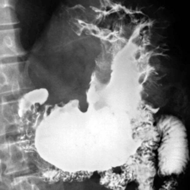
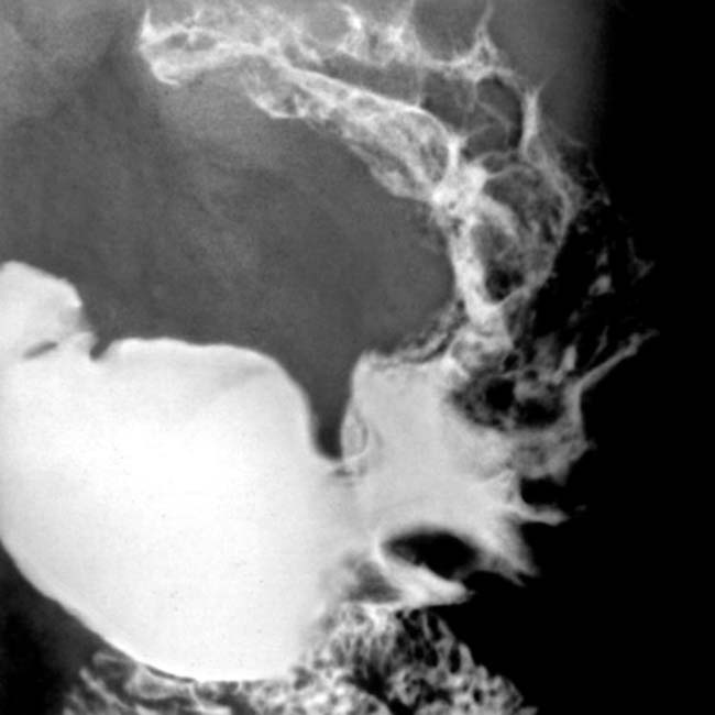
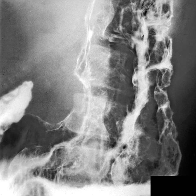
in the gastric fundus and body, along with engorged gastric vessels image. The thick, tortuous folds resemble cerebral convolutions.
image
(Left) Film from an upper GI series shows massive fold thickening of the gastric fundus and body, with sparing of the antrum. Note the poor coating of the gastric mucosa with barium to the surface of the stomach, reflecting the excessive mucus discharge of the gastric glands.

image
(Right) In the same patient, CECT shows marked thickening of the gastric mucosa and submucosa image
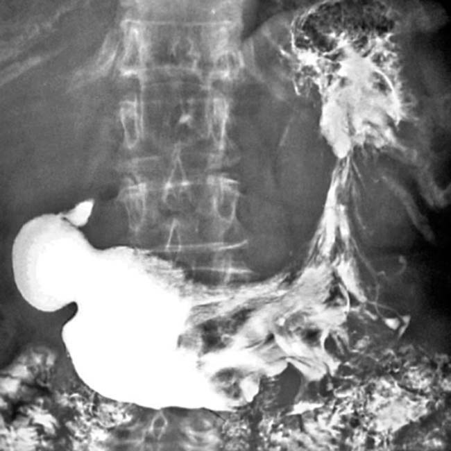
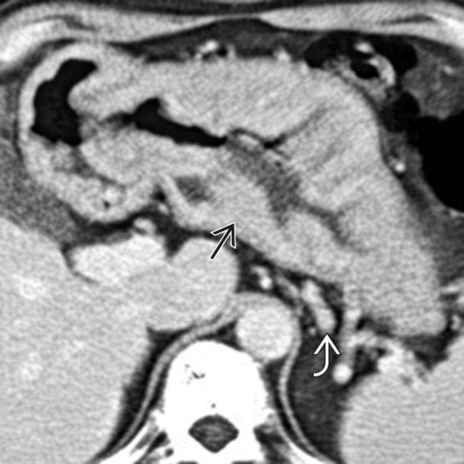
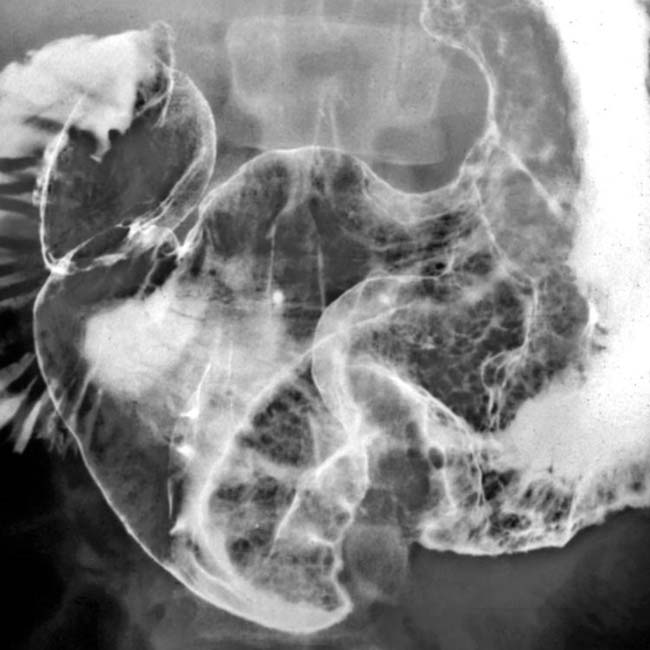
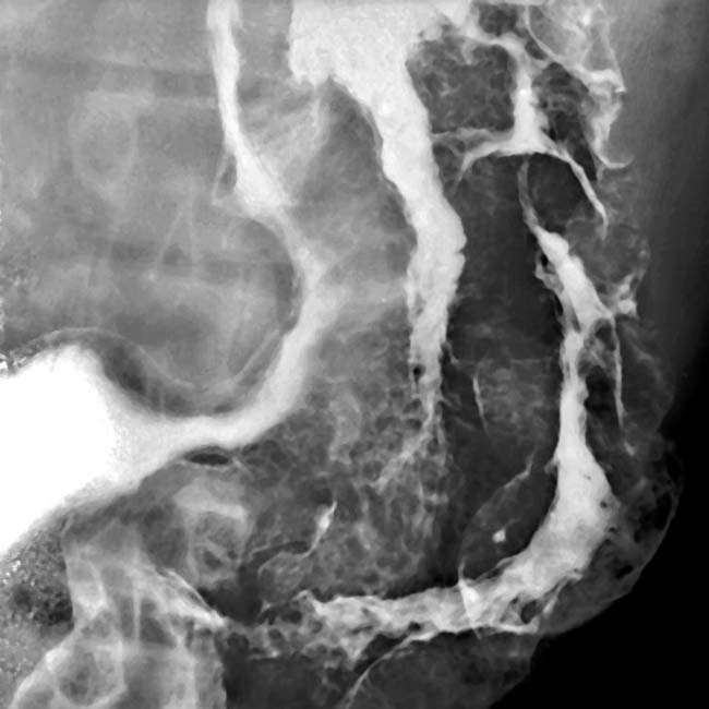
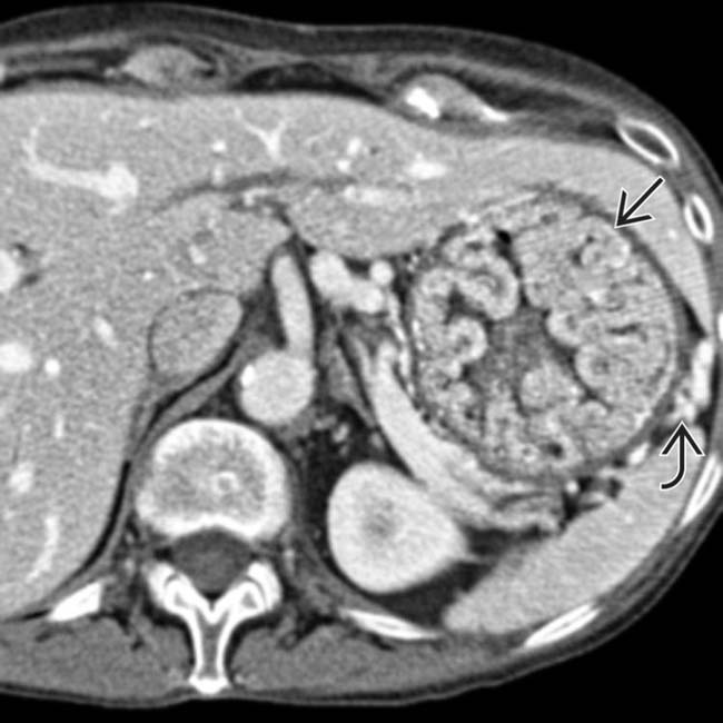
, but there is no sign of extension into the perigastric tissues. The gastric arterial and venous branches are engorged image, indicating hyperemia of the stomach.

TERMINOLOGY

Synonyms

• Hyperplastic gastropathy, protein-losing gastropathy

Definitions

• Rare acquired condition characterized by hyperproliferative protein-losing gastropathy of gastric foveolar epithelium

IMAGING

General Features

• Best diagnostic clue

image Grossly thickened, lobulated folds in gastric fundus and body with poor barium coating
• Other general features

image Rare condition of unknown cause
image Characterized by

– Enlarged, tortuous gastric rugal folds
– Marked foveolar hyperplasia (mucin production)

image Hypoproteinemia: Protein loss
– Hypochlorhydria (HCL output ↓ in 75% of cases)

Radiographic Findings

• Upper GI series

image Grossly thickened, lobulated folds in gastric fundus and body with relative sparing of antrum
image May show thickened gastric folds even in antrum
image Focal area of rugal hypertrophy on greater curvature
image Giant, mass-like, tortuous folds resemble cerebral convolutions
image Stomach remains pliable and distensible
image Excess mucus may dilute barium and ↓ mucosal coating

CT Findings

• Massive thickening of mucosa and submucosa
• Engorged gastric arteries and veins
• No extension into perigastric tissues

Imaging Recommendations

• Upper GI series, CECT

DIFFERENTIAL DIAGNOSIS

Gastritis

• From other causes, such as Helicobacter pylori
• Thickened, lobulated folds favor antrum
• Diagnosis: Endoscopic biopsy, culture, urease test

Zollinger-Ellison Syndrome

• Also causes thick folds and increased fluid in stomach
• Multiple ulcers, pancreatic tumor (gastrinoma) are not found in Ménétrier

Gastric Metastases and Lymphoma

• Thickened gastric folds similar to Ménétrier
• No excess fluid in stomach
• Submucosal soft tissue rather than edema
• Associated extragastric tumor or nodes

Gastric Carcinoma

• More often causes large discrete mass
• Diffuse infiltration limits distensibility
• Submucosal soft tissue density tumor (not edema)

PATHOLOGY

General Features

• Etiology

image Unknown
image Mucosal thickening (massive foveolar hyperplasia)
• Associated abnormalities

image Generalized signs of edema, hypoproteinemia (e.g., ascites, pleural effusion)

Gross Pathologic & Surgical Features

• Large, thickened, tortuous gastric mucosal folds
• Mucosal inflammation

Microscopic Features

• Cystic dilatation, elongated gastric mucous glands
• Massive ↑ in foveolar mucin-producing cells
• Atrophy and ↓ number of chief and parietal cells (→ decreased acid production)

CLINICAL ISSUES

Presentation

• Most common signs/symptoms

image Epigastric pain, vomiting, diarrhea, weight loss
image Occasionally peripheral edema (hypoproteinemia)
• Lab: ↓ albumin, ↓ or absent HCL, ± fecal occult blood
• Diagnosis: Endoscopic full thickness biopsy

Demographics

• Age

image Bimodal age distribution

– Children (usually boys)

image Has been associated with cytomegalovirus infection
image Relatively mild and self-limited disease
– Adults (mean age: 55 years); M > F
• Epidemiology

image Rare (< 1 in 200,000 people)

Natural History & Prognosis

• Complications

image Gastric carcinoma may have ↑ prevalence (controversial)
image ↑ risk of deep venous thrombosis
image Risk of atrophic gastritis, gastric ulcer, GI bleeding
• Prognosis

image Prolonged and progressive illness in most adults
image Children may have spontaneous remission

Treatment

• Medical therapy: Anticholinergic agents, antibiotics, proton pump inhibitors

image Octreotide (somatostatin analog)
image High-protein diet
image Monoclonal antibody (cetuximab) to epidermal growth factor receptor (EGFR)
• Total gastrectomy (unresponsive cases)
image
Upper GI series shows massive fold thickening in gastric fundus and body due to Ménétrier disease.

image
Upper GI series shows gross, tortuous gastric fold thickening in fundus and body with poor gastric coating by the barium.
image
Upper GI series shows massive fold thickening, sparing only the antrum.
image
Upper GI series shows massive gastric fold thickening and poor coating by barium.

SELECTED REFERENCES

1. Fiori, R, et al. Ménétrier’s disease diagnosed by enteroclysis CT: a case report and review of the literature. Abdom Imaging. 2011; 36(6):689–693.

Lambrecht, NW. Ménétrier’s disease of the stomach: a clinical challenge. Curr Gastroenterol Rep. 2011; 13(6):513–517.

Friedman, J, et al. Ménétrier disease. Radiographics. 2009; 29(1):297–301.

Rothenberg, M, et al. Successful use of octreotide to treat Ménétrier’s disease: a rare cause of abdominal pain, weight loss, edema, and hypoalbuminemia. Dig Dis Sci. 2009; 54(7):1403–1407.

Narumi, S, et al. Correspondence between computed tomography and endoscopy in Menetrier’s disease. Pediatr Int. 2008; 50(2):245–247.

Fishman, EK, et al. CT of the stomach: spectrum of disease. Radiographics. 1996; 16(5):1035–1054.

Wolfsen, HC, et al. Menetrier’s disease: a form of hypertrophic gastropathy or gastritis? Gastroenterology. 1993; 104(5):1310–1319.

REESE, DF, et al. Giant hypertrophy of the gastric mucosa (Menetrier’s disease):a correlation of the roentgenographic, pathologic, and clinical findings. Am J Roentgenol Radium Ther Nucl Med. 1962; 88:619–626.