21 Maxillary Block
Perspective
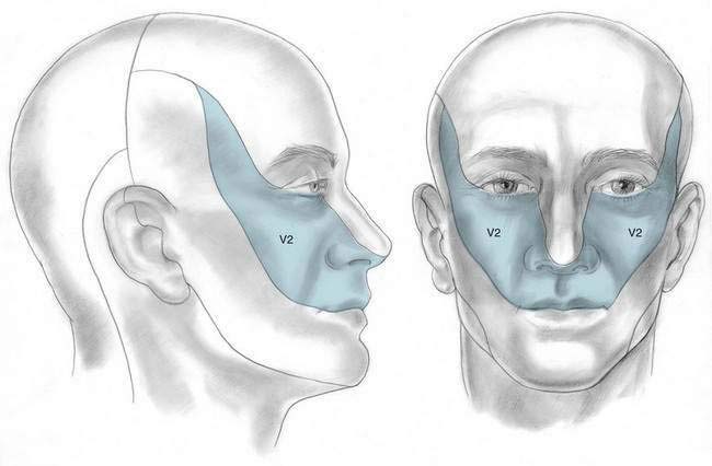
Local anesthetic block of the maxillary nerve in its peripterygoid location is most commonly used to evaluate facial neuralgia. However, it can be used to facilitate surgical procedures in the nerve’s cutaneous distribution (Fig. 21-1). Injection of neurolytic solution from the lateral approach to the maxillary nerve in its peripterygoid location should be undertaken with extreme caution owing to its location near the orbit.
Placement
Anatomy
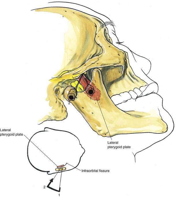
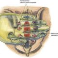
The maxillary nerve is entirely sensory and passes through the foramen rotundum to exit from the cranium. The nerve passes through the pterygopalatine fossa, medial to the lateral pterygoid plate, on its way to the infraorbital fissure. As illustrated in Figure 21-2, it is accessible to the anesthesiologist through a lateral approach as it passes into the pterygopalatine fossa.
Needle Puncture
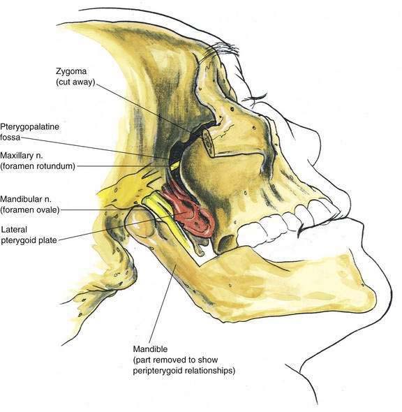
A 22-gauge, 8-cm needle is inserted through the mandibular notch in a slightly cephalomedial direction, as illustrated in Figure 21-3. This allows the needle to impinge on the lateral pterygoid plate at a depth of approximately 5 cm (needle position 1). The needle is then withdrawn and redirected in a stepwise manner toward position 2 (the pterygopalatine fossa). The needle should not be advanced more than 1 cm past the depth of initial contact with the pterygoid plate. As the needle is “walked off” the pterygoid plate, a sense of walking into the pterygopalatine fossa should be appreciated. Once the needle is adequately positioned, 5 mL of local anesthetic is injected.