MANAGEMENT OF COAGULATION DISORDERS IN THE SURGICAL INTENSIVE CARE UNIT

Published on 20/03/2015 by admin

Filed under Critical Care Medicine

Last modified 22/04/2025

Print this page

rate 1 star rate 2 star rate 3 star rate 4 star rate 5 star
Your rating: none, Average: 0 (0 votes)

This article have been viewed 1570 times

CHAPTER 89 MANAGEMENT OF COAGULATION DISORDERS IN THE SURGICAL INTENSIVE CARE UNIT

Surgeons commonly encounter coagulation disorders in the course of caring for patients, especially those with serious injury and those undergoing or recovering from surgery. Whereas bleeding is a condition well known to man since the beginnings of time, understanding the pathophysiology of bleeding and coagulation and developing effective therapies for them have come relatively recently and continue to undergo change as more is learned about the complex mechanism of blood coagulation and fibrinolysis. The ability to treat hemorrhage effectively had to await the discovery of blood types A, B, and O by Karl Landsteiner in 1900 and the AB blood type by Alfred Decastello and Adriano Sturli in 1902.

It would be nearly 40 years before the first blood bank was established in the United States in 1937. The development of reliable techniques of cross-matching, anticoagulation, and storage of blood was followed by the introduction of plastic bags for storage and devices for plasmapheresis making component therapy possible. The discovery of blood coagulation pathways and the development of reliable tests of coagulation made it possible to provide treatment for a variety of coagulation disorders, including those encountered as a result of the newfound ability to keep humans alive by the infusion of blood and the surgical control of bleeding.

The ability to replace blood loss is critically important in modern surgical practice and in trauma care. Equally important is the ability to provide therapy to patients who need individual blood components. Effective use of the precious resource that blood and its products represents is increasingly important as problems of supply continue to exist even while demand increases. The purpose of this chapter is to familiarize the practicing surgeon with the types of coagulation disorders encountered in critically ill or injured patients, reliable ways of diagnosing these disorders, and effective therapeutic strategies for treating them.

INCIDENCE

Congenital Bleeding Disorders

Von Willebrand Disease

Von Willebrand disease (vWD) is the most common inherited bleeding disorder, occurring in 1/100 to 1/1000 live births via autosomal inheritance. The disease consists of deficiency or dysfunction of von Willebrand factor (vWf), which promotes platelet adhesion to damaged endothelium and stabilizes factor VIII. There are three types of vWD. Knowing the specific type is important to direct therapy. In type 1, a deficiency of vWf exists. In type 3, vWf is absent. The main subtypes of type 2, 2a, and 2b, both consist of a qualitative functional defect in vWf.

Diagnosis of vWD is supported by prolonged partial thromboplastin time (PTT), and in types 1 and 3 reduced levels of vWf antigen. Factor VIII activity may be reduced, and bleeding time or other platelet functional assays may be abnormal. The ristocetin cofactor assay is a test that measures the ability of vWf to induce platelet aggregation.

1-deamino-8-D-arginine vasopressin (DDAVP) may be used to stimulate production of vWf and increase factor VIII levels in type 1 and type 2a disease. It is ineffective in type 3, however, and contraindicated in type 2b due to risk of thrombocytopenia and increased bleeding. Concentrates of factor VIII vWf are virus inactivated and are used commonly in types 2 and 3, but also in type 1 that is unresponsive to DDAVP. Cryoprecipitate contains vWf and factor VIII, and may be used in all types of vWD. However, it is pooled and not virus inactivated. It is only recommended as a third-line therapy. Antifibrinolytic amino acids, such as aminocaproic acid and tranexamic acid, are used as adjuvant therapy in all types of vWD along with the previously cited treatments.

Acquired Bleeding Disorders

Coagulopathy of Hemorrhagic Shock

Hemorrhagic shock causes a complex coagulopathy whose etiology is multifactorial, and widely misunderstood. Misinterpretation of clinical and laboratory data may lead clinicians to incorrectly label this coagulopathy as disseminated intravascular coagulation (DIC) or dilutional coagulopathy, which may misdirect treatment. In hemorrhagic shock, blood loss and tissue hypoperfusion result in acidosis from anaerobic metabolism—leading to the generation of lactate. Decreased ATP production from tissue ischemia contributes to hypothermia and inability to maintain core temperature. Coagulopathy is the result, which exacerbates bleeding and perpetuates the “bloody vicious cycle.” Resuscitation with room-temperature fluids worsens hypothermia. In massive resuscitation from hemorrhagic shock, variable degrees of dilution of coagulation factors occur.

Hypothermia and acidosis are the two major contributors to the coagulopathy of hemorrhagic shock, and are discussed in more detail in material following. In the operating room and postoperatively in the intensive care unit (ICU), multiple treatments are obviously conducted simultaneously. However, the priorities in general are to stop the bleeding, resuscitate with crystalloid and blood to reverse ischemia and acidosis, and prevent and treat hypothermia. Because of the overwhelming influence of hypothermia and acidosis, coagulopathy is primarily that of ineffective clotting. This is in contrast to DIC, which implies an overactivated coagulation system with unregulated microvascular thrombosis. Attributing the bleeding to DIC may lead one to focus therapy on providing clotting factors with fresh frozen plasma (FFP), or the rarely needed cryoprecipitate, when time is much better spent on adequate resuscitation and rewarming.

Dilutional coagulopathy (the idea that microvascular bleeding can result from dilution of clotting factors) has limited scientific support. Clotting factor concentrations as low as 30% of normal are sufficient for hemostasis, as are fibrinogen levels greater than 75 mg/dL. Even replacement of an entire blood volume leaves one with about a third of the normal coagulation factor concentration. This is probably the minimum volume of transfusion that can lead to a true dilutional coagulopathy. Although dilution of factors may result in abnormalities in laboratory measures of coagulation such as PT and PTT, these alterations do not necessarily affect hemostasis in vivo. Furthermore, platelet count cannot reliably be predicted based on volume of blood loss. Formula-based replacement (X units of FFP and platelets for every Y units of blood transfused) has little rationale, and should be discouraged. In the perioperative hemorrhagic shock patient, factor replacement with FFP should be based primarily on clinical evidence of microvascular (nonsurgical) bleeding to target clinical hemostasis while efforts to correct hypothermia and acidosis are optimized.

Hypothermia

Hypothermia is often seen in the critical care setting in association with the systemic inflammatory response syndrome (SIRS), sepsis, and shock, in which decreased oxygen consumption prevents maintenance of core body temperature. It routinely accompanies major surgery for hemorrhagic shock, in which it exacerbates the coagulopathy and should prompt a “damage control” strategy. In addition, heat loss from hemorrhage is compounded by the administration of room-temperature fluids and blood products. In trauma patients, temperatures less than 32° C are associated with 100% mortality.

Hypothermia slows the rate of reaction of the proteolytic enzymes of coagulation, resulting in impaired hemostasis. Both coagulation enzyme activity and platelet function are impaired at temperatures below 34° C in trauma patients. Platelet dysfunction is multifactorial, and is caused by defective adhesion and aggregation and decreased thromboxane production.

Prompt and efficient rewarming is essential in the hypothermic coagulopathic surgical patient. Although controlled hypothermia has proven beneficial in other conditions, such as cardiac arrest, no clear benefit has been proven in trauma or general surgery. The priority of therapy is to treat the underlying cause, whether by stopping any ongoing surgical bleeding, evacuating an undrained abscess, treating infection, or debriding necrotic tissue. External rewarming methods, although slow and inefficient, help to prevent further heat loss. Ambient room temperature should be raised, and warm air blankets and fluid pads applied to the patient (including the head). Core rewarming is far more efficacious than external techniques. At the very least, all infused fluids and blood products should be run through a fluid warmer, and warm humidified air given via the mechanical ventilator. When available, the more aggressive rapid technique of continuous arteriovenous rewarming may be used. A randomized prospective study suggests improved early survival and reduced fluid resuscitation requirements with this method when compared with slower methods.

Thrombocytopenia

Thrombocytopenia is generally defined as a platelet count lower than 100,000/mm3. Counts of 50,000/mm3 to 100,000/mm3 increase risk of bleeding with surgery or major trauma, and spontaneous bleeding is a risk below 10,000/mm3 to 20,000/mm3. Thrombocytopenia in the ICU setting has a lengthy differential diagnosis, but its etiology can be broadly divided into three categories: decreased production of platelets, consumption or sequestration of platelets, and dilution. Malignancies or chemotherapy may affect platelet production, and massive transfusion and fluid resuscitation can lead to dilution of the total platelet count. In critically ill surgical patients, sepsis can cause a consumptive coagulopathy that in its most severe form manifests as DIC. Platelet consumption also occurs through immune mechanisms (antibodies to platelet glycoproteins), most notably in response to certain drugs. The list of such drugs includes heparin, H2 antagonists, sulfa, rifampin, quinidine, hypoglycemics, and gold salts.

Heparin-induced thrombocytopenia is a rare but highly morbid condition associated with a greatly increased risk of thrombosis. Dilutional thrombocytopenia may occur with massive transfusion because stored blood contains negligible levels of platelets. However, the decrease in platelet count is not proportional to the volume of blood transfusion. Thus, simple dilution is unlikely to be the sole determinant of the low platelet count. Release of platelets from the spleen and bone marrow may partly account for this variability. As with coagulation factors, dilutional thrombocytopenia alone does not account for microvascular bleeding. Treatment and transfusion guidelines are discussed later in this chapter.

Disseminated Intravascular Coagulation

DIC is a syndrome involving diffuse systemic hypercoagulation and fibrinolysis that occurs in response to specific clinical conditions. Disorders associated with DIC in the surgical ICU include sepsis, trauma, severe pancreatitis, malignancies, fulminant liver failure, and transfusion reactions—among others. The syndrome involves excessive fibrin deposition in the microvasculature, with platelet aggregation and microvascular thrombosis. The pathophysiology of DIC is linked to the inflammatory cascade and TF pathway, and is reviewed in more detail elsewhere. The condition ranges in severity from a subclinical low-grade acceleration of thrombosis and fibrinolysis to overt pathologic bleeding. Fulminant DIC is associated with multiple-organ dysfunction and death.

Diagnosis of DIC is made with a few laboratory tests in the proper clinical setting, after other causes of coagulopathy have been excluded. Scoring systems and algorithms have been proposed to aid the diagnosis. However, treatment is mainly supportive and targets the underlying cause, clinical endpoints, and associated laboratory abnormalities. Given the nonspecific nature of DIC, setting a defined threshold for making the diagnosis in the clinical setting is unnecessary—whereas set criteria are still needed for therapeutic trials and research. In addition, the label of DIC is often applied to patients receiving massive transfusion and resuscitation when their coagulopathy stems from other more common and reversible causes. It has also been observed that trauma patients with DIC have a thrombotic and fibrinolytic profile distinct from the usual hemostatic response to trauma.

DIC may be suspected in the setting of a generalized coagulopathy and clinical microvascular bleeding associated with an underlying process such as those described previously. The laboratory profile includes a low platelet count, prolonged PT and PTT, and elevated fibrin split products. D-dimer levels are increased in up to 94% of patients diagnosed with DIC, and the D-dimer assay is the most sensitive test for this condition. Fibrinogen levels may be maintained except in severe forms of DIC.

Therapy for DIC centers on treatment of the underlying disease process to remove the proinflammatory stimulus of the syndrome. Clinical hemostasis is the goal. Platelet counts and the PT/PTT are used to guide response to therapy, but are not endpoints themselves. FFP and platelet transfusion are indicated in patients with active bleeding and those with significant laboratory derangements undergoing surgery or procedures. Cryoprecipitate may be considered to replace fibrinogen if fibrinogen levels fall below 100 mg/dl and are not corrected with FFP infusion.

Many other therapeutic agents have been investigated, but to date no specific treatment has proven successful in improving outcome in patients with DIC. Anticoagulation has been used to attempt to control the hypercoagulation in DIC, and although improvement in certain lab parameters has been reported no survival benefit has been demonstrated with low-molecular-weight heparin, thrombin inhibitors, or antifibrinolytics.

Severe Sepsis

Research in recent years continues to elucidate the complex interrelationship of the inflammatory process and the coagulation mechanism. The initial manifestation of this relationship leads to a hypercoagulable state. Inflammation in sepsis induces tissue factor (TF) expression on circulating monocytes, tissue macrophages, and the endothelial surface—and fibrinolysis is inhibited. As fibrinolysis is impaired, fibrin deposition in the microvasculature proceeds unchecked. In addition, most patients with severe sepsis have low levels of the natural anticoagulants protein C and antithrombin III. Diffuse thrombosis leads to tissue ischemia and the multiple-organ dysfunction syndrome (MODS).

Coagulopathy in sepsis is multifactorial. Sepsis-induced thrombocytopenia occurs through immune mechanisms, platelet sequestration on activated endothelium, and consumption in DIC. Extensive thrombin generation consumes clotting factors, and fibrinogen is often reduced (although levels may be normal due to its generation as an acute-phase reactant). Pathologic bleeding may occur due to lack of circulating clotting factors and platelets that have been consumed, but this is relatively uncommon. Although DIC is estimated to occur in 15%–30% of patients with severe sepsis, the incidence of serious bleeding episodes in a recent study of septic patients was only 5%.

Transfusion of FFP or platelets in septic patients is indicated for active bleeding, or those at high risk for bleeding. As mentioned previously, transfused factors and platelets usually have only a transient impact because they are depleted by the ongoing consumption in the microvasculature. However, in the face of active bleeding aggressive therapy is warranted while every effort is made to treat or remove the source of the sepsis.

Cirrhosis and End-Stage Liver Disease

Severe liver disease is associated with abnormal coagulation from multiple hemostatic defects. The diseased liver’s ability to synthesize coagulation factors is impaired, and fibrinogen levels are low in end-stage liver disease (ESLD) and decompensated cirrhosis but may be normalized by its acute-phase reaction to inflammation. Thrombocytopenia may be present due to decreased production of thrombopoietin in the liver, and platelets may be destroyed or sequestered. Platelet function may be altered as well, by an excess of circulating inhibitors of platelet aggregation such as nitrous oxide. Systemic fibrinolysis occurs, in part by reduced clearance in the liver of profibrinolytic enzymes. Patients with ESLD may appear to have a baseline low-grade DIC (e.g., elevated fibrin split products) and are at higher risk of declining into overt DIC. The frequency and severity of DIC generally advance with the disease stage of the liver.

Cirrhotic patients who require surgery pose a significant challenge to the surgeon. Morbidity and mortality are increased in such patients, especially for emergent operations (for which mortality may reach 50%). In one study, patients undergoing trauma laparotomy with intraoperatively diagnosed cirrhosis had 45% mortality compared to 24% in injury severity-matched controls. Postoperative ICU stay was significantly longer as well. Patients with ESLD undergoing surgery may have an enhanced fibrinolytic response due to release of tissue plasminogen activator (tPA) and other factors, hindering stable clot formation. Compounding the risk of bleeding from coagulopathy in ESLD is the presence of large intraabdominal and abdominal wall varices, which can make even the laparotomy incision itself a daunting task. Given all of these obstacles, the decision to undertake any invasive procedure on a patient with cirrhosis or ESLD must be made with the utmost discretion.

The goal of treatment of coagulopathy in ESLD should be clinical hemostasis, and not complete normalization of laboratory values (which is often not possible). Mild aberrations in lab assays are frequent and may not result in a bleeding diathesis. FFP is used for factor and fibrinogen replacement, but cryoprecipitate may be necessary if fibrinogen levels are lower than 100 mg/dl. Due to the short half-life of some clotting factors, large volumes of FFP may be needed to maintain the hemostatic state. Continuous FFP infusion is sometimes warranted, and can be titrated to clinical endpoints. In cases of life-threatening bleeding or the need for emergency surgery, recombinant human factor VIIa (rFVIIa) may be used to correct the INR acutely. However, its short half-life may necessitate repeated dosing after a few hours to maintain hemostasis. Transfusion guidelines for thrombocytopenia are the same as described elsewhere in this chapter. However, patients with splenomegaly may sequester transfused platelets and the rise in platelet count may be less than expected. The presence of microvascular bleeding with a normal platelet count may indicate platelet dysfunction, and a platelet function test may be considered. However, transfusion in these cases may result in brief or no improvement in hemostasis unless the underlying cause of the thrombocytopathy has been corrected. Administration of DDAVP may be considered, but its efficacy is unproven in this setting.

Despite an underlying coagulopathy, risk for thrombosis remains. Cirrhotic patients should not be presumed protected by an “auto-anticoagulation.” In fact, hepatic and portal vein thromboses are common in these patients, especially in advanced disease. The INR may be misleading, in that an elevated INR in ESLD does not necessarily correlate with the same level of anticoagulation as if that value were achieved with warfarin therapy. Deficient factor VII synthesis may produce a measurable abnormality in laboratory tests due to its short half-life, but clinical clotting abnormalities may not be apparent. Maintenance of normal fibrinogen levels is usually sufficient to aid in coagulation, except in late stages.

DIAGNOSIS

Clinical Evaluation

Bleeding in a critically ill patient should be evaluated in a systematic fashion to detect the cause and direct the treatment of the bleeding (Figure 1). In the surgical ICU, the first critical decision in a bleeding patient is to differentiate surgical bleeding from nonsurgical coagulopathic microvascular bleeding. This may be one of the most difficult decisions a surgeon can face. A nontherapeutic operation on a coagulopathic patient may exacerbate the vicious cycle, but leaving a surgically correctable source of bleeding untouched can prove fatal.

The evaluation begins with a detailed history, especially review of operative notes if the patient has had surgery or invasive procedures. Physical examination may reveal blood in operative wounds, tubes, or drains that indicate a source of bleeding requiring reoperation. Conversely, oozing of blood from multiple sites or seemingly minor wounds (e.g., intravenous catheter sites) may indicate coagulopathy. All recently administered medications should be reviewed for drugs that may affect hemostasis, in addition to reviewing the patient’s medical history.

Postoperative bleeding may be considered in the broad categories of loss of surgical hemostasis versus coagulation disorders. Loss of surgical hemostasis is bleeding at the operative site, which may be due to technical problems such as slipped ligature or inadequate hemostasis from the procedure. During an operation, vasoconstriction may prevent visible bleeding—but with warming and resuscitation bleeding resumes. Loss of surgical hemostasis usually requires definitive control through reoperation. Postoperative surgical bleeding may be associated with signs and symptoms ranging from hypovolemia to hemorrhagic shock. The physician should intervene based on early signs of shock (tachycardia, restlessness, anxiety, pallor, oliguria), and not wait until shock is glaringly obvious. Anxiety or agitation in a postoperative surgical patient should prompt first an assessment of perfusion and oxygenation, before analgesics or sedatives are given. Hypotension is a late sign of hemorrhage, indicating severe volume deficit.

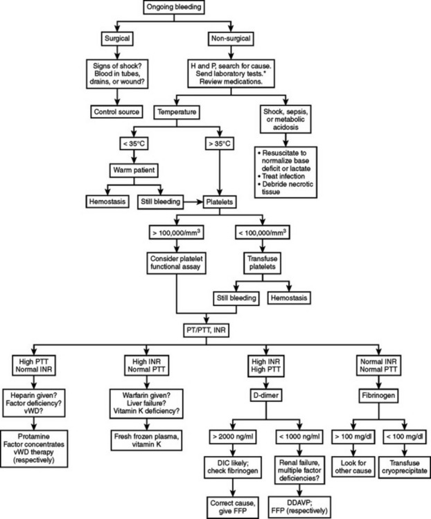
Coagulation disorders may be grouped into those affecting primary hemostasis (formation of initial platelet plug) or secondary hemostasis (clotting factors and the coagulation cascade). These groups may be subdivided into qualitative defects (e.g., dysfunctional platelets, factor inhibition by heparin) or quantitative defects (e.g., thrombocytopenia, factor deficiencies). Furthermore, these conditions may be congenital or acquired. The algorithm presented in Figure 1 represents one example of a systematic approach that can help guide therapy in most surgical patients, even if the exact cause of the coagulopathy is not evident. It is intended to aid rapid assessment and initiation of treatment in the ICU, rather than as a definitive guide to diagnosis of specific bleeding disorders.

A few basic laboratory tests are helpful in guiding diagnosis and treatment of coagulopathy. It is worthwhile first to reiterate that the primary goal of therapy is clinical hemostasis, and not complete normalization of every clotting parameter. Platelet count, PT, INR, and PTT are the minimum basic lab tests needed to help differentiate problems with primary or secondary hemostasis. A baseline hematocrit level should be checked, keeping in mind that acute hemorrhage will not be reflected by a change in hematocrit until dilution of the intravascular space occurs from fluid shifts and intravenous fluid administration. Thromboelastography (TEG) is a global test of coagulation that may help define the etiology of a coagulopathy. Fibrin split products, D-dimer, and fibrinogen levels are rarely necessary in the setting of hemorrhagic shock-induced coagulopathy but may help confirm a clinical diagnosis of DIC. Each test is discussed in more detail in the following section.

Laboratory Tests of Coagulation

PT: This test is done by adding a thromboplastin containing TF, phospholipid, and calcium to citrated plasma and measuring the time in seconds until a fibrin clot is formed compared to a control. The PT measures the activity of the extrinsic pathway (factor VII) and the common pathway (fibrinogen, factors II, IX, and X). It is used to monitor warfarin therapy, and is affected by depletion of the vitamin-K–dependent factors (factors II, VII, IX, and X, and proteins C and S).

INR: The INR is used to adjust for individual lab variation in the PT, using the formula INR = (log patient PT/log control PT) to the power of “c,” where c is the international sensitivity index (ISI). The thromboplastin used in individual laboratories is thus calibrated against a reference thromboplastin. The INR was developed to monitor the degree of warfarin anticoagulation.

PTT: The PTT is done by adding a partial thromboplastin (mixture of phospholipids), an activating substance, and calcium chloride to citrated plasma. It measures the activity of the intrinsic pathway (HMW kininogen, prekallikrein, and factors VIII, IX, XI, and XII) and the common pathway (fibrinogen, factors II, IX, and X). Only factor VII activity is not measured by the PTT.

Bleeding time: The bleeding time is a test of platelet function and primary hemostasis. However, due to variation in the performance of the test it is relatively insensitive and nonspecific in identifying platelet function abnormalities and may not predict surgical bleeding.

Platelet function tests: Several tests of platelet function are available through the lab or as point-of-care tests. Our hospital has abandoned the bleeding time in favor of the PFA-100 (Platelet Function Analyzer, Dade-Behring). The PFA-100 measures platelet function by the time it takes whole blood to occlude an aperture in a filter as it flows under high shear conditions. It is a global test of primary hemostasis that may detect platelet dysfunction due to certain disorders or medications, and congenital diseases such as vWD, but its role has not yet been completely defined. Other tests measure the percentage of platelets working normally to determine the functional platelet count, and are used often during cardiac surgery. Several point-of-care tests are available to assess platelet inhibition by drugs such as aspirin or GPIIb/IIIa inhibitors. Platelet aggregation tests use several agonists in different concentrations to induce aggregation in platelet-rich plasma, and will reveal quantitative or qualitative defects. It is a gold standard test but takes hours to perform, making it less useful in acute coagulopathy management.

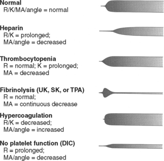
TEG: TEG is reported as a graph of clot formation in a sample of whole blood (Figure 2). The TEG tracing is drawn based on several factors, including rate of clot formation, fibrin cross-linking, and platelet-fibrin interaction. By measuring various parameters of the tracing, TEG provides an assessment of platelet function, coagulation enzyme activity, and the overall degree of coagulability. It can identify conditions such as primary fibrinolysis, consumptive coagulopathy, anticoagulant therapy, and the effect of hypothermia. TEG is used frequently during cardiopulmonary bypass, liver transplantation, and in intensive care settings due to its rapid availability and ability to assess the components of coagulation in an integrated fashion.

image

Figure 2 Thromboelastogram (TEG).

(Adapted from Kaufmann CR, et al: Usefulness of thromboelastography in assessment of trauma patient coagulation. J Trauma 42:716, 1997.)

TT: The TT is done by adding thrombin to citrated plasma +/– calcium. The TT measures the time for conversion of fibrinogen to fibrin, which is induced by thrombin. It is prolonged when fibrinogen is deficient (<100 mg/dl) or abnormal, in the presence of circulating anticoagulants (including fibrin split products [FSPs] and heparin), and during excessive fibrinolysis. Its high sensitivity to exogenous anticoagulants such as heparin limit its usefulness in hospitalized patients, but it can be used to detect low levels of circulating heparin that do not cause changes in the PTT.

Fibrinogen: Fibrinogen is a large protein that is cleaved by thrombin to produce fibrin monomers, which cross-link to form a fibrin clot in the presence of factor XIII. Fibrinogen levels may fall with the excess clotting seen in consumptive coagulopathy or with overanticoagulation by thrombolytic agents. It is also an acute-phase reactant, increasing in response to physiologic stress.

FSPs: FSP’s are fragments of the fibrin molecule that result from breakdown of fibrin by plasmin. The test is nonspecific, but elevated levels may indicate fibrinolysis and support a clinical picture of consumptive coagulopathy. The D-dimer is a specific form of FSP that is most closely associated with DIC.

Factor assays: Specific coagulation factor levels can be used to help diagnose certain diseases or deficiencies (for example, factor VIII for hemophilia A and factor IX for hemophilia B). Other assays may detect deficiencies in factors V, VII, X, XI, and XII (Hagemann factor), prekallikrein, and HMWK, all of which are very rare. Factor assays are used infrequently in the ICU setting.

MANAGEMENT

Blood Product Transfusion

Platelets

Treatment of thrombocytopenia or platelet dysfunction centers on the underlying cause and the patient’s clinical condition. In the absence of active bleeding or imminent surgery, platelet counts above 10,000/mm3 do not require treatment—whereas counts below 10,000/mm3 warrant platelet transfusion to prevent spontaneous bleeding. Patients with microvascular bleeding and thrombocytopenia may benefit from platelet transfusion after excluding hypothermia because the transfused platelets will not function properly at low temperatures. Evidence-based guidelines are lacking for surgical patients with platelet counts between 50,000/mm3 and 100,000/mm3, and therapy should take into account the patient’s condition, risk of significant bleeding, and plans for surgery or high-risk invasive procedures (e.g., ventriculostomy).

In general, platelet transfusion is not indicated at these levels in the absence of microvascular bleeding. Patients with consumptive coagulopathy rarely benefit from platelet transfusion because the same process consumes newly transfused platelets. However, microvascular bleeding in the presence of sepsis or DIC usually warrants treatment. If surgery or invasive procedures are necessary in the presence of a consumptive process and low platelet count, platelet transfusion should be given just before or during the procedure to maximize the number of circulating platelets available for hemostasis.

Different platelet concentrates are available. The traditional “six-pack” from random donor concentrates contains platelets from multiple individuals and equals six units of platelet concentrates. One unit of single-donor platelets, also called apheresis platelets, contains roughly the same volume of platelets as 6 random donor units but has the advantage of originating from one person and thereby exposing the recipient to only one set of antigens. One can expect a rise in platelet count by about 30,000/mm3 for each unit of single-donor platelets and for each 6 units of random donor. Repeated platelet transfusion may lead to alloantibody formation in some patients, making them refractory to further platelet transfusions. HLA-matched or cross-matched platelets may be required in such cases. The use of single-donor platelet transfusions has been adopted by many institutions to minimize antibody formation and preserve the response to a platelet transfusion for as long as possible.

Reversal of Warfarin

The prevalence of preinjury warfarin use among trauma patients increases with age. The effect of this drug on morbidity and mortality in trauma is variable, but potentially significant. Emergent reversal of warfarin anticoagulation is occasionally required in patients with TBI or serious injury associated with hemorrhage, and slower reversal is often used for patients with increased risk of bleeding due to trauma or perioperative status. Before initiating therapy, several factors should be considered—including urgency of warfarin reversal, expected length of time until re-anticoagulation, and cardiac function of the patient (i.e., tolerance of volume loading).

Reversal of warfarin is guided by the INR and is best managed by standardized evidence-based guidelines. Vitamin K takes 8–12 hours to take effect, and is the first-line choice for nonemergent treatment of a high INR. However, it has a long half-life and high or repeated doses should be avoided if re-anticoagulation with warfarin is anticipated in the next several days. Oral vitamin K is preferred for nonemergent reversal of warfarin, whereas the subcutaneous route is not recommended because of inefficient absorption. Patients receiving intravenous vitamin K should have continuous cardiac monitoring due to the risk of anaphylaxis. FFP is the standard therapy for patients with a high INR and significant bleeding or need for invasive procedures.

Many elderly patients on warfarin have concomitant heart disease, and caution must be used to avoid precipitating congestive heart failure with overly aggressive fluid loading. In our experience, an INR greater than 2 is rarely normalized with only one or two units of FFP. In addition, patient factors vary considerably—resulting in an unpredictable and nonlinear dose-response relationship. Recombinant activated factor VII (rFVIIa) may be used when immediate reversal of anticoagulation is required in emergent situations such as severe TBI or life-threatening bleeding. However, data on its proper use in these conditions is limited. rFVIIa’s half-life is only a fraction of that of warfarin, and thus it must be used in conjunction with FFP and vitamin K to maintain normal coagulation. Table 1 is an example of a management scheme for patients on warfarin with an elevated INR and risk of bleeding.

Table 1 Management Options for Patients with Warfarin Anticoagulation and Bleeding Risk

Clinical Context Treatment Options
INR < 5, no significant bleeding Hold warfarin
INR > 5, no significant bleeding Vitamin K 1–5 mg orally
INR > 1.5, bleeding or high risk of bleeding, nonemergent FFP in 2- to 4-unit doses, recheck INR after each dose, until bleeding stopped or INR < 1.5
Consider vitamin K 5 mg orally
INR > 1.5, life-threatening bleeding or emergent surgery or invasive procedure required FFP in 4-unit doses, recheck INR after each dose, and vitamin K 10 mg slow IV infusion
Consider factor 7a (repeat in 2–3 hours if still bleeding)

Reversal of Heparin

Unfractionated heparin (UFH) and low-molecular-weight heparin (LMWH) are used commonly in the surgical ICU for venous thromboembolism prophylaxis or treatment of other conditions. Although risk of major bleeding events with prophylactic doses is low, full anticoagulation is associated with higher risk. When nonsurgical bleeding occurs in patients anticoagulated with heparin, reversal of the drug’s effects may be necessary. The half-life of UFH is about an hour, and thus most treatment doses are reversed by holding the infusion for 6 hours. When immediate reversal is desired, protamine may be used. Protamine binds heparin and neutralizes its effects. The dose is 1 mg of protamine for each 100 units of heparin given. The half-life of heparin must be taken into account when calculating the protamine dose, such that the dose of heparin must be halved for each hour since its injection. If a continuous infusion has been used, the cumulative dose must be estimated. Protamine administration carries risks of hypotension, which may be avoided by slow injection over 10 minutes, and a 1% risk of anaphylaxis in patients who have had previous exposure to protamine or NPH insulin.

The half-life of LMWH varies with the particular agent used, but in general ranges from 2 to 5 hours. LMWH is only partially neutralized by protamine, which reverses most of the anti-IIa (thrombin) activity but only some of the anti-Xa activity. The reversal is based on the level of anti-Xa activity, in a dose of 1 mg protamine per 100 anti-Xa units.

Recombinant Activated Factor VIIa

rFVIIa is a synthetic form of coagulation factor VII, intended to promote hemostasis. It is an FDA-approved drug for bleeding in hemophilia patients with inhibitors, but has also been used in a variety of other conditions. The primary mechanism of action has been debated. When bound to exposed TF in the subendothelium, rFVIIa can activate factors X and IX—which then promote thrombin formation. This mechanism would explain its localized activity at sites of injury. Other data suggest that high-dose rFVIIa acts independently of TF by activating factor Xa on the platelet surface.

rFVIIa has proven efficacious in reducing blood loss and improving survival in multiple animal studies of its use for the coagulopathy of hemorrhagic shock, and in reducing blood loss and operative time in humans undergoing radical prostate surgery. In blunt trauma patients, rFVIIa reduces the need for blood transfusion and for massive transfusion. A similar significant benefit was not seen in patients with penetrating trauma, however. Initial concerns about an increased risk of thrombosis with rFVIIa have not been borne out. Studies have revealed no evidence of systemic thrombi or increased risk of thrombotic complications in animals or humans.

The optimal dose of rFVIIa for surgical patients has not yet been determined. Doses ranging from 20 to 200 micrograms/kg have been used successfully in clinical trials. Due to the drug’s short half-life, certain conditions such as severe coagulopathy may require a second or third dose within a few hours of the first to maintain hemostasis while other contributing factors are aggressively treated. Until specific guidelines are developed through future large multicenter trials, use of rFVIIa should be directed in accordance with local hospital policies, economic considerations, and specific patient variables.

The current expense of the drug precludes its routine use in most bleeding conditions. We have employed a multidisciplinary approach to develop guidelines for use of rFVIIa on our Trauma Service, and have limited prescribing authority to certain specialists. Our guidelines promote use of rFVIIa in two specific conditions: severe hepatic trauma requiring surgery and coagulopathy from hemorrhagic shock (as diagnosed by operating surgeon in the presence of microvascular bleeding) that is unresponsive to standard therapy. Its use in other conditions, such as TBI, remains unspecified.

SUGGESTED READINGS

Aird WC. Sepsis and coagulation. Crit Care Clin. 2005;21:417.

Ansell J, et al. The pharmacology and management of the vitamin K antagonists: the seventh ACCP conference on antithrombotic and thrombolytic therapy. Chest. 2004;126(Suppl 3):204S.

Bernard GR, et al. Safety and dose relationship of recombinant human activated protein C for coagulopathy in severe sepsis. Crit Care Med. 2001;29:2051.

Boffard KD, et al. Recombinant factor VIIa as adjunctive therapy for bleeding control in severely injured trauma patients: two parallel randomized, placebo-controlled, double-blind clinical trials. J Trauma. 2005;59:8.

Boccardo P, et al. Platelet dysfunction in renal failure. Semin Thromb Hemost. 2004;30(5):579.

Cable R, et al. Practice guidelines for blood transfusion: a compilation from recent peer-reviewed literature. American National Red Cross, 2002. http://www.redcross.org/services/biomed/profess/pgbtscreen.pdf

Committee on Trauma of the American College of Surgeons. Advanced Trauma Life Support Course for Doctors, 7th ed., Chicago: American College of Surgeons; 2004:69.

Cosgriff N, et al. Predicting life-threatening coagulopathy in the massively transfused trauma patient: hypothermia and acidoses revisited. J Trauma. 1997;42:857.

Demetriades D, et al. Liver cirrhosis in patients undergoing laparotomy for trauma: effect on outcomes. J Am Coll Surg. 2004;199:539.

Fakhry SM, et al. Hematologic principles in surgery. In: Townsend CM, Beauchamp RD, Evers BM, Mattox KL, editors. Sabiston Textbook of Surgery: The Biological Basis of Modern Surgical Practice. 16th ed. Philadelphia: WB Saunders; 2001:68.

Forsythe SM, Schmidt GA. Sodium bicarbonate for the treatment of lactic acidosis. Chest. 2000;117:260.

Gando S. Disseminated intravascular coagulation in trauma patients. Semin Thromb Hemost. 2001;27(6):585.

Gando S, et al. Coagulofibrinolytic changes after isolated head injury are not different from those in trauma patients without head injury. J Trauma. 1999;46:1070.

Gentilello LM, et al. Is hypothermia in the victim of major trauma protective or harmful? A randomized, prospective study. Ann Surg. 1997;226(4):439.

Holcomb JB. Use of recombinant activated factor VII to treat the acquired coagulopathy of trauma. J Trauma. 2005;58:1298.

Jurkovich GJ, et al. Hypothermia in trauma victims: an ominous predictor of survival. J Trauma. 1987;27:1019.

Kashuk JL, et al. Major abdominal vascular trauma—a unified approach. J Trauma. 1982;22:672.

Kujovich JL. Hemostatic defects in end stage liver disease. Crit Care Clin. 2005;21:563.

Levi M. Disseminated intravascular coagulation: what’s new? Crit Care Clin. 2005;21:449.

Mannucci PM. Treatment of von Willebrand’s disease. N Engl J Med. 2004;351:683.

Mina AA, et al. Complications of preinjury warfarin use in the trauma patient. J Trauma. 2003;54:842.

Oung CM, et al. In vivo study of bleeding time and arterial hemorrhage in hypothermic versus normothermic animals. J Trauma. 1993;35:251.

Owings JT, Gosselin RC. Bleeding and transfusion. In: Fink MP, Jurkovich GJ, Kaiser LR, Pearce WH, Pemberton J, Soper NJ, editors. ACS Surgery: Principles & Practice. New York: Web MD Corp, 2003. www.acssurgery.com

Rodgers RPC, Levin J. A critical reappraisal of the bleeding time. Semin Thromb Hemost. 1990;16:1.

Stein SC, Smith DH. Coagulopathy in traumatic brain injury. Neurocrit Care. 2004;1:479.

The American Society of Anesthesiologists Task Force on Blood Component Therapy. practice guidelines for blood component therapy. Anesthesiology. 1996;84(3):732.

Valeri CR, et al. Hypothermia induced reversible platelet dysfunction. Ann Surg. 1987;205:175.

Watts DD, et al. Hypothermic coagulopathy in trauma: effect of varying levels of hypothermia on enzyme speed, platelet function, and fibrinolytic activity. J Trauma. 1998;44:846.

Wolberg AS, et al. A systematic evaluation of the effect of temperature on coagulation enzyme activity and platelet function. J Trauma. 2004;56:1221.

Wojcik R, et al. Preinjury warfarin does not impact outcome in trauma patients. J Trauma. 2001;51:1147.