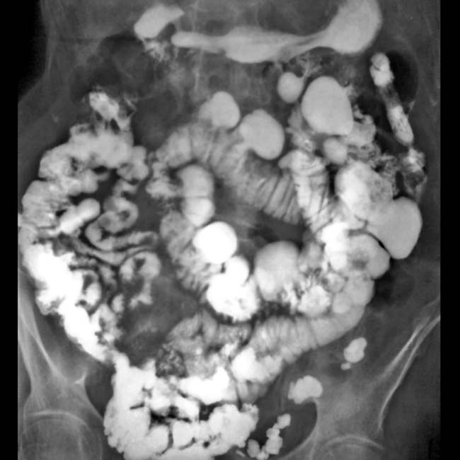
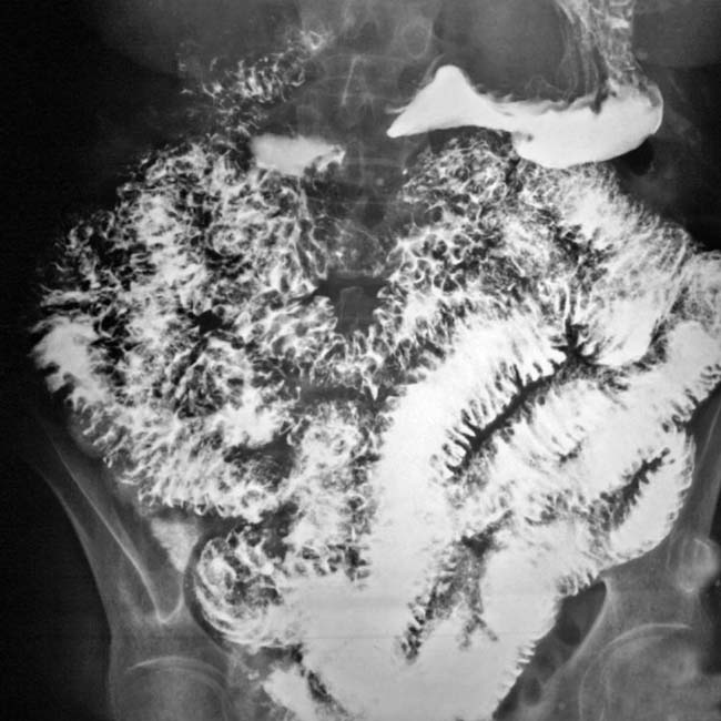
(Left) Small bowel (SB) follow through in a 56-year-old man with adult onset of malabsorption condition illustrates segments of jejunum that are featureless and devoid of valvulae conniventes, reminiscent of normal loops of ileum.
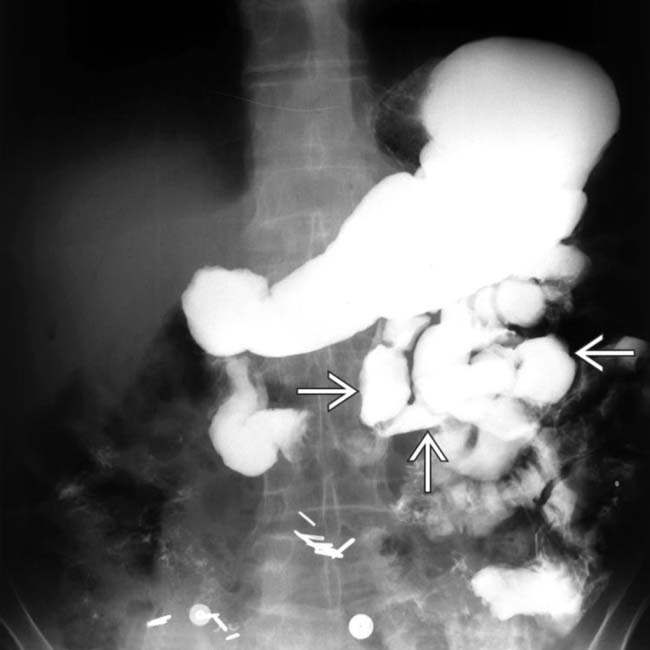
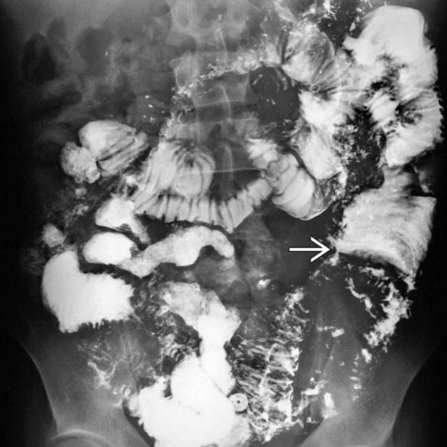
(Right) Small bowel follow-through in the same patient demonstrates the ileum to have a fold pattern reminiscent of the normal jejunum . This reversal of the fold pattern is characteristic of adult celiac disease.
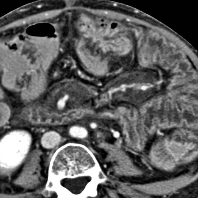
(Left) Axial CECT in a 61-year-old man with chronic diarrhea and recent weight loss shows marked dilation of the duodenal and jejunal lumen with a very diminished fold pattern.
(Right) Axial CECT in the same patient conversely illustrates abnormally prominent ileal folds , the “fold reversal” pattern characteristic of celiac-sprue. Focal segmental luminal spasm and dilation are also evident, along with excess fluid within the SB.
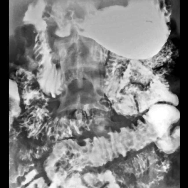
Small bowel follow through (SBFT) in a 24 year old with diarrhea shows dilated bowel with segments of spasm . Note the barium dilution due to increased intestinal fluid and the irregular fold thickening, findings constituting the “malabsorption” pattern.
Enteroclysis shows symmetrical fold thickening due to hypoproteinemia.
SBFT shows distorted nodular SB folds and diluted barium in this patient with dysgammaglobulinemia.
Axial CECT shows SB fold thickening, excess fluid in the lumen, and mesenteric engorgement due to primary lymphangiectasia.
SBFT shows a nodular SB fold pattern with dilated lumen in this patient with Waldenström macroglobulinemia.
SBFT in a patient with sprue shows segmental dilation with spasm of the bowel, dilution, and flocculation of barium. Also, note the transient intussusception of the jejunum with a “coiled spring” appearance.
SBFT shows numerous large diverticula throughout the jejunum, which can lead to stasis, bacterial overgrowth, and malabsorption.


 that are featureless and devoid of valvulae conniventes, reminiscent of normal loops of ileum.
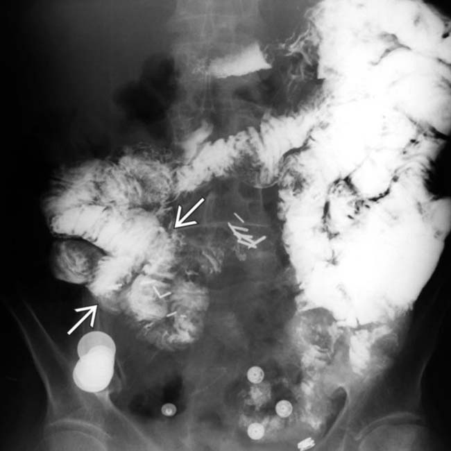
 . This reversal of the fold pattern is characteristic of adult celiac disease.
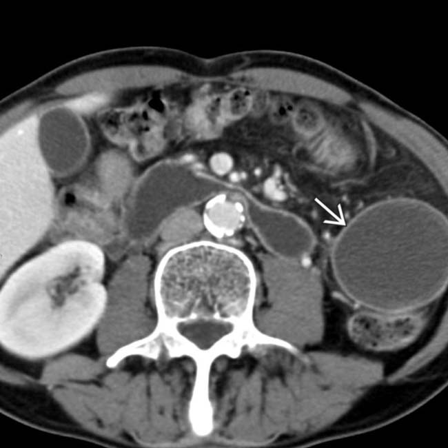
 with a very diminished fold pattern.
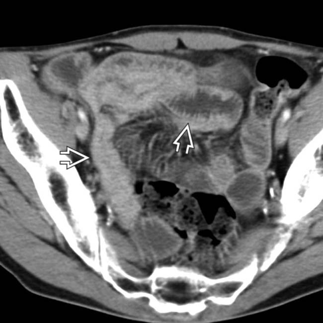
 , the “fold reversal” pattern characteristic of celiac-sprue. Focal segmental luminal spasm and dilation are also evident, along with excess fluid within the SB.
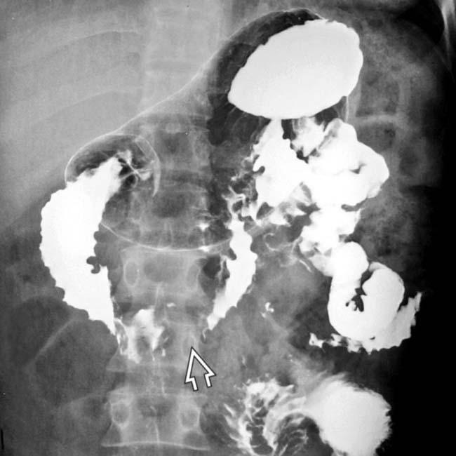
 . Note the barium dilution due to increased intestinal fluid and the irregular fold thickening, findings constituting the “malabsorption” pattern.
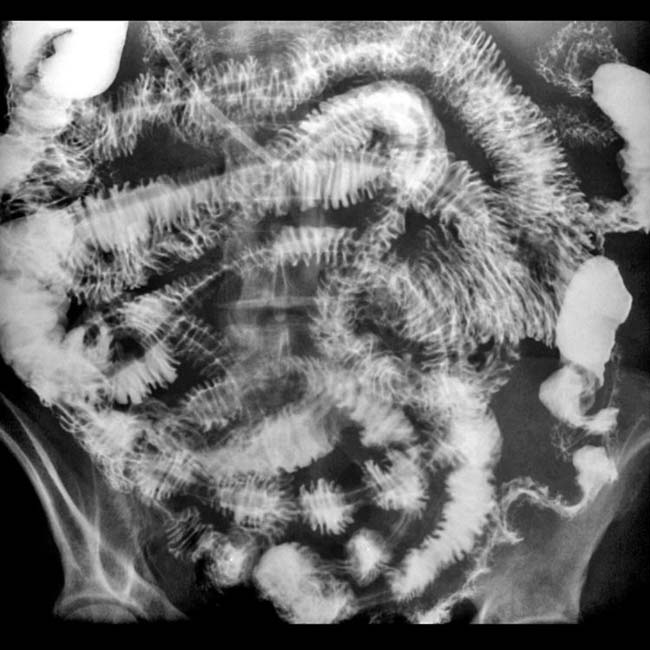
 of the jejunum with a “coiled spring” appearance.