Chapter 11 Lymph glands, lymphatics and tumours
Positron Emission Tomography Imaging
Positron emission tomography (PET) imaging is a technique used to detect and accurately stage malignant disease, to differentiate benign and malignant tissue, and to assess response to treatment. Until recently, PET imaging availability was restricted due to high capital cost and logistics of radiopharmaceutical supply. It uses short-lived cyclotron-produced radionuclides such as 18Fluorine, 11Carbon, 13Nitrogen and 15Oxygen with half-lives of 110, 20, 10 and 2 min respectively. 18Fluorine is the only one of these that has a half-life long enough to allow it to be produced off-site. This does permit 2-[18F]fluoro-2-deoxy-d-glucose (18F-FDG), the single most important PET radiopharmaceutical, to be used by sites without their own cyclotron.
The widespread acceptance of PET as a major advance is due to two major factors:
2-[18F]FLUORO-2-DEOXY-D-GLUCOSE (18F-FDG) PET SCANNING
Indications (oncology)
General
Patient preparation
Technique
British Nuclear Medicine Society Web Site guidelines –. www.bnms.org.uk, 2008.
Cook G.J., Wegner E.A., Fogelman I. Pitfalls and artifacts in 18FDG PET and PET/CT oncologic imaging. Semin. Nucl. Med.. 2004;34(2):122-133.
Kapoor V., McCook B.M., Torok F.S. An introduction to PET CT imaging. RadioGraphics. 2004;24(2):523-543.
Rohren E.M., Turkington T.G., Coleman R.E. Clinical applications of PET in oncology. Radiology. 2004;231(2):305-332.
von Schulthess G.K., Steinert H.C., Hany T.F. Integrated PET/CT: current applications and future directions. Radiology. 2006;238(2):405-422.
Gallium Radionuclide Tumour Imaging
This is rarely used, having almost entirely been superseded by cross-sectional techniques and PET scanning.1 The main disadvantages are the high radiation dose, the extended nature of the investigation, its non-specific nature, and difficulties in interpretation in the abdomen due to normal bowel activity.
Indications
Equipment
Images
Bombardieri E., Aktolun C., Baum R.P., et al. 67Ga scintigraphy: procedure guidelines for tumour imaging. Eur. J. Nucl. Med. Mol. Imaging.. 2003;30(12):BP125-131.
Front D., Bar-Shalom R., Israel O. The continuing clinical role of gallium 67 scintigraphy in the age of receptor imaging. Semin. Nucl. Med.. 1997;27(1):68-74.
Radioiodine Metaiodobenzylguanidine Scan
Metaiodobenzylguanidine (MIBG) is a noradrenaline (norepinephrine) analogue. It is taken up actively across cell membranes of sympathetic and adrenal medullary tissue into intracellular storage vesicles. There is no further metabolism, and it remains sequestered and localized in the storage vesicles of catecholamine-secreting tumours and tumours of neuroendocrine origin.1
Radiopharmaceuticals
Patient preparation
Technique
Additional techniques
1 Ilias I., Pacak K. Current approaches and recommended algorithm for the diagnostic localization of pheochromocytoma. J. Clin. Endocrinol. Metab.. 2004;89(2):479-491.
2 Solanki K.K., Bomanji J., Moyes J., et al. A pharmacological guide to medicines which interfere with the biodistribution of radiolabelled meta-iodobenzylguanidine (MIBG). Nucl. Med. Commun.. 1992;13(7):513-521.
Somatostatin Receptor Imaging
Somatostatin is a physiological neuropeptide which has biological effects including inhibition of growth hormone release, and suppression of insulin and glucagon excretion. Octreotide (a long-acting analogue of the human hormone, somatostatin) can be used therapeutically to inhibit hormone production by carcinoids, gastrinomas and insulinoma, etc. A number of tumours, particularly those of neuroendocrine origin, express neuroendocrine receptors. Imaging after the administration of radionuclide-labelled somatostatin analogues such as octreotide, therefore, allows their localization.1
Radiopharmaceuticals
111Indium (In) pentetreotide (a DTPA conjugate of octreotide) 220 MBq i.v. (ED 17 mSv).
Patient preparation
Lymph Node Imaging
ULTRASOUND
MAGNETIC RESONANCE LYMPHANGIOGRAPHY
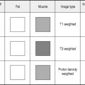
Currently under assessment is MR lymphangiography. High-resolution T2*-weighted MR scans are obtained prior to and post the injection of lymphotropic superparamagnetic nanoparticles. Normal nodes are high signal on the pre-contrast scan, but lose signal on the post contrast signal due the T2* effect from the iron content in the particles taken up by normal reticuloendothelial cells. Nodes containing malignant tissue retain their high signal intensity on the post contrast scans and small tumour deposits can be detected in even normal-sized nodes.1
RADIONUCLIDE LYMPHOSCINTIGRAPHY
Indications
Radiopharmaceuticals
Technique
1 Harisinghani M.G., Barentsz J., Hahn P.F., et al. Noninvasive detection of clinically occult lymph-node metastases in prostate cancer. New Engl. J. Med.. 2003;348(25):2491-2499.
2 Krynyckyi B.R., Kim C.K., Goyenechea M.R., et al. Clinical breast lymphoscintigraphy: optimal techniques for performing studies, image atlas, and analysis of images. RadioGraphics. 2004;24(1):121-145.
3 Witte C.L., Witte M.H., Unger E.C., et al. Advances in imaging of lymph flow disorders. RadioGraphics. 2000;20(6):1697-1719.
Radionuclide Imaging of Infection and Inflammation
A number of radionuclide techniques exist for this, the most commonly used of which is radionuclide-labelled leucocyte imaging.1,2 The ready availability and sensitivity for collections and inflammation of anatomical imaging techniques such as US and CT has, however, reduced the demand for radionuclide procedures.