Chapter 4 Liver, biliary tract and pancreas
Methods of imaging the hepatobiliary system
PLAIN FILMS
May be useful to demonstrate air within the biliary tree or portal venous system, opaque calculi or pancreatic calcification.
ULTRASOUND OF THE LIVER
Indications
Technique
Additional views
Spleen
The spleen size should be measured in all cases of suspected liver disease or portal hypertension. 95% of normal adult spleens measure 12 cm or less in length, and less than 7×5 cm in thickness. The spleen size is commonly assessed by ‘eyeballing’ and measurement of the longest diameter.1 In children, splenomegaly should be suspected if the spleen is more than 1.25 times the length of the adjacent kidney,1 normal ranges have also been tabulated according to age and sex.2
1 Loftus W.K., Metreweli C. Ultrasound assessment of mild splenomegaly: spleen/kidney ratio. Pediatr. Radiol.. 1998;28(2):98-100.
2 Megremis S.D., Vlachonikolis I.G., Tsilimigaki A.M. Spleen length in childhood with US: normal values based on age, sex, and somatometric parameters. Radiology. 2004;231(1):129-134.
Albrecht T., Hohmann J., Oldenburg A., et al. Detection and characterisation of liver metastases. Eur. Radiol.. 2004;8(14 Suppl):25-33.
Kim T.K., Jang H.J., Burns P.N., et al. Focal nodular hyperplasia and hepatic adenoma: differentiation with low-mechanical-index contrast-enhanced sonography. Am. J. Roentgenol.. 2008;190(1):58-66.
Kono Y., Mattrey R.F. Ultrasound of the liver. Radiol. Clin. North Am.. 2005;43(5):815-826.
Shapiro R.S., Wagreich J., Parsons R.B., et al. Tissue harmonic imaging sonography: evaluation of image quality compared with conventional sonography. Am. J. Roentgenol.. 1998;171:1203-1206.
Wilson S.R., Burns P.N. An algorithm for the diagnosis of focal liver masses using microbubble contrast-enhanced pulse-inversion sonography. Am. J. Roentgenol. 2006;186(5):1401-1412.
Ultrasound of the Gallbladder and Biliary System
Technique
Additional views
Assessment of gallbladder function
Extrahepatic bile ducts
Ultrasound of the Pancreas
Technique
The pancreatic duct should not measure more than 3 mm in the head or 2 mm in the body.
Endoscopic US (see p. 81) and intra-operative US are useful adjuncts to transabdominal US. EUS may be used to further characterize and biopsy pancreatic mass lesions. Intra-operative US is used to localize small lesions (e.g. islet cell tumours prior to resection).
Eloubeidi M.A., Jhala D., Chhieng D.C., et al. Yield of endoscopic ultrasound-guided fine-needle aspiration biopsy in patients with suspected pancreatic carcinoma. Cancer. 2003;99(5):285-292.
Rizk M.K., Gerke H. Utility of endoscopic ultrasound in pancreatitis: a review. World J. Gastroenterol.. 2007;13(47):6321-6326.
Computed Tomography of the Liver and Biliary Tree
Indications
Multi-phasic contrast-enhanced CT
The fast imaging times of helical/multi-slice CT enable the liver to be scanned multiple times after a single bolus injection of contrast medium. Most liver tumours receive their blood supply from the hepatic artery, unlike the hepatic parenchyma, which receives 80% of its blood supply from the portal vein. Thus liver tumours (particularly hypervascular tumours) will be strongly enhanced during the arterial phase (beginning 20–25 s after the start of a bolus injection) but of similar density to enhanced normal parenchyma during the portal venous phase. Some tumours are most conspicuous during early-phase arterial scanning (25 s after the start of a bolus injection), others later, during the late arterial phase 35 s after the start of a bolus injection. Thus a patient who is likely to have hypervascular primary or secondary liver tumours should have an arterial phase scan as well as a portal venous phase CT scan (see above). Early and late arterial phase with portal venous phase is appropriate for patients with suspected hepatocellular cancer (triple phase). In general, late arterial and portal venous scans are appropriate to investigate suspected hypervascular metastases, although an alternative strategy would be to perform an unenhanced scan followed by a portal venous phase scan.
1 Hashimoto M., Itoh K., Takeda K., et al. Evaluation of biliary abnormalities with 64-channel multidetector CT. Radiographics. 2008;28(1):119-134.
2 Schindera S.T., Nelson R.C., Paulson E.K., et al. Assessment of the optimal temporal window for intravenous CT cholangiography. Eur. Radiol.. 2007;17(10):2531-2537.
Francis I.R., Cohan R.H., McNulty N.J., et al. Multidetector CT of the liver and hepatic neoplasms: effect of multiphasic imaging on tumor conspicuity and vascular enhancement. Am. J. Roentgenol.. 2003;180(5):1217-1224.
Erratum in. Am. J. Roentgenol.. 2003;181(1):283.
Oto A., Tamm E.P., Szklaruk J. Multidetector row CT of the liver. Radiol. Clin. North Am.. 2005;43(5):827-848.
Computed Tomography of the Pancreas
Technique
Fletcher J.G., Wiersema M.J., Farrell M.A., et al. Pancreatic malignancy: value of arterial, pancreatic, and hepatic phase imaging with multi-detector row CT. Radiology. 2003;229(1):81-90.
Goshima S., Kanematsu M., Kondo H., et al. Pancreas: optimal scan delay for contrast-enhanced multi-detector row CT. Radiology. 2006;241(1):167-174.
Magnetic Resonance Imaging of the Liver
Indications
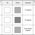
Magnetic resonance (MRI) is rapidly emerging as the imaging modality of choice for detection and characterization of liver lesions. There is high specificity with optimal lesion-to-liver contrast and characteristic appearances on differing sequences and after contrast agents. Focal lesionsmay be identified onmost pulse sequences. Most metastases are hypo- to isointense on T1 and iso- to hyperintense on T2-weighted images. However, multiple sequences are usually necessary for confident tissue characterization. The timing, degree and nature of tumour vascularity form the basis for liver lesion characterization based on enhancement properties. Liver metastases may be hypo or hyper-vascular.
Magnetic resonance imaging pulse sequences
A further breath-hold technique with very short sequential image acquisition.
This sequence can be obtained rapidly following i.v. gadolinium.
T2-weighted fast spin-echo (FSE; General Electric) or turbo spin-echo (TSE; Siemens)
Compared with conventional T2-weighted SE images, FSE/TSE images show:
Fat suppression is also used to allow better delineation of fluid-containing structures.
Contrast-enhanced magnetic resonance liver imaging
Liver-specific contrast agents
High signal contrast can be seen in the bile ducts. These agents are also excreted by the kidneys. Further details may be found in Chapter 2.
Catalano O.A., Sahani D.V., Kalva S.P., et al. MR imaging of the gallbladder: a pictorial essay. Radiographics. 2008;28(1):135-155.
Dromain C., de Baere T., Baudin E., et al. MR imaging of hepatic metastases caused by neuroendocrine tumors: comparing four techniques. Am. J. Roentgenol.. 2003;180(1):121-128.
Lutz A.M., Willmann J.K., Goepfert K., et al. Hepatocellular carcinoma in cirrhosis: enhancement patterns at dynamic gadolinium – and superparamagnetic iron oxide-enhanced T1-weighted MR imaging. Radiology. 2005;237(2):520-528. Epub 2005 Sep 28
Onishi H., Murakami T., Kim T., et al. Hepatic metastases: detection with multi-detector row CT, SPIO-enhanced MR imaging, and both techniques combined. Radiology. 2006;239(1):131-138. Epub 2006 Feb 16
Rappeport E.D., Loft A., Berthelsen A.K., et al. Contrast-enhanced FDG-PET/CT vs. SPIO-enhanced MRI vs. FDG-PET vs. CT in patients with liver metastases from colorectal cancer: a prospective study with intraoperative confirmation. Acta Radiol.. 2007;48(4):369-378.
Ward J., Robinson P.J., Guthrie J.A., et al. Liver metastases in candidates for hepatic resection: comparison of helical CT and gadolinium – and SPIO-enhanced MR imaging. Radiology. 2005;237(1):170-180. Epub 2005 Aug 26
Magnetic Resonance Cholangiopancreatography (MRCP)
Indications
Technique
MRCP is a non-invasive technique which uses heavily T2-weighted images to demonstrate the intra- and extra-hepatic biliary tree and pancreatic duct. Most commonly used to demonstrate the presence of stones and the level and cause of obstruction, especially combined with cross-sectional MRI, in cases of tumour or suspected tumour.
MAGNETIC RESONANCE IMAGING OF THE PANCREAS
ENDOSCOPIC RETROGRADE CHOLANGIOPANCREATOGRAPHY
Technique
The pharynx is anaesthetized with 50–100mg Xylocaine spray and the patient is sedated until conscious sedation is achieved. The patient then lies on the left side and the endoscope is introduced. The ampulla of Vater is located and the patient is turned prone. A polythene catheter prefilled with contrast medium is inserted into the ampulla, having ensured that all air bubbles are excluded. A small test injection of contrast under fluoroscopic control is made to determine the position of the cannula. It is important to avoid over-filling of the pancreas. If it is desirable both to opacify the biliary tree and the pancreatic duct, the latter should be cannulated first. A sample of bile should be sent for culture and sensitivity if there is evidence of biliary obstruction.
Intra-Operative Cholangiography
Indications
During cholecystectomy or bile duct surgery, to avoid surgical exploration of the common bile duct. This technique has been replaced in some centres by pre-operative MRCP.
Films
The criteria for a normal operative choledochogram were given by Le Quesne1 as:
Post-Operative (T-Tube) Cholangiography
Indications
Technique
PERCUTANEOUS TRANSHEPATIC CHOLANGIOGRAPHY
Indications
Technique
Films
Complications
BILIARY DRAINAGE
EXTERNAL DRAINAGE
This is achieved following transhepatic cannulation of the biliary tree as described above. The procedure may be performed to improve jaundice or sepsis prior to surgery or as a further percutaneous intervention.
INTERNAL DRAINAGE
Patient preparation, see percutaneous transhepatic cholangiography above
Technique
Transhepatic
Burke D.R., Lewis C.A., Cardella J.F., et alSociety of Interventional Radiology Standards of Practice Committee. Quality improvement guidelines for percutaneous transhepatic cholangiography and biliary drainage. J. Vasc. Interv. Radiol.. 2003;14(9, part 2):S 234-S 236.
Percutaneous Extraction of Retained Biliary Calculi (Burhenne Technique)
Contrast medium
HOCM or LOCM 150. (Low-density contrast medium is used to avoid obscuring the calculus.)
Technique
ANGIOGRAPHY
RADIONUCLIDE IMAGING OF LIVER AND SPLEEN
Indications
Radionuclide Hepatobiliary and Gallbladder Radionuclide Imaging
Radiopharmaceuticals
These 99mTc-labelled IDA derivatives are rapidly cleared from the circulation by hepatocytes and secreted into bile in a similar way to bilirubin;4 this allows the assessment of biliary drainage and gallbladder function. A number have been developed with similar kinetics, but the later ones, such as TBIDA, have high hepatic uptake and low urinary excretion, giving better visualization of the biliary tract at high bilirubin levels than the early agents.
Films
Additional techniques
Cholecystokinin (CCK) and morphine provocation1
Pharmacological intervention can be used in combination with TBIDA scanning to improve diagnosis of diseases affecting the gallbladder, common bile duct or sphincter of Oddi. CCK causes gallbladder contraction and sphincter of Oddi relaxation. An i.v. infusion of CCK is given over 2–3 min when the gallbladder is visualized 30–45 min after TBIDA administration. Dynamic imaging is continued for a further 30–40 min.
Quantitative measures of gallbladder ejection fraction and emptying rate can be calculated. It has been suggested that a slow CCK infusion over 30–60 min may improve specificity.5
Morphine causes sphincter of Oddi contraction. In a clinical setting of suspected acute cholecystitis, if the gallbladder is not observed by 60 min, an infusion of 0.04 mg kg−1 over 1 min can be given and imaging continued for a further 30 min. Continued non-visualization of the gallbladder up to 90 min is considered to confirm the diagnosis. Morphine provocation has also found success in diagnosis of elevated sphincter of Oddi basal pressure.2
1 Krishnamurthy S., Krishnamurthy G.T. Cholecystokinin and morphine pharmacological intervention during 99mTc-HIDA cholescintigraphy: a rational approach. Semin. Nucl. Med.. 1996;26:16-24.
2 Thomas P.D., Turner J.G., Dobbs B.R., et al. Use of 99mTc-DISIDA biliary scanning with morphine provocation for the detection of elevated sphincter of Oddi basal pressure. Gut. 2000;46(6):838-841.
3 Rayter Z., Tonge C., Bennett C., et al. Ultrasound and HIDA: scanning in evaluating bile leaks after cholecystectomy. Nucl. Med. Commun.. 1991;12:197-202.
4 Krishnamurthy G.T., Turner F.E. Pharmacokinetics and clinical application of technetium 99m-labeled hepatobiliary agents. Semin. Nucl. Med.. 1990;20:130-149.
5 Ziessman H.A. Cholecystokinin cholescintigraphy: victim of its own success? J. Nucl. Med.. 1999;40:2038-2042.
Investigation of Specific Clinical Problems
THE INVESTIGATION OF JAUNDICE
The aim is to separate haemolytic causes of jaundice from obstructive jaundice or hepatocellular jaundice. Clinical history and examination are followed by biochemical tests of blood and urine, and haematological tests.
THE INVESTIGATION OF PANCREATITIS
CT is the next investigation. It should be performed initially without oral or i.v. contrast enhancement to look for the presence of calcification within the pancreas itself and to look for small gallstones, which can be obscured by the presence of oral contrast medium. Scans of the pancreas should then be obtained during portal venous phase enhancement. This will enable identification of non-perfused areas of pancreas, and the presence of pseudocysts, abscesses and phlegmons should be sought.
Burns P.N., Wilson S.R. Focal liver masses: enhancement patterns on contrast-enhanced images – concordance of US scans with CT scans and MR images. Radiology. 2007;242(1):162-174. Epub 2006 Nov 7
Hussain S.M., Semelka R.C. Hepatic imaging: comparison of modalities. Radiol. Clin. North Am.. 2005;43(5):929-947.
Rubens D.J. Hepatobiliary imaging and its pitfalls. Radiol. Clin. North. Am.. 2004;42(2):257-278.
Siddiqi A.J., Miller F. Chronic pancreatitis: ultrasound, computed tomography, and magnetic resonance imaging features. Semin. Ultrasound CT MR. 2007;28(5):384-394. Review
Wilson S.R., Kim T.K., Jang H.J., et al. Enhancement patterns of focal liver masses: discordance between contrast-enhanced sonography and contrast-enhanced CT and MRI. Am. J. Roentgenol.. 2007;189(1):W7-W12.