Leukemia

Published on 06/06/2015 by admin

Filed under Pediatrics

Last modified 22/04/2025

Print this page

rate 1 star rate 2 star rate 3 star rate 4 star rate 5 star
Your rating: none, Average: 1 (1 votes)

This article have been viewed 2026 times

55 Leukemia

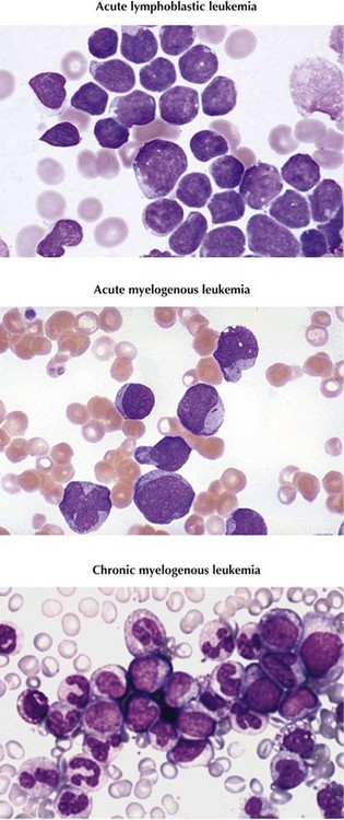
Leukemia is the most common type of childhood cancer, accounting for more than 3000 new cases annually and 25% of all malignancies diagnosed in patients younger than 20 years in the United States. Subtypes and prevalence include acute lymphoblastic leukemia (ALL), 75%; acute myelogenous leukemia (AML), 20%; and chronic myelogenous leukemia (CML), less than 5% (Figure 55-1). Other types of chronic leukemia, including those of lymphocytic and myelomonocytic cell lineages, are extremely rare in childhood.

Cure rates of pediatric leukemia have improved drastically over the past 50 years as knowledge of genetic and molecular factors of the disease have increased, supportive care has improved, and treatment strategies have become more sophisticated. For pediatric patients, 80% to 85% of patients with ALL and 50% to 60% of patients with AML are cured of their disease.

Etiology And Pathogenesis

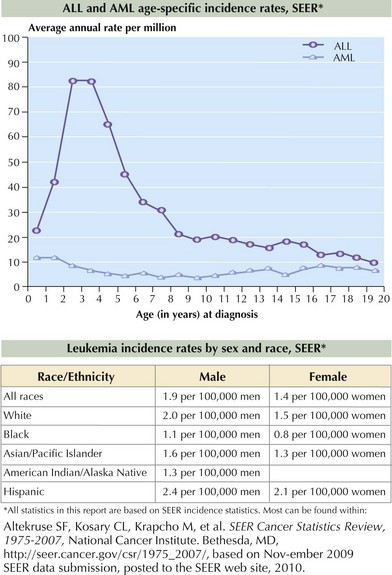
ALL occurs more frequently in boys and Caucasian children. The incidence peaks at 2 to 5 years, as shown in Figure 55-2. AML affects boys and girls equally, and pediatric incidence peaks in the neonatal and late adolescent periods (see Figure 55-2). In the United States, Hispanic and African American children are diagnosed with AML slightly more often than Caucasian children.

Leukemic cells are derived from hematopoietic stem cells that acquire genetic alterations that affect their ability to mature or undergo apoptosis, leading to perpetual self-renewal. The etiology of most of these mutations is unknown; however, certain environmental exposures, familial factors, genetic syndromes, and infectious diseases have been investigated as predisposing agents in childhood leukemia.

Studies of twins, neonatal blood spots (Guthrie cards), and cord blood are beginning to provide insight into the development of acute leukemia. In twin studies, concordance for all leukemias in monozygotic pairs is 5% to 25%; 10% in ALL and approaching 100% in infant leukemia. The extraordinarily high twin concordance rate for infant leukemia is attributed to blood chimerism of monochorionic twins. It is thought that the leukemia cells are passed from one twin to the other via a shared blood supply. This chimerism also occurs through placental vascular anastomoses in approximately 8% of dichorionic twin pairs.

Upon review of Guthrie cards of children who later developed leukemia, genetic mutations unique to leukemic clones were found to be present at birth, suggesting some leukemia may originate in utero. Because not all children with these mutations at birth develop leukemia, the mutations are believed to be necessary but not sufficient to induce leukemogenesis. Clearly, a second mutation and probably multiple sequences of mutations are required. Based on twin concordance studies, these latter mutations most likely occur postnatally.

Prenatal and postnatal genetic insults resulting in leukemia can be extrapolated to nontwin patients. A survey of hundreds of cord blood samples revealed that 1% had a functional TEL-AML1 gene (a common chromosomal translocation in pre B-cell ALL). This rate is 100 times that of clinically diagnosed ALL in the pediatric population. This serves as additional evidence that the genetic abnormalities found in cord blood at birth are not sufficient alone to result in the development of ALL.

Several environmental factors are associated with pediatric leukemia. These include ionizing radiation and chemotherapeutic agents, such as topoisomerase II inhibitors and alkylating agents. Other suspected environmental exposures include hydrocarbons such as benzene and pesticides leading to AML. Children with inherited genetic syndromes, including trisomy 21, Fanconi anemia, ataxia-telangiectasia, Wiskott-Aldrich syndrome, and neurofibromatosis type I, have an increased risk of developing acute leukemia.

Infectious agents have been proposed as playing a role in the development of leukemia, especially ALL. The peak age of incidence correlates with the age of first exposure to many infections for children in developed countries. Additionally, pediatric leukemia is more common in industrial regions of developed countries than in developing countries. It is thought that the later onset of exposure to infectious agents seen in developed countries results in an abnormally rapid immune cell proliferation and dysregulation. This hypothesis is also supported by several studies that compared infectious exposures of children who did and did not attend daycare early in life. The results showed that children with early daycare attendance and earlier exposure to infectious agents were less likely to develop ALL.

Genetics

Although little is known about the cause of initial mutations in the hematopoietic stem cell DNA of leukemia cells, the resultant genetic abnormalities are well studied. Leukemia is caused by multiple disruptions in cell DNA leading to (1) impaired maturation, (2) unregulated proliferation, and (3) lack of programmed cell death (apoptosis). In combination, this leads to the abnormal survival of mutated hematopoietic progenitor cells and unregulated growth of dysfunctional lymphoblasts or myeloblasts.

Some of the mutations are caused by chromosomal translocations resulting in fusion proteins that bring activated kinases and altered transcription factors together inappropriately. TEL-AML1 is the most commonly identified chromosomal translocation in pediatric ALL. TEL-AML1 t(8;21) is the product of the TEL gene responsible for recruiting progenitor stem cells into the bone marrow and the AML1 gene that plays a central role in hematopoietic cell differentiation. The Philadelphia chromosome is the product of a translocation of chromosomes 9 and 22, which is found in 95% of patients with CML and 2% of those with ALL. The translocation results in a fusion protein, BCR-ABL, which encodes a constitutively active tyrosine kinase protein. The tyrosine kinase activates proteins responsible for signaling within the cell cycle, therefore inducing uncontrolled cell proliferation, reducing apoptosis, and inhibiting DNA repair mechanisms (Figure 55-3). MLL, found on chromosome 11q23, partners with more than 40 genes and is found in childhood AML, secondary (or therapy-induced) AML, and the majority of infant ALL. Because the MLL gene arrangement is associated with several types of leukemia, specifically secondary AML after prior topoisomerase II inhibitor exposure, investigators are evaluating environmental topoisomerase II exposures that might be responsible for in utero mutations resulting in infant leukemia.

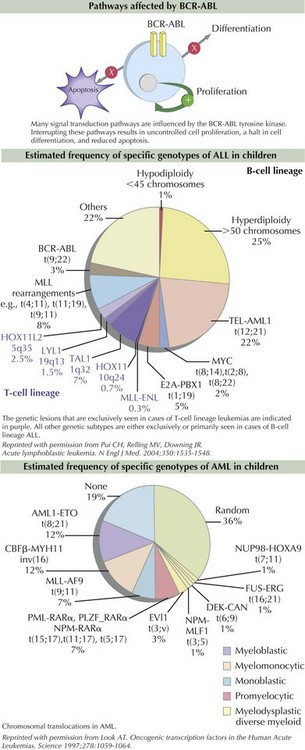
The process of cytogenetic profiling involves evaluating leukemic blasts for both the number of chromosomes and specific translocations (see Figure 55-3). This information is used to classify leukemia subtypes and give prognostic information and is often part of treatment risk stratification algorithms. For example, in pre–B-cell ALL, hyperdiploidy (>50 chromosomes) is associated with a good prognosis. However, hypodiploidy (<45 chromosomes) signifies a poor prognosis. Patients with AML are considered to have good-risk disease if the leukemia cell contains t(8;21) and inv(16) mutations. Other genetic abnormalities such as monosomy 5 and 7 and abnormalities of 3q are considered unfavorable. As many as 40% of patients with AML have activating mutations within the FLT3 gene, which are known as internal tandem duplications (ITDs). Both increased number of ITDs and location within the FLT3 gene are associated with poor clinical outcome.

High-resolution genetic analysis using single nucleotide polymorphism (SNP) array studies allow for more detailed profiling of additional genetic alterations. SNP arrays reveal mutations of genes involved in cell pathways such as lymphoid development, cell cycle regulation, apoptosis, and drug responsiveness. Clinically, this provides insight into the genotype and phenotype of leukemia and the underlying biology of the disease and can be associated with prognosis. SNP analysis can also follow the progression of genetic alterations during the disease course, which may predict response or resistance to therapy. SNP arrays have shown that genetic alterations in 60% of pre–B-cell ALL cases involve transcription factors that control B-cell differentiation. SNP analyses are increasingly important in providing clinical information about leukemia and have tremendous potential for discovering new molecular targets to treat specific leukemias.

Clinical Presentation

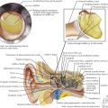
Children with leukemia primarily present with symptoms of bone marrow failure. The leukemia cells overpopulate the marrow and prevent normal growth of other hematopoietic cells. Signs and symptoms of anemia include fatigue, headache, pallor, and tachycardia. Thrombocytopenia often presents with petechiae and easy bruising, and leukopenia results in infection and fevers. The crowding of the bone marrow can also cause bony pain, often mistaken for “growing pains” in school-aged children (Figure 55-4).

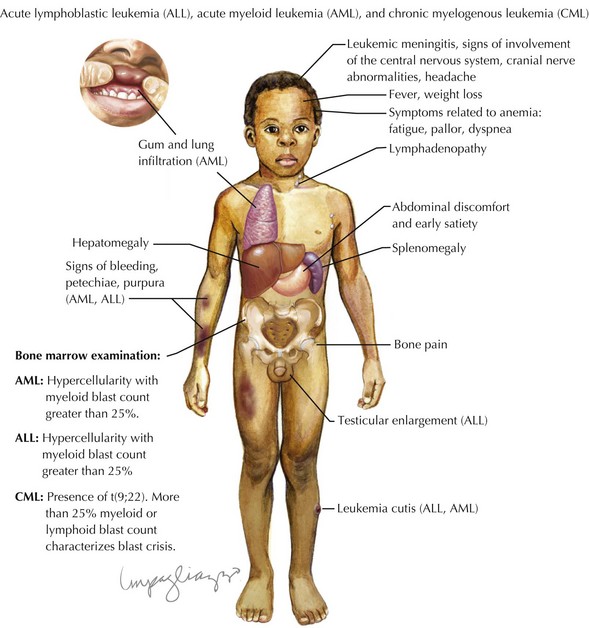
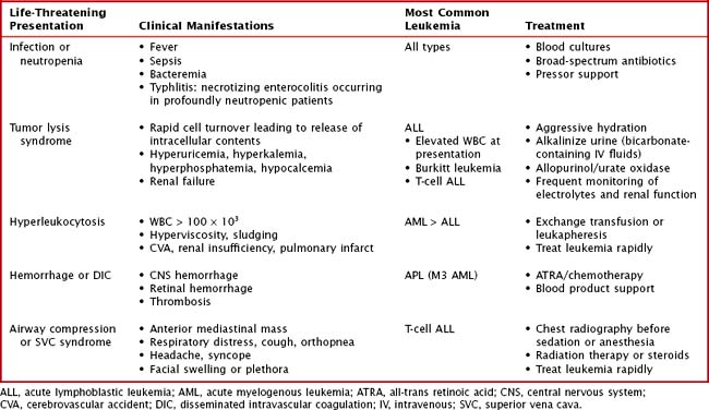
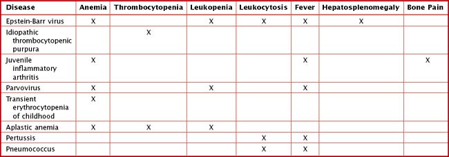
Other findings on physical examination are caused by leukemic infiltrate into normal tissues, such as the liver, spleen, lymph nodes, thymus, or testicles. The testicle is a common extramedullary site for ALL and may present as an enlargement of one or both testes. Leukemia cutis, or leukemic infiltration of the skin, has a varied presentation and may appear as single or multiple lesions commonly described as violaceous or hemorrhagic. Leukemia cutis most commonly presents in infant ALL and AML. Central nervous system (CNS) involvement at diagnosis is most often asymptomatic, but children with CNS disease may present with symptoms of increased intracranial pressure, visual disturbances, cranial nerve palsies, or gait disturbance. Other rare CNS effects not directly related to leukemic infiltration include intracranial hemorrhage and infarction. The presentation of CML can be very nonspecific and is often only suspected when a high white blood cell (WBC) count is seen on the complete blood count (CBC). Table 55-1 lists some of the life-threatening complications of leukemia at presentation and during therapy. Childhood leukemia presents similarly to many other common childhood illnesses (Table 55-2), and a bone marrow aspirate is necessary to make a definitive diagnosis.

Evaluation

The initial laboratory evaluation should include a CBC with manual differential and basic metabolic panel including calcium and phosphorus, prothrombin time, partial thromboplastin time, fibrinogen, uric acid, and lactate dehydrogenase. Automated differentials are inadequate because myeloblasts are often identified as monocytes and lymphoblasts as atypical lymphocytes. Further laboratory testing may be necessary based on presenting signs and symptoms. Children with a suspected diagnosis of leukemia should be referred to a pediatric cancer center for stabilization, preparation for diagnostic procedures, and molecular testing.

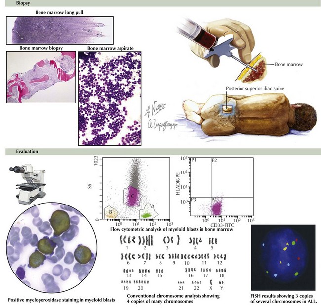
Chest radiography should be performed before sedation or anesthesia is used to determine the presence of a mediastinal mass. A bone marrow aspirate and biopsy must be reviewed for morphology, immunohistochemistry, and cytogenetics (Figure 55-5). A lumbar puncture is also performed at diagnosis to determine the presence of leukemia within the CNS.

Morphologic review delineates the cell lineage and characteristics to classify the type of leukemia. Immunohistochemistry identifies specific cell surface cluster of differentiation (CD) markers, which increases the accuracy of morphologic diagnosis. For example, B-cell precursor ALL blasts are known to express CD10, CD19, and CD20; AML blasts express CD13 and CD33. In some cases, morphology appears varied, and immunophenotyping may indicate a biphenotypic blast population with both lymphoid and myeloid blasts present. Identification of specific translocations and gene copy number alterations via karyotyping, fluorescence in situ hybridization, and SNP arrays indicate the cytogenetic profile of blast cells.

Specific molecular probes to known blast signatures have enabled monitoring of very small numbers of blast cells, or minimal residual disease (MRD), throughout therapy. Blasts may be detected at a level of 0.01%. This information is collected at regular intervals and used to tailor therapy or to predict relapse. MRD is a relatively new tool in leukemia therapy, and there is continued investigation into its clinical uses. The continued presence of MRD after the first 12 weeks of therapy is associated with a very poor prognosis.

Classification

ALL is classified according to cell lineage as either B- or T-cell disease. B-cell disease is further categorized based on the level of differentiation of the B-cell involved (Table 55-3). Historically, the French-American-British (FAB) classification system divided ALL into three subtypes. Because this classification did not account for more sophisticated methods of immunophenotyping and cytogenetic profiling, it is no longer used. The FAB classification of AML is still in use and consists of subtypes M0 to M7 based on cell type and differentiation (Table 55-4).

Table 55-3 Acute Lymphoblastic Leukemia Subtypes and Frequency

Subtype Frequency (%)
Early precursor B-cell 60-65
Precursor B-cell 20
Mature B-cell “Burkitt leukemia” 3
T-cell 15

Table 55-4 Acute Myelogenous Leukemia Classification Based on Cell Type

Subtype Cell type Details
M0 Undifferentiated stem cells Very rare in children
M1 Immature myeloblasts  
M2 Slightly matured myeloblasts Accounts for 25%-30% pediatric AML
M3 Promyelocytes (mature myeloblasts) Also known as acute promyelocytic leukemia
M4 Immature monoblasts Also known as acute myelomonocytic leukemia; most common in children younger than 2 years of age
M5 Monoblasts Also known as acute monocytic leukemia; more common in children younger than 2 years of age
M6 Erythroblasts Also known as acute erythroblastic leukemia; very rare in children
M7 Megakaryoblasts Also known as acute megakaryoblastic leukemia

AML, acute myelogenous leukemia.

CML classification is based on disease phase: chronic phase, accelerated phase, or blast crisis. Each phase requires specific monitoring and therapy. Most pediatric patients are diagnosed in the chronic phase, often resulting in mild symptoms. If untreated, the disease will progress to the accelerated phase. The accelerated phase is established based on one of the following parameters: thrombocytopenia, refractory thrombocytosis, increasing splenomegaly or leukocytosis, new cytogenetic abnormalities, or increasing blast load (10%-19%). The accelerated phase indicates progression of disease and impending blast crisis. Blast crisis occurs when there are more than 20% blasts in peripheral blood or bone marrow or when an extramedullary leukemic infiltrate develops.

Management And Prognosis

Acute Lymphoblastic Leukemia

As the survival rate of children with ALL improves, the goals of therapy include decreasing the toxicity of treatment for low-risk patients and escalating or targeting therapy for high-risk patients. Patients are considered high risk based on clinical characteristics, which are WBC greater than 50 × 103 or age older than 10 years at diagnosis. All others are defined as standard risk. Children younger than 1 year of age are considered to have infant ALL and undergo more intensive chemotherapy than their older counterparts. CNS or testicular involvement at diagnosis and slow early response to treatment are high-risk features, and intensified therapy is recommended for these groups. Molecular features, including immunophenotypic and cytogenetic profile, and MRD further define risk, prognosis, and need for treatment intensification.

ALL therapy in the United States generally consists of five phases: induction, consolidation, interim maintenance, delayed intensification, and maintenance, for a total duration of 2.5 to 3 years depending upon gender. ALL induction therapy has an excellent rate of remission (>99%) and a very low mortality rate from toxicity (<1%). Induction therapy is a 4-week phase and includes a steroid, vincristine, and asparaginase. Anthracycline therapy is added for high-risk patients. Methotrexate, given intrathecally, is used for CNS chemoprophylaxis in all patients with ALL. Cranial irradiation is added for high-risk patients and those with CNS involvement. Intensive outpatient chemotherapy continues for the first 6 to 8 months. The mainstay of ALL maintenance chemotherapy is low-dose oral 6-mercaptopurine and methotrexate as well as vincristine associated with a 5-day steroid pulses.

The 5-year survival rate for ALL is 80% to 85%. The prognosis depends on the factors that determine risk and the response to appropriate risk-stratified therapy.

Suggested Readings

Bailey LC, Lange BJ, Rheingold SR, et al. Bone-marrow relapse in paediatric acute lymphoblastic leukaemia. Lancet Oncol. 2008;9(9):873-883.

Belson M, Kingsley B, Holmes A. Risk factors for acute leukemia in children: a review. Environ Health Perspect. 2007;115(1):138-145.

Campana D. Status of minimal residual disease testing in childhood haematological malignancies. Br J Haematol. 2008;143(4):481-489.

Gilham C, Peto J, Simpson J, et al. Day care in infancy and risk of childhood acute lymphoblastic leukaemia: findings from UK case-control study. BMJ. 2005;330(7503):1294.

Greaves MF, Maia AT, Wiemels JL, et al. Leukemia in twins: lessons in natural history. Blood. 2003;102(7):2321-2333.

Kantarjian H, Schiffer C, Jones D, et al. Monitoring the response and course of chronic myeloid leukemia in the modern era of BCR-ABL tyrosine kinase inhibitors: practical advice on the use and interpretation of monitoring methods. Blood. 2008;111:1774-1780.

Mullighan CG. Genomic analysis of acute leukemia. Int J Lab Hematol. 2009;31(4):384-397.

Pui CH, Robison LL, Look AT. Acute lymphoblastic leukaemia. Lancet. 2008;371(9617):1030-1043.

Pui CH, Relling MV, Downing JR. Acute lymphoblastic leukemia. N Engl J Med. 2004;350(15):1535-1548.

Pui CH, Schrappe M, Ribeiro RC, et al. Childhood and adolescent lymphoid and myeloid leukemia. Hematol Am Soc Hematol Educ Program. 2004;Jan:118-145.

Rubnitz JE, Gibson B, Smith FO. Acute myeloid leukemia. Pediatr Clinic of North Am. 2008;55(1):21-51.

Smith MA, Ries LA, Gurney JG, et al. Leukemia. In: Ries LAG, Smith MA, Gurney JG, et al, editors. Cancer Incidence and Survival Among Children and Adolescents: United States SEER Program 1975-1995, NIH Pub. No. 99-4649. Bethesda, MD: National Cancer Institute, SEER Program, 1999.