Leptospira

Published on 25/03/2015 by admin

Filed under Pediatrics

Last modified 22/04/2025

Print this page

rate 1 star rate 2 star rate 3 star rate 4 star rate 5 star
Your rating: none, Average: 2 (1 votes)

This article have been viewed 3770 times

Chapter 212 Leptospira

Leptospirosis is a common and widespread zoonosis in the world and is caused by spirochetes of the genus Leptospira.

Clinical Manifestations

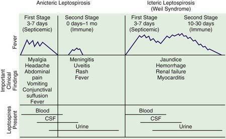
The spectrum of human leptospirosis ranges from asymptomatic infection (most cases) to severe disease with multiorgan dysfunction and death. The onset is usually abrupt, and the illness tends to follow a biphasic course (Fig. 212-1). After an incubation period of 7-12 days, there is an initial or septicemic phase lasting 2-7 days, during which leptospires can be isolated from the blood, cerebrospinal fluid (CSF), and other tissues. This phase may be followed by a brief period of well-being before onset of a second symptomatic immune or leptospiruric phase. This phase is associated with the appearance of circulating antibody, disappearance of organisms from the blood and CSF, and appearance of signs and symptoms associated with localization of leptospires in the tissues. Despite the presence of circulating antibody, leptospires can persist in the kidney, urine, and aqueous humor. The immune phase can last for several weeks. Symptomatic infection may be anicteric or icteric.

image

Figure 212-1 Stages of anicteric and icteric leptospirosis. Correlation between clinical findings and presence of leptospires in body fluids, CSF, cerebrospinal fluid.

(Reprinted with permission from Feigin RD, Anderson DC: Human leptospirosis, CRC Crit Rev Clin Lab Sci 5:413–467, 1975. Copyright CRC Press, Inc, Boca Raton, FL.)

Anicteric Leptospirosis

The septicemic phase of anicteric leptospirosis has an abrupt onset with flulike signs of fever, shaking chills, lethargy, severe headache, malaise, nausea, vomiting, and severe debilitating myalgia most prominent in the lower extremities, lumbosacral spine, and abdomen. Bradycardia and hypotension can occur, but circulatory collapse is uncommon. Conjunctival suffusion with photophobia and orbital pain (in the absence of chemosis and purulent exudate), generalized lymphadenopathy, and hepatosplenomegaly may also be present. A transient (<24 hr) erythematous maculopapular, urticarial, petechial, purpuric, or desquamating rash occurs in 10% of cases. Rarer manifestations include pharyngitis, pneumonitis, arthritis, carditis, cholecystitis, and orchitis. The second or immune phase can follow a brief asymptomatic interlude and is characterized by recurrence of fever and aseptic meningitis. CSF abnormalities include a modest elevation in pressure, pleocytosis with early polymorphonuclear leukocytosis followed by mononuclear predominance rarely exceeding 500 cells/mm3, normal or slightly elevated protein levels, and normal glucose values. Although 80% of infected children have abnormal CSF profiles, only 50% have meningeal manifestations. Encephalitis, cranial and peripheral neuropathies, papilledema, and paralysis are uncommon. A self-limited unilateral or bilateral uveitis can occur during this phase, rarely resulting in permanent visual impairment. Central nervous system symptoms usually resolve spontaneously within 1 wk, with almost no mortality.

Diagnosis

Leptospirosis should be considered in the differential diagnosis of acute flulike febrile illnesses with a history of direct contact with animals or with soil or water contaminated with animal urine. This disease may be difficult to distinguish clinically from dengue or malaria.

Use of Warthin-Starry silver staining, polymerase chain reaction, and immunofluorescent and immunohistochemical methods permits diagnosis of leptospirosis from infected tissue or body fluids. The diagnosis is most often made by serologic testing and less often by isolation of the infecting organism from clinical specimens. Serologic tests for Leptospira include genus-specific and serogroup-specific tests. The reference method is the microscopic agglutination test, a serogroup-specific assay using live antigen suspension of leptospiral serovars and dark-field microscopy for agglutination. A 4-fold or greater increase in titer in paired sera confirms the diagnosis. Agglutinins usually appear by the 12th day of illness and reach a maximum titer by the 3rd wk. Low titers can persist for years. Approximately 10% of infected persons do not have detectable agglutinins, presumably because available antisera do not identify all Leptospira serotypes. Enzyme-linked immunosorbent assay (ELISA) methods, including an immunoglobulin M-specific dot-ELISA test, are also available. Phase-contrast and dark-field microscopy are insensitive for spirochete detection.

Unlike other pathogenic spirochetes, leptospires can be recovered from the blood or CSF during the first 10 days of illness and from urine after the 2nd week by repeated culture of small inoculum (i.e., 1 drop of blood or CSF in 5 mL of medium) on commercially available media-containing rabbit serum or bovine serum albumin and long-chain fatty acids. However, the inoculum in clinical specimens is small, and growth can take up to 13 wk.