[level-membership-for-anesthesiology-category]
14 Lateral Femoral Cutaneous Block
Perspective
Pharmacologic Choice
The same concerns about local anesthetic choice that were outlined for sciatic and femoral blocks (Chapters 12 and 13, respectively) apply to the lateral femoral cutaneous block. If multiple lower extremity blocks are being used, the operator must consider the total dosage being administered. Because the lateral femoral cutaneous nerve does not have motor components, a lower concentration of 10 to 15 mL of local anesthetic is effective.
Placement
Anatomy
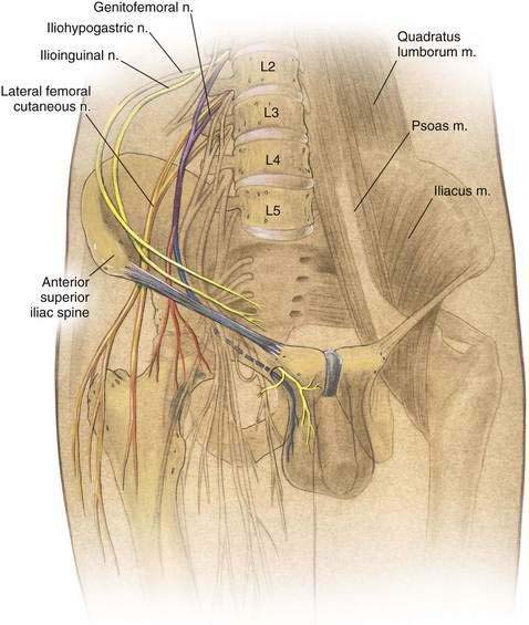
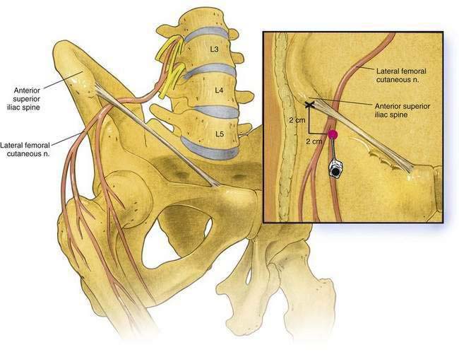
As shown in Figure 14-1, the lateral femoral cutaneous nerve emerges along the lateral border of the psoas muscle immediately caudad to the ilioinguinal nerve. It courses deep to the iliac fascia and anterior to the iliacus muscle to emerge from the fascia immediately inferior and medial to the anterior superior iliac spine, as shown in Figure 14-2. After passing beneath the inguinal ligament, it crosses or passes through the origin of the sartorius muscle and travels beneath the fascia lata, dividing into anterior and posterior branches at variable distances below the inguinal ligament. The anterior branch supplies the skin over the anterolateral thigh, whereas the posterior branch supplies the skin over the lateral thigh from the greater trochanter to the midthigh.
Needle Puncture
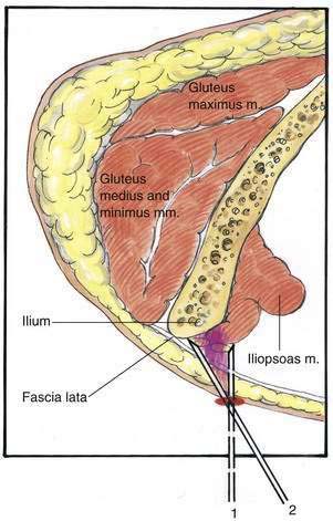
The anterior superior iliac spine is marked in the supine patient, and a 22-gauge, 4-cm needle is inserted at a site 2 cm medial and 2 cm caudal to the mark (see Fig. 14-2). As shown in Figure 14-3, the needle is advanced until a “pop” is felt as the needle passes through the fascia lata. Local anesthetic is then injected in a fanlike manner above and below the fascia lata, from medial (position 1) to lateral (position 2), as illustrated in Figure 14-3.
[/level-membership-for-anesthesiology-category][not-level-membership-for-anesthesiology-category]
14 Lateral Femoral Cutaneous Block
Perspective
Pharmacologic Choice
The same concerns about local anesthetic choice that were outlined for sciatic and femoral blocks (Chapters 12 and 13, respectively) apply to the lateral femoral cutaneous block. If multiple lower extremity blocks are being used, the operator must consider the total dosage being administered. Because the lateral femoral cutaneous nerve does not have motor components, a lower concentration of 10 to 15 mL of local anesthetic is effective.
