Laser Injury (Photothermal and Photomechanical)
Clinical Features:
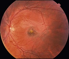
The affected region of the retina is typically the central macula. In the acute setting, laser injuries produce a yellow subretinal lesion that can be of varied appearance (Fig. 20.1.1). Within a short time, the affected area becomes pigmented and over time this appearance can resolve leaving more subtle retinal pigment epithelium disturbances.
OCT Features:
A significant photothermal laser injury causes retinal trauma identical to photocoagulation. On OCT, this is seen as localized outer retinal, IS–OS/ellipsoid zone, and RPE disruption in the acute setting (Figs 20.1.2 and 20.1.3). The inner retina may also be affected (Fig. 20.1.4). With mild exposure, the findings may be very subtle (Fig. 20.1.5). The abnormalities tend to fade quickly, in most cases, over weeks to months (Figs 20.1.6 and 20.1.7).
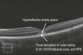
Figure 20.1.2 OCT (corresponding to Figure 20.1.1) shows focal disruption of the outer retina, ELM, IS–OS/ellipsoid zone, and RPE underlying the fovea. There is also a thin hyporeflective empty space above the focal disruption.
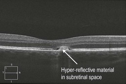
Figure 20.1.3 OCT of a different accidental high-powered laser pointer injury also shows focal disruption of the outer retina, ELM, IS–OS/ellipsoid zone, and RPE with a collection of hyper-reflective material under the retina.
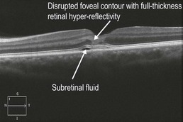
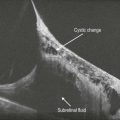
Figure 20.1.4 OCT of an accidental military defense laser injury shows an abnormal hyper-reflective signal involving the full thickness of the retina in the fovea. There is also a tiny pocket of subretinal fluid adjacent to the central abnormality.
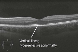
Figure 20.1.5 OCT of the fellow eye of the patient in Figure 20.1.2 shows a subtle abnormality due to limited exposure of this eye to the laser beam. There is a vertical, linear, hyper-reflective abnormality underneath the center of the fovea that spans from the RPE to the external limiting membrane.
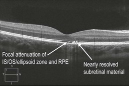
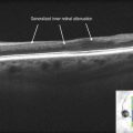
Figure 20.1.6 One month following the injury (see Figure 20.1.3), OCT shows near resolution of the focal outer retinal disruption and subretinal material. The IS–OS/ellipsoid zone and RPE are still somewhat attenuated underneath the fovea.
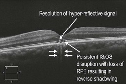
Figure 20.1.7 One month following the injury (see Figure 20.1.4), OCT shows shrinking of the focal outer retinal disruption underneath the fovea and the inner retinal hyper-reflective signal has resolved.