Laparoscopic Nissen Fundoplication and Heller Myotomy

Published on 11/04/2015 by admin

Filed under Surgery

Last modified 22/04/2025

Print this page

rate 1 star rate 2 star rate 3 star rate 4 star rate 5 star
Your rating: none, Average: 0 (0 votes)

This article have been viewed 2041 times

CHAPTER 7 Laparoscopic Nissen Fundoplication and Heller Myotomy

LAPAROSCOPIC NISSEN FUNDOPLICATION

BACKGROUND

Some degree of reflux from the stomach into the esophagus is normal, particularly after meals, and is easily cleared by esophageal peristalsis. The high-pressure zone created by the lower esophageal sphincter (LES), diaphragmatic contraction at the esophageal hiatus during periods of increased abdominal pressure (e.g., coughing and bending), and the intra-abdominal positive pressure exerted on the distal esophagus serve to impede reflux. In contrast, migration of the distal esophagus and LES into the chest, as in a hiatal hernia, transient relaxation of the LES, and inhibitors of LES contraction, including caffeine, smoking, and alcohol, may exacerbate reflux. Repetitive injury of the esophageal mucosa by gastric acid results in gastroesophageal reflux disease (GERD) characterized by chronic symptoms (e.g., substernal chest pain and regurgitation) and, sometimes, complications such as Barrett’s esophagus and stricture formation.

Surgical therapy for the treatment of GERD is a relatively recent innovation. Indeed, the gastric fundoplication operations still performed today were introduced in the 1950s by Nissen, Belsey, and others. The application of minimally invasive techniques has led to more rapid recovery and reduced morbidity after antireflux surgery. As a result, the indications for surgery have broadened despite the availability of relatively effective medical therapies. In most cases, the antireflux operation of choice is the Nissen fundoplication, which involves a 360-degree wrap of the fundus of the stomach around the distal esophagus. In selected circumstances (e.g., when esophageal motility is markedly abnormal), a partial wrap may be preferable. Laparoscopic Nissen fundoplication is the focus of the first part of this chapter.

INDICATIONS FOR SURGERY

There are few absolute indications for antireflux surgery. A number of relative indications have emerged and must be weighed against associated risks.

COMPONENTS OF THE PROCEDURE AND APPLIED ANATOMY

See Figure 7-1.

III. Port Placement

A. A 10-mm port is placed 15 cm inferior to the xiphoid process (Fig. 7-2), just to the left of the midline. Either an open technique or a Veress needle technique may be used to access the peritoneal cavity; however, the former should always be used if the patient has had previous abdominal surgery. After port insertion, pneumoperitoneum is established with insufflation of CO2.
B. Four additional ports (see Fig. 7-2) are placed under laparoscopic visualization. The left and right subcostal ports, placed in the midclavicular lines, function as the primary operative ports and correspond to the surgeon’s left and right hands, respectively. A lateral right subcostal port is placed to allow for the insertion of a self-retaining liver retractor, and a lateral left subcostal port provides access through which an assistant can retract.

IV. Exposure

image image

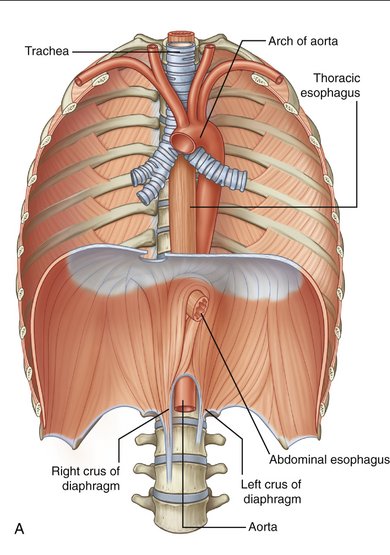
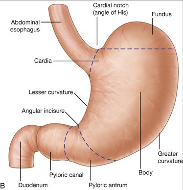
Figure 7-1 Anatomy of the esophagus (A) and stomach (B).

(From Drake RL, Vogl W, Mitchell AWM: Gray’s Anatomy for Students. Philadelphia, Churchill Livingstone, 2005.)
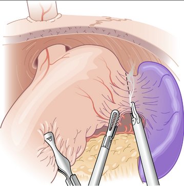
image

Figure 7-3 Exposure of the right and left crura and mobilization of the esophagus.

(From Townsend CM, Beauchamp RD, Evers BM, Mattox KL [eds]: Sabiston Textbook of Surgery: The Biological Basis of Modern Surgical Practice, 17th ed. Philadelphia, Saunders, 2004.)

image

Figure 7-6 Fundoplication. The fundus is wrapped around the distal esophagus loosely enough to allow for passage of a 52 French bougie.

(From Townsend CM, Beauchamp RD, Evers BM, Mattox KL [eds]: Sabiston Textbook of Surgery: The Biological Basis of Modern Surgical Practice, 17th ed. Philadelphia, Saunders, 2004.)

COMPLICATIONS

LAPAROSCOPIC HELLER MYOTOMY

BACKGROUND

Achalasia is a rare disorder characterized by esophageal aperistalsis, high resting LES pressures, and failure of LES relaxation. Although the etiology of achalasia is unknown and is likely multifactorial, the pathogenesis involves loss of inhibitory ganglion cells in the esophageal body and LES. Typical symptoms include dysphagia and regurgitation; however, the onset of symptoms is often insidious. Early in the disease course, dysphagia is more pronounced with liquids than with solids. Patients with achalasia often eat slowly and contort their bodies to propel food through the esophagus. Ultimately, as esophageal dysfunction progresses, food cannot transit through the LES and is regurgitated. Malnutrition, weight loss, and recurrent pulmonary complications, such as pneumonia, bronchiectasis, and lung abscess, may ensue.

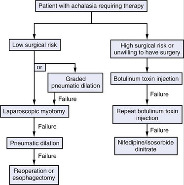
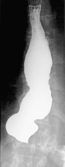
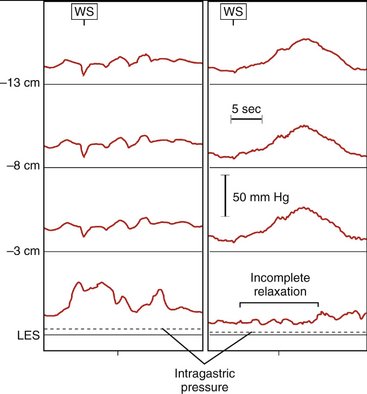
Surgical intervention should be considered for all patients with achalasia; however, two effective nonsurgical therapies are available: pneumatic dilation and botulinum toxin injection. Pneumatic dilation involves passage of a balloon dilator to the level of the LES and disruption of the muscular fibers of the LES through balloon inflation under endoscopic guidance. The vast majority of patients experience symptomatic relief after balloon dilation, but it is frequently short-lived. Additionally, pneumatic dilation is associated with a 5% risk of esophageal perforation. Botulinum toxin inhibits acetylcholine release from the excitatory neurons in the LES, thereby reducing the resting pressure. Injection of botulinum toxin in the area of the LES results in symptomatic relief in the majority of patients, but like pneumatic dilation, this therapy is often transient. Generally, patients at lower surgical risk should be offered surgical myotomy or pneumatic dilation. Patients at high surgical risk should be offered botulinum toxin injection and medical therapies (e.g., calcium channel blockers) aimed at reducing LES pressure (Fig. 7-7).

image

Figure 7-7 Management of patients with previously untreated achalasia.

(Modified from Richter JE: Comparison and cost analysis of different treatment strategies in achalasia. Gastrointest Endosc Clin North Am 11:359, 2001.)

PREOPERATIVE EVALUATION

image

Figure 7-8 Radiographic appearance of the esophagus in a patient with achalasia. A barium esophagogram reveals a dilated esophagus ending in a so-called pointed bird’s beak at the nonrelaxing lower esophageal sphincter.

(From Feldman M, Friedman LS, Brandt JL [eds]: Sleisenger and Fordtran’s Gastrointestinal and Liver Disease, 8th ed. Philadelphia, Saunders, 2006.)

COMPONENTS OF THE PROCEDURE AND APPLIED ANATOMY