Laboratory Investigations in Diagnosis and Management of Neurological Disease

Published on 12/04/2015 by admin

Filed under Neurology

Last modified 22/04/2025

Print this page

rate 1 star rate 2 star rate 3 star rate 4 star rate 5 star
Your rating: none, Average: 0 (0 votes)

This article have been viewed 2587 times

Chapter 31 Laboratory Investigations in Diagnosis and Management of Neurological Disease

The history and examination are key to making the diagnosis in a patient with neurological disease (see Chapter 1). Laboratory investigations are becoming increasingly important in diagnosis and management, however, and are discussed in some detail in later chapters on the specific disorders. A test may be diagnostic (e.g., the finding of cryptococci in the cerebrospinal fluid [CSF] of a patient with a subacute meningitis, a low vitamin E level in a patient with ataxia and tremor, a low serum vitamin B12 level in a patient with a combined myelopathy and neuropathy).

Laboratory tests should be directed to prove or disprove the hypothesis that a certain disease is responsible for the condition in the patient. They should not be used as a “fishing expedition.” Sometimes, a physician who cannot formulate a differential diagnosis from the clinical history and examination is tempted to order a wide range of tests to see what is abnormal. In addition to the high costs involved, this approach is likely to add to the confusion because “abnormalities” may be found that have no relevance to the patient’s complaints. For instance, many patients are referred to neurologists to determine whether they have multiple sclerosis (MS) because their physicians requested magnetic resonance imaging (MRI) of the brain for some other purpose such as the investigation of headaches. If the MRI shows small T2-weighted abnormalities in the centrum semiovale (changes that are seen in a proportion of normal older adults and in those with hypertension and diabetes), the neuroradiologist will report that the differential diagnosis includes MS, despite the fact that the patient has no MS symptoms.

Moreover, neuroimaging modalities have expanded remarkably in the past decade, and the neurologist ordering these tests should be familiar with each one, so that appropriate sequences and methods are used to address the particular question presented by the patient’s history. Also, because of the increasing use of pacemakers, deep brain stimulators, and other devices, the neurologist should be aware that certain precautions must be taken before MRI scans are ordered; in many instances, computed tomography (CT) scans or alternative investigations must be used to avoid potential danger to the patient.

Results of laboratory tests can be used to determine response to treatment. For instance, the high erythrocyte sedimentation rate (ESR) typical with cranial arteritis falls with corticosteroid treatment and control of the condition. A rising ESR as the corticosteroid dosage is reduced indicates that the condition is no longer adequately controlled and that headaches and the risk of loss of vision will soon return.

It is important to use laboratory tests judiciously and to understand their sensitivity, specificity, risks, and costs. The physician must understand how to interpret the hematological, biochemical, and bacteriological studies and the specific neurodiagnostic investigations. The latter studies include clinical neurophysiology, neuroimaging, and the pathological study of biopsy tissue. Knowledge of the various DNA tests available and their interpretation is critical before they are ordered; their results may have far-reaching implications not only for the patient but for all other family members. The neurologist also must have a working knowledge of several related disciplines that provide specific investigations to aid in neurological diagnosis. These include neuropsychology, neuro-ophthalmology, neuro-otology, uroneurology, neuroepidemiology, clinical neurogenetics, neuroimmunology and neurovirology, and neuroendocrinology. Chapter 34, Chapter 35, Chapter 36, Chapter 37, Chapter 38, Chapter 39, Chapter 40, Chapter 41, Chapter 42 describe these disciplines and the investigations they offer.

Biopsy of skeletal muscle or peripheral nerve may be needed to diagnose neuromuscular diseases. A brain biopsy may be needed to diagnose a tumor, infection, vasculitis, or (rarely) degenerative disease of the nervous system.

The investigations used to diagnose neurological disease change rapidly. Genetic studies of DNA mutations in the blood now allow the diagnosis of Huntington disease (HD), a growing number of spinocerebellar ataxias and parkinsonian disorders, a form of autosomal dominant dystonia (DYT1), Duchenne and other muscular dystrophies, many forms of Charcot-Marie-Tooth disease, Rett syndrome, fragile X premutation, and a variety of other neurogenetic disorders (see http://www.ncbi.nlm.nih.gov/sites/GeneTests/?db=GeneTests; http://www.genetests.org; http://www.geneclinics.org; http://www.ncbi.nlm.nih.gov/entrez/query.fcgi?db=OMIM). Blood tests for human immunodeficiency virus infection (HIV), Lyme disease, and other infections and for various paraneoplastic syndromes affecting the nervous system also can be diagnostic. For example, three types of anti–Purkinje cell antibodies are recognized: anti-Yo (PCA-1), seen with tumors of breast, ovary, and adnexa; atypical anti–cytoplasmic antibody (anti-Tr or PCA-Tr), seen with Hodgkin disease and tumors of the lung and colon; and PCA-2, identified mostly with lung tumors. In addition, three antineuronal antibodies can be detected: anti-Hu (ANNA-1), seen in conjunction with encephalomyelitis, small cell lung tumor, and tumors of breast, prostate, and neuroblastoma; anti-Ri (ANNA-2), found with tumors of breast and ovary; and atypical anti-Hu, seen with tumors of lung, colon, adenocarcinoma, and lymphoma. Anti-CV2 (CRMP) antibody, expressed by oligodendrocytes, is associated with a syndrome of ataxia and optic neuritis and has been seen with small cell lung carcinoma. The role of antineuronal antibodies, such as those presumably directed to components of the basal ganglia, is not established and may be of doubtful pathogenic relevance.

Antibodies directed to a serum protein, Ma (anti-Ma1 and anti-Ma2), have been seen in patients with limbic encephalitis associated with testicular and other tumors. Antibodies directed to amphiphysin have been detected in patients with a cerebellar syndrome and small cell lung carcinoma. Antibodies against a glutamate receptor are seen in rare patients with a pure cerebellar syndrome associated with cancer and a variety of autoimmune diseases. Antibodies against glutamic acid decarboxylase (anti-GAD) have been seen in patients with the stiff person syndrome and in patients with ataxia in a setting of an autoimmune disease such as diabetes, thyroid disease, or vitiligo. Antigliadin antibodies are helpful in evaluating patients with unexplained ataxia. As a result of advances in laboratory technology, genetic, immunological, and other blood tests are expanding the ability of clinicians to confirm the diagnosis of an increasing number of neurological disorders, obviating more invasive studies.

MRI has replaced CT for most conditions, and MR angiography and venography have largely replaced conventional catheter-based blood vessel imaging studies. In general, older, more invasive tests are now used for therapy rather than diagnostics. For example, the diagnosis and cause of an acute stroke may be determined by MRI, but catheter angiography is used to deliver intraarterial tissue plasminogen activator (tPA) or perform embolectomies. The neurologist must know enough about each laboratory test to request it appropriately and to interpret the results intelligently. As a rule, it is inappropriate to order a laboratory test if the result will not influence diagnosis or management. Tests should be used to diagnose and treat disease, not to protect against litigation. When used judiciously, laboratory investigations serve both purposes; when ordered indiscriminately, they serve neither.

Interpretation of Results of Laboratory Investigations

Every biological measurement in a population varies over a normal range, which usually is defined as plus or minus 2 or 3 standard deviations (SDs) from the mean value; 2 SDs encompass 96%, and 3 SDs encompass 99% of the measurements from a normal population. Even with 3 SDs, 1 normal person in 100 has a value outside the normal range. Therefore, an abnormal result may not indicate the presence of a disease. It also is important to know the characteristics of the normal population used to standardize a laboratory test. Ranges that were normalized using adults are almost never correct for newborns and children. Ranges normalized using a hospitalized population may not be accurate for ambulatory people.

An abnormal test result may not be caused by the disorder under investigation. For example, an elevated serum creatine kinase (CK) concentration can result from recent exercise, electromyography (EMG) or intramuscular injection, liver disease, or myocardial infarction (MI), as well as from a primary muscle disease. A common problematic finding for pediatric neurologists is centrotemporal spikes on the electroencephalogram (EEG) in a child with headache or learning disability who has never had a seizure. The EEG should not have been ordered in the first place, and to give such a patient antiepileptic drugs would compound poor judgment in diagnosis with worse judgment in management.

The neurologist should personally review test results that are ordered. In most instances, the actual imaging studies should be reviewed in addition to the report, and when appropriate, the neuroradiologist should participate. Similarly, for neurologists experienced in pathology, biopsy findings may be reviewed with the neuropathologist. The neurologist who knows the patient may be of great help in interpreting imaging or pathological studies.

Risk and Cost of Investigations

If two different tests provide equivalent information, the physician should choose the one that causes less pain and risk to the patient. The costs of the two tests also should be considered. The diagnostic capability of two tests may not be identical, and the more expensive test may not be better. The cost of a test must be considered in the context of the total cost of the illness. An expensive test that shortens a hospital stay may be cost-effective. The selection of laboratory tests and the sequence in which performed are important components of good medical practice.

Risk-to-Benefit Analysis

The neurologist makes judgments about the risk-to-benefit ratio of tests every day. The following examples can help clarify the principles used in making these decisions.

Lumbar Puncture

The risks and benefits of LP must be weighed in every patient. The LP may yield a specific diagnosis such as subarachnoid hemorrhage or bacterial meningitis. It may help confirm the diagnosis, such as by showing raised intracranial pressure (ICP) in benign intracranial hypertension. The LP may yield information that is not specific but aids in confirming the diagnosis. A fourfold increase in the CSF protein concentration (without an increase in the cell count) suggests one of the following diagnoses: an acute or chronic inflammatory demyelinative polyradiculoneuropathy, schwannoma or meningioma within the CSF pathways, or spinal compression that obstructs the flow of CSF (Froin syndrome). A moderately increased number of lymphocytes, an increased g-globulin concentration, and oligoclonal bands in the CSF point to an immunological process in the central nervous system (CNS), such as MS.

LP carries significant risks, the most disastrous being cerebral or cerebellar herniation. The LP may suddenly release elevated CSF pressure produced by an expanding supratentorial lesion and may force the medial temporal lobe through the tentorium cerebelli to compress the midbrain. In the case of an expanding infratentorial lesion, it may cause the cerebellar tonsils to herniate through the foramen magnum and compress the cervicomedullary junction (see Chapter 50B). These herniations can be fatal, so never perform an LP in a patient with a possible space-occupying lesion without first examining the optic fundi for evidence of papilledema or consulting a recent head CT or MRI.

LP is justified in some situations despite increased ICP. The prime example is acute meningitis, in which CSF examination is essential to establish the diagnosis and identify the organism. Other risks associated with LP include the production of meningitis as a result of contamination of the needle, a post-LP headache (from low CSF pressure), a spinal epidural hematoma in a patient with a coagulopathy, and the later development of an implantation dermoid (if the needle is inserted without the trocar).

Cerebral Arteriography

The question of whether to request percutaneous cerebral arteriography (see Chapter 33B) entails analysis of the risks and benefits for each patient. In a patient with cerebrovascular disease, the study may show thrombotic or embolic occlusion of arteries and abnormalities of the arterial wall, including arteriosclerotic plaques, fibromuscular hyperplasia, medial dissection, and arteritis. It also may demonstrate an intracranial aneurysm or arteriovenous malformation (AVM). Any of these findings can clarify the diagnosis, treatment, and prognosis. Many of these diagnostic outcomes can be identified with advanced MRI or a combination of MRI, CT, and ultrasound strategies. Thus, it is critical that the attending neurologist have a specific hypothesis in mind before ordering such examinations, as well as understand the relative ability of noninvasive versus invasive tests to demonstrate specific abnormalities in the blood vessels. If treatment (e.g., aneurysm occlusion) can be combined with a diagnostic procedure (e.g., catheter angiography), this may also alter the decision-making outcome.

Invasive studies such as arteriography have risks. These include thrombosis of the artery at the site of puncture, dissection of the vessel wall, allergic reactions to contrast, and cerebral infarction from thrombosis, embolism, or dissection. The likelihood that a patient being considered for cerebral arteriography will experience a particular complication is influenced by patient-specific factors including age and the presence of arteriosclerosis and other diseases. Traditionally noninvasive tests such as MRI and CT can also have risks related to preexisting patient conditions (e.g., contrast for either procedure can damage the kidney in patients with prior renal disease or diabetes). These patient-specific probabilities of risk must be balanced against the potential benefits the angiographic information may provide, specifically the likelihood of demonstrating a treatable condition. The likelihood of risk also varies with the skill, experience, and judgment of the physician performing these procedures. As such, the neurologist requesting invasive procedures should have an accurate estimate of the physician-specific risk factors. The final decision will need to consider the combined risk probabilities, including both patient- and physician-specific factors.

Arteriography is definitely indicated in a previously healthy 55-year-old woman with an acute transient right hemiplegia and aphasia and a left carotid artery bruit, especially when carotid ultrasound and/or MRI studies suggest a 75% internal carotid artery stenosis. Invasive angiography clearly is not indicated in a 75-year-old woman with unstable congestive cardiac failure and advanced carcinoma of the breast who suffers a similar transient ischemic attack (TIA). Noninvasive techniques may be adequate for revealing the cause of the patient’s symptoms, thereby avoiding the risks of catheter cerebral angiography. Carotid Doppler ultrasound and transcranial Doppler studies can be as reliable as angiography for demonstrating extracranial occlusive disease. MR angiography, a technique that images the main extracranial and intracranial vessels noninvasively, may obviate invasive angiography in patients with extracranial occlusive disease, AVMs or a family history of intracranial aneurysms.

Brain Biopsy

Brain biopsy carries significant risks that always necessitate discussion of the risk-to-benefit ratio with the patient and family. The four main situations in which a brain biopsy may be considered are intraparenchymal brain tumor, intraparenchymal infectious lesion, cerebral vasculitis, and in special circumstances, cerebral degenerative disease. The risk-to-benefit analysis is influenced by the availability of computer-assisted stereotactic technology to obtain a biopsy through a burr hole, reducing the risk of obtaining tissue for pathological and bacteriological study.

Open craniotomy for brain biopsy is significantly more risky. The patient’s age, presence of other diseases, lesion location, and the patient’s wishes all must be taken into account when open brain biopsy is considered. Hemorrhage, infection, post-biopsy epileptic seizures, and the production of a neurological deficit are the main risks associated with the procedure. The risk of a permanent neurological deficit is reduced if the biopsy specimen is from certain areas of the brain, such as the nondominant frontal or temporal lobes. The procedure carries a high risk of worsening the neurological deficit (unless that deficit is already total) if the lesion is located in the sensorimotor cortex, the Broca speech area, the internal capsule, or optic radiations.

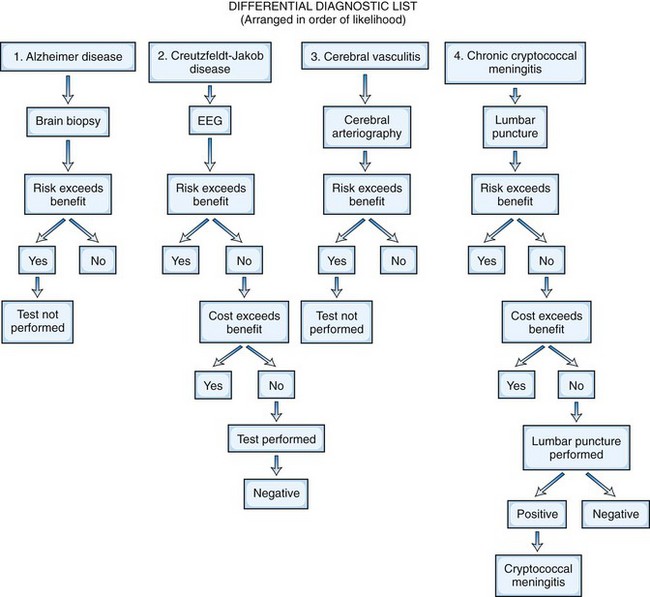
The treatability of the possible cause of the disease is the crucial benefit to consider in the risk-to-benefit analysis. If the neuroimaging study suggests a malignant glioma, for which treatment is likely to be ineffective, biopsy alone may not be considered justified, although resection and tissue diagnosis would be. If it suggests a primary lymphoma of the brain, which is likely to respond to radiotherapy, then confirmatory biopsy may be recommended. If the differential diagnosis in a patient with acquired immunodeficiency syndrome includes toxoplasmosis or lymphoma, it may be reasonable to give anti-Toxoplasma therapy rather than perform a brain biopsy; biopsy would be needed only if the lesions do not respond to 2 to 3 weeks of treatment. Figure 31.1 presents a risk-to-benefit analysis and prioritization of investigations for an 80-year-old man with possible cerebral degeneration.

Research Investigations and Teaching Hospitals

Because many of our readers are neurologists in training, here we briefly mention the use of investigations in teaching and research centers. Clinical research is closely regulated in most parts of the world, and research investigations cannot be performed until the protocol is approved by an institutional review board or an ethics-in-research committee. The peer review process is designed to ensure that the risks of the research study are justified, taking into account the patient’s particular disease and the likely benefits of the research. The institutional review board ensures that the patient receives full information contained in an informed consent form and understands the risks of the study and what is likely to be learned from the research. Special policies and procedures also apply to minors, patients with cognitive dysfunction, those in emergency situations, or those with alterations in consciousness. No patient should be coerced, knowingly or unknowingly, into participating in a research procedure. Once the institutional review board gives permission for a research project, it continues to monitor the study to ensure that the research conforms to the protocol.

In a teaching hospital, the attending or consultant physician is legally and ethically responsible for the care provided to a patient by physicians in training. The attending neurologist must ensure that every investigation is justified for diagnostic and management purposes. All physicians are legally and ethically bound to ensure that the patient understands the reason for each investigation and gives informed consent. The neurologist in training must learn to use tests judiciously and not perform them simply for curiosity or education. The two-way discussion with more senior neurologists about the rationale, risk-to-benefit ratio, and cost-to-benefit ratio of each investigation is an important part of the learning process.

Role of Laboratory Investigations in Neurological Disease Management

The standard neurological examination is designed more to detect abnormal function for diagnostic purposes than to quantify the neurological abnormalities. When possible, therefore, laboratory investigations are used to measure the response of the disease to treatment. Laboratory investigations usually are quantitative and may be helpful in managing disease. Generally, abnormal laboratory values return toward normal as a disease resolves or become increasingly abnormal as it worsens. The vital capacity in a patient with Guillain-Barré syndrome is an example of a measurement that improves as the disease improves. This is not always the case, however. In Duchenne muscular dystrophy, the serum CK concentration decreases as the disease worsens, because fewer muscle fibers remain to release enzyme into the serum. In myasthenia gravis, the patient’s condition can go from minimal weakness to total paralysis unrelated to the titers of acetylcholine receptor antibodies in the blood. Therefore, monitoring laboratory values cannot always be used as an index of disease severity or response to treatment. Other limitations on the use of laboratory tests to monitor disease progression include sampling errors and test sensitivity and specificity.

Quantitative tools provide important information for measuring a patient’s status objectively during the course of a disease. They can be as simple as visual acuity measurement, how many serial numbers from 1 to 100 a patient can count on a single breath, or the frequency and severity of headaches each month. Alternatively, they can be sophisticated measurements such as the force of maximum voluntary muscle contraction or the temperature perception threshold for an area of skin. They can be summated scores of semiquantitative assessments, such as the Kurtzke scale devised to follow the clinical course in patients with MS, the Norris score for amyotrophic lateral sclerosis, or the z-scores of muscle strength. Quantitative measures of neurological function allow much better assessment of the response of a disease to treatment than does the routine neurological examination.