Emboli to SMA from cardiac sources most commonly


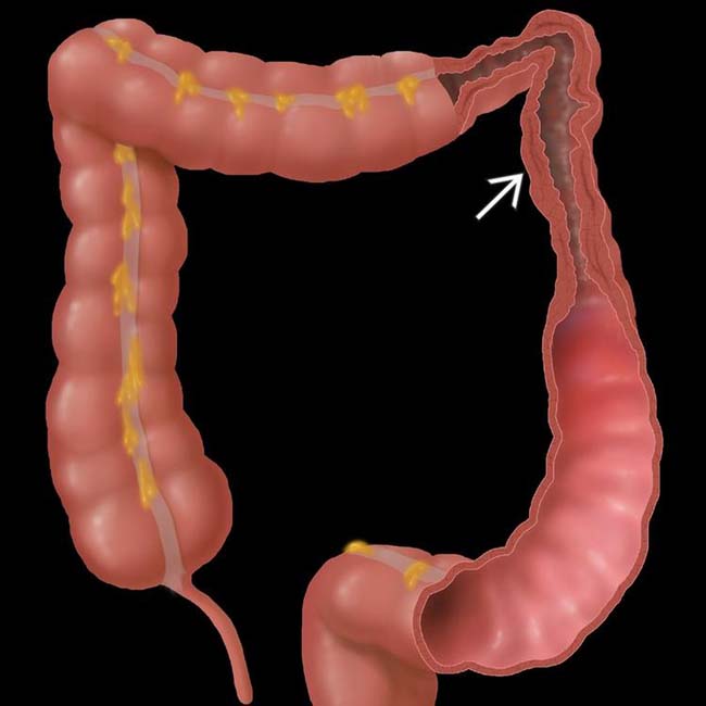
 near the splenic flexure, a “watershed” area between the vascular distribution of the SMA and IMA.
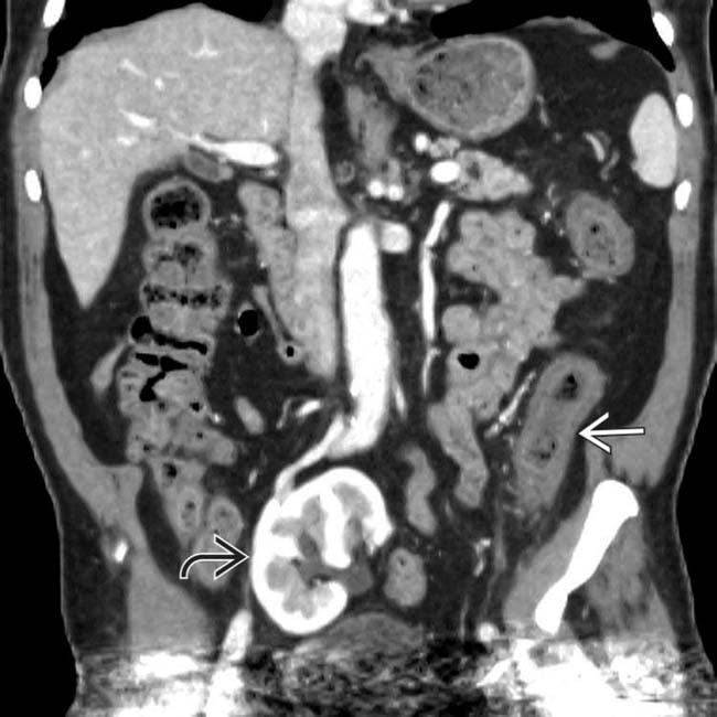
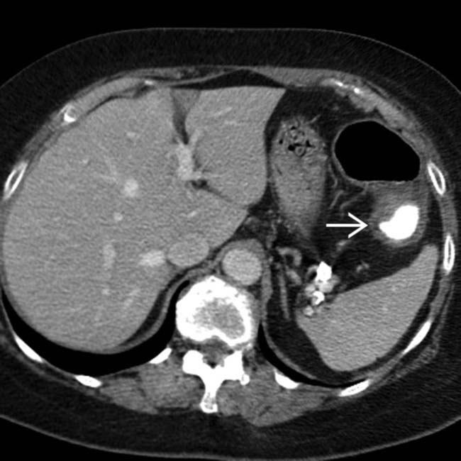
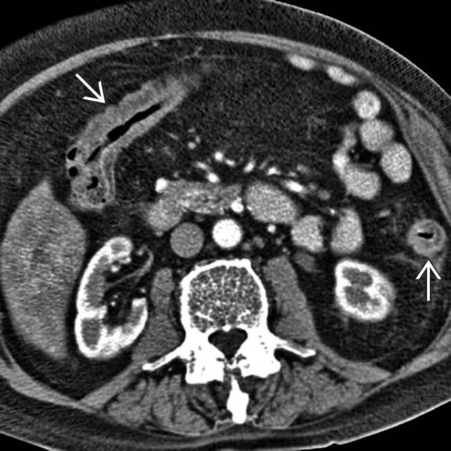
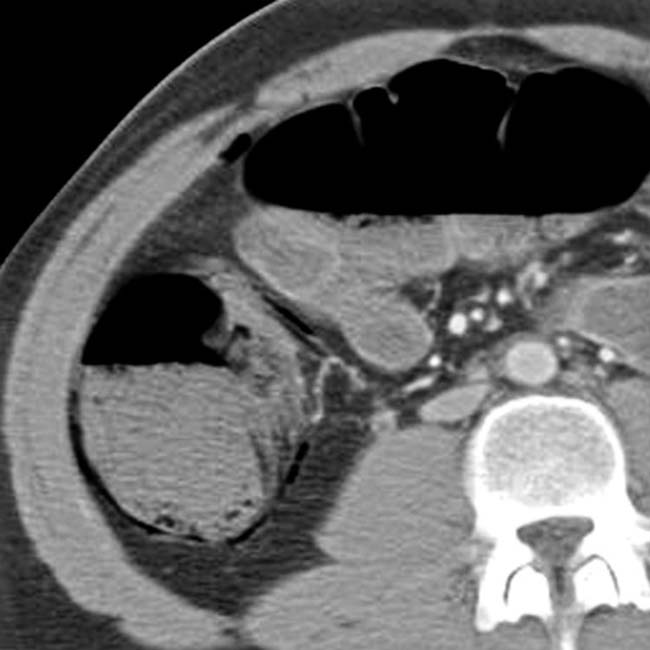
 . Incidental renal allograft noted
  .
.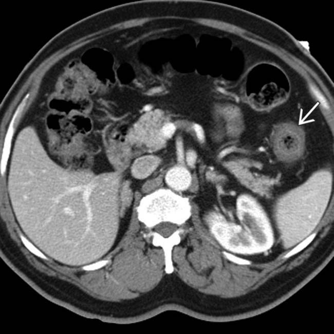
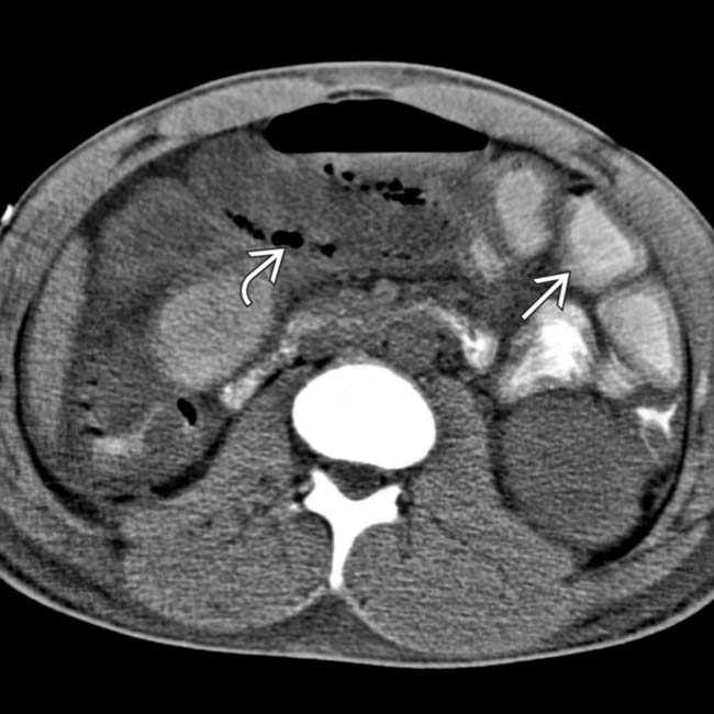
 . The SMA and SMV are patent.
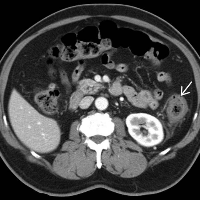
 , whereas the remaining colon is normal. The rectum (not shown) was normal. These are classic clinical and CT features of ischemic colitis due to a hypotensive episode in an elderly patient.
IMAGING
General Features


 , whereas the more proximal colon is gas-distended with a normal wall.
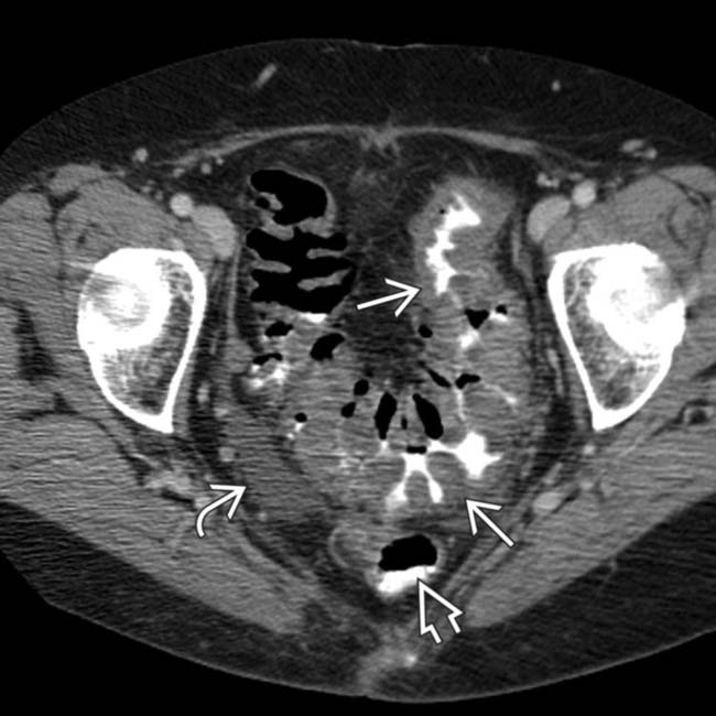
 . The rectum
  is spared. Ascites
  is noted. These are classic clinical and CT features of ischemic colitis.
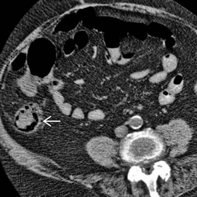
 , which is otherwise relatively normal in appearance.
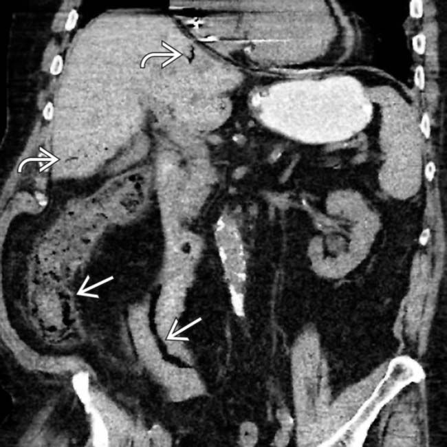
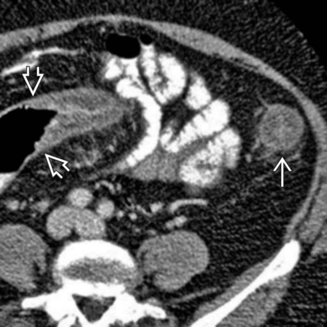
 as well as the ascending colon. Portal venous gas
  is also present. These are typical features of thromboembolic occlusion of the SMA.
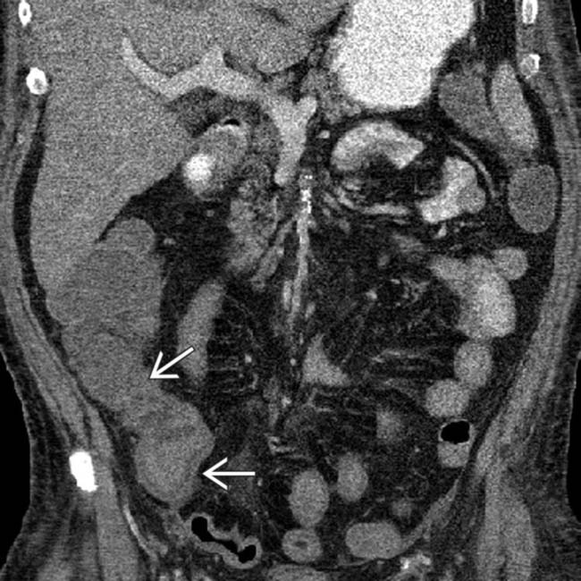
 and transverse colon (not shown).
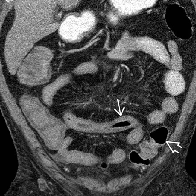
 , whereas the distal colonic wall
  is normal. This distribution of ischemic injury likely resulted from a combination of atherosclerotic narrowing of the SMA and the hypotensive episode.
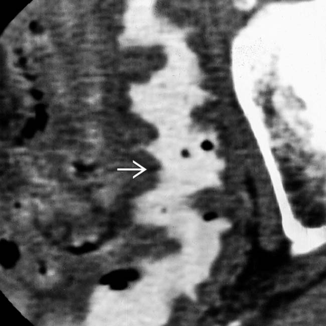
 due to submucosal hemorrhage.
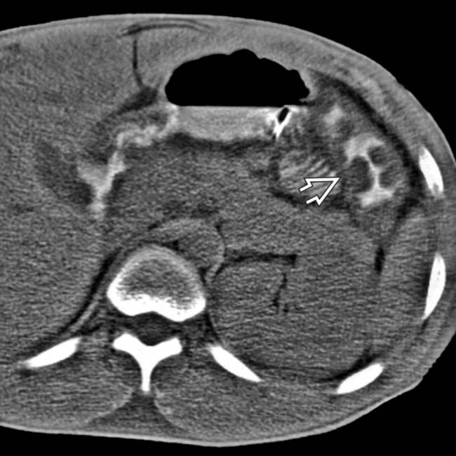
 .
.
 and focal pneumatosis
  .
.
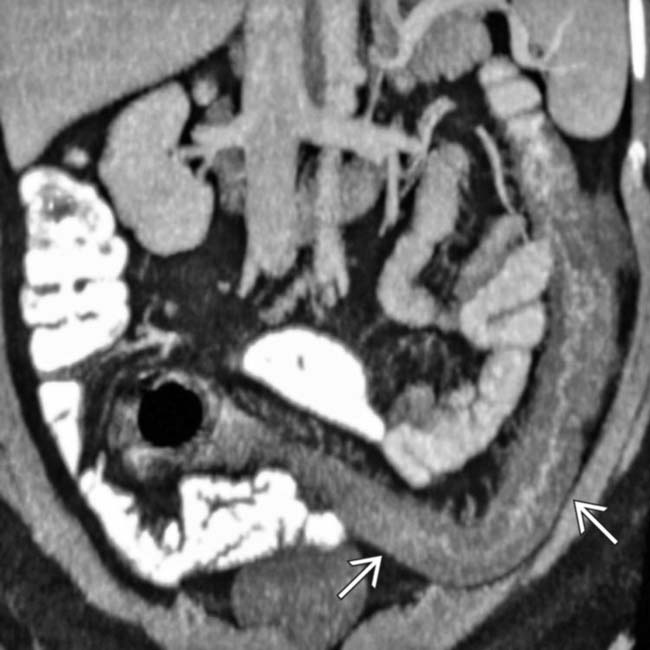
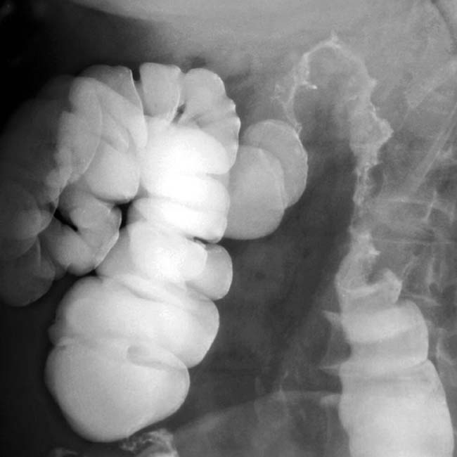
 . Note the abrupt transition to normal sigmoid colon
  .
.
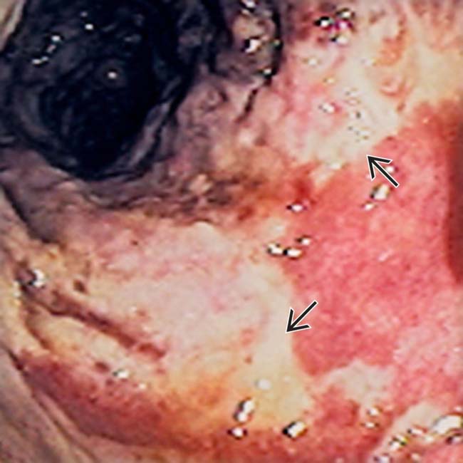
 . Ischemia was confirmed endoscopically.
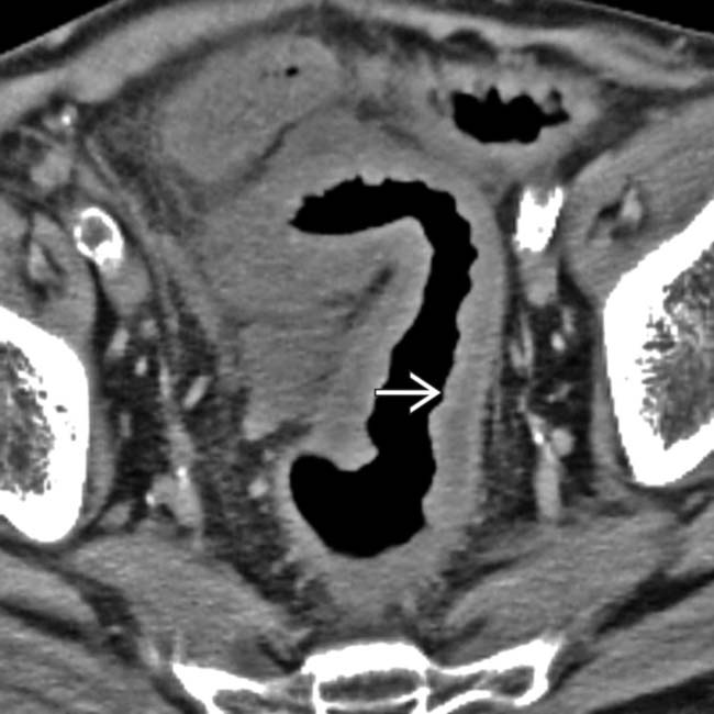
 .
.
 .
.
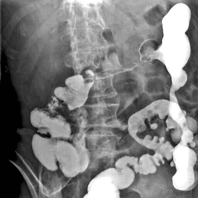
 , submucosal edema, and luminal narrowing of the colon.
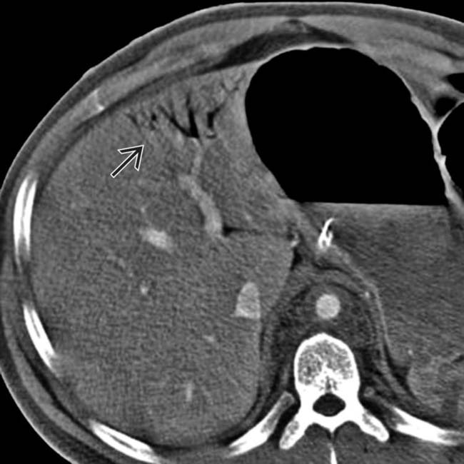
 .
.
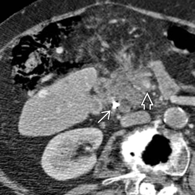
 . The superior mesenteric artery and vein
  are encased and narrowed. Gas is present in the colon wall.
 gas present due to colon infarction.


