Intracerebral Hemorrhage

Published on 03/03/2015 by admin

Filed under Neurology

Last modified 22/04/2025

Print this page

rate 1 star rate 2 star rate 3 star rate 4 star rate 5 star
Your rating: none, Average: 4.5 (2 votes)

This article have been viewed 12995 times

58 Intracerebral Hemorrhage

Clinical Vignette

A 72-year-old woman with history of hypertension noticed tingling in the right arm. Within 30 minutes, her right leg buckled and she fell. Her husband helped her up and she was able to walk without support. She rested, but within an hour, she developed speech difficulties and more definite right-sided weakness.

She was brought to the emergency department (ED) and was noted to have mild right arm weakness and some word-finding difficulties. Her blood pressure was 140/80 mm Hg. She got up to go to the bathroom, walked approximately 10 ft, and collapsed on the floor. She was then globally aphasic, with left gaze deviation (i.e., paralysis of gaze to the right) and right hemiplegia. Within 10 minutes, she became gradually unresponsive.

She was intubated for airway protection and taken for brain CT scanning, which demonstrated a large left putaminal hemorrhage. Soon thereafter, she had bilaterally dilated fixed pupils. The vestibular-ocular reflexes were lost. Within another 10 minutes, she was determined brain dead.

The preceding vignette demonstrates the classic presentation of a primary intracerebral hemorrhage (ICH with a rapidly evolving focal neurologic deficit). Its progressively increasing size led to increased intracranial pressure with coma from downward herniation of the uncus onto the brainstem and ultimately irreversible neurologic damage. The only means to prevent ICH is appropriate therapy of its major risk factor, hypertension.

There are two forms of intrinsic cerebral hemorrhage, primary ICH, which has a predilection to affect the striatum, thalamus, midbrain, pons, and cerebellum, and subarachnoid hemorrhage (Chapter 57). ICH comprises approximately 10% of all strokes in the Caucasian population and up to 20% in the Asian population. Over the past several years, improved treatment of hypertension has decreased the number of patients experiencing ICH.

Hypertension is a major risk factor for intracerebral hemorrhages in patients between the ages of 40 and 70. A small proportion of intracerebral hemorrhages in patients older than age 60 years is not directly related to hypertension and is primarily caused by amyloid angiopathy. Furthermore, the increased use of oral anticoagulant therapy in the elderly population has led to higher rates of warfarin-associated ICH. Warfarin anticoagulation increases the risk of intracerebral hemorrhage fivefold, and some studies have estimated that 18% of intracerebral hemorrhages admitted to the hospital are related to its use. Of interest is that most bleeds in patients on warfarin occur when the INR is within the recommended therapeutic limits. Secondary, less common causes of intracerebral hemorrhages include primary and metastatic tumors of the brain and in younger individuals cerebral hemorrhage associated with underlying arterial venous malformation or cavernous angioma.

Pathophysiology of Hypertensisve Primary Ich

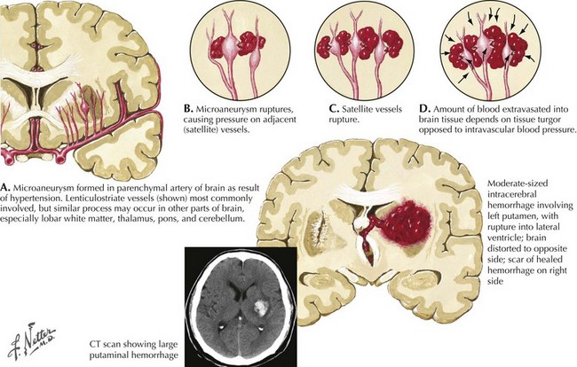
Intracranial hemorrhage is a rapidly evolving process that may progress over hours or days. The pressure effects of the initial hemorrhage lead to mechanical disruption and tearing of surrounding vessels with subsequent gradual expansion of the hematoma out from the original center. Rebleeding is the most feared early complication of ICH and occurs in approximately 40% of patients. Rebleeding usually occurs within the first 24 hours but, on occasion, has been reported up to a week later. The underlying pathological mechanism of primary hypertensive ICH is attributable to either the formation of miliary microaneurysms or primary arteriolar degeneration (lipohyalinosis and weakening of the blood vessel intima and media wall layers) (Fig. 58-1). The presence of miliary aneurysms is directly related to hypertension but is not necessarily the initial site of bleeding, and cases of hypertensive ICH outside the areas of microaneurysms have been noted. This suggests that degeneration of the arteriolar smooth muscle wall is likely an important factor in the evolution of ICH. Hypertensive intracerebral hemorrhages have a predilection to occur in the basal ganglia and the thalamus. The arterioles in these structures are likely more vulnerable to degenerative changes brought on by diffuse, large pressure pulses over time.

Although hypertension is considered an important risk factor for ICH, studies are inconsistent in estimating the exact risk of ICH in hypertensive patients. It has been noted in some studies that up to 90% of patients with ICH have evidence of hypertension at the time of presentation. Other important risk factors include smoking, alcohol consumption, black race, and low total serum cholesterol levels. The latter is likely to be an anomaly of population studies. Patients with ICH and concomitantly low serum cholesterol levels tend to be older than age 80 years and, on average, have higher diastolic pressures. The effect of lower cholesterol levels, particularly LDL, on increasing the incidence of ICH is likely small and the mechanism remains unclear; whether it is causative or an epiphenomenon of another process warrants further investigation.

Clinical Presentation

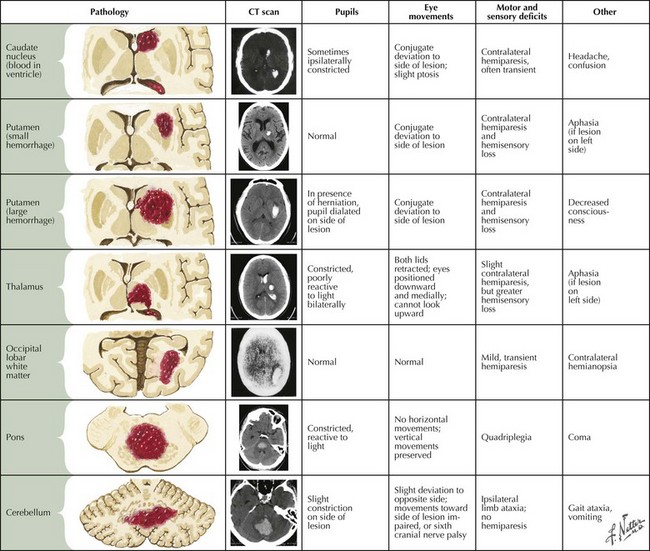
Intraparenchymal hemorrhages vary in presentation depending on the site of the bleeding (Fig. 58-2). In approximately 60% of patients, neurologic symptoms develop gradually or stepwise over a period of hours. To some extent, the location and size of the hematoma predict clinical outcome.

Headache occurs at presentation in approximately 40% of patients with ICH. Less commonly, headache develops within a few days after the ictus. Intracerebral hemorrhages presenting with headache are often located at the brain surface or within the cerebellum. Depression in the level of consciousness and vomiting occur in 50% of patients, particularly those with large cerebellar bleeds. Seizures occur at onset in up to 10% and are seen most commonly with lobar bleeds in the anterior circulation. There are rare incidences of patients with deep hemorrhages having seizures. The subsequent risk for seizures in ICH patients is up to 29% for those with lobar hemorrhages but only 4% for those with deep hemorrhages. Other symptoms seen in association with ICH include low-grade fever without obvious infection, cardiac arrhythmias, and dysautonomia, especially with pontine bleeds. A description of some of the most common symptoms at different sites of ICH follows.

Deep Supratentorial Hemorrhage

Putaminal Hemorrhages

The most common site of ICH is the putamen, and these are classified into anterior, middle, and posterior lesions. Anterior putaminal hemorrhage often causes motor weakness due to compression of the anterior limb of the internal capsule. If the ICH is on the left, abulia and aphasia are common accompaniments. When the lesion occurs on the right, significant behavioral changes can occur, including disinhibition, poor insight and judgment, and occasionally violent behavior. Caudate hemorrhages are associated with similar behavioral and cognitive changes. Studies suggest that these behaviors result from frontal lobe disconnection. Often, deficits from small anterior putaminal hemorrhages are reversible.

A hemorrhage within the midputamen, however, results in severe deficits, often with poor recovery. In this case, ICH compresses and undercuts nearby cortical structures, causing global aphasia if involving the left hemisphere and severe neglect if involving the right. With posterior putaminal hemorrhages, a combination of sensory-motor deficits, visual field difficulties, limb ataxia, and behavioral changes often results. Some putaminal hemorrhages not extending into the globus pallidus present with short-lived hemichorea or hemiballismus, although a variety of other abnormal involuntary movements have been described. Large or medially located putaminal hemorrhages and head of the caudate hemorrhages can dissect toward the ventricle, with resultant intraventricular hemorrhage and the development of acute obstructive hydrocephalus with rapid deterioration due to increased intracranial pressure. Primary intraventricular hemorrhage, in contrast, does not affect surrounding brain tissue, and most cases present as a nonlocalizing rapidly progressive syndrome of nausea, vomiting, stupor, and seizure. In less acute cases, the patient presents with headaches, confusion, and somnolence.

Thalamic Hemorrhages

Clinically, thalamic ICHs are classified into posterior–inferior, posterior–lateral, and dorsal–medial. Somnolence is one of the most common presentations of medial–posterior and inferior thalamic bleeds and can be profound as a result of bilateral disruption of the rostral reticular activating system. If the hemorrhage dissects anteriorly, often persistent hypokinetic behavior results from disconnection of the frontal lobe. With inferior–lateral thalamic hemorrhage, there is weakness and clumsiness and, occasionally, tremors and choreoathetoid movements. Tremors are likely related to disruption of projections from the cerebellum and dentate nucleus. Disruption of the fibers of the ansa lenticularis are likely responsible for the choreoathetoid movements.

More lateral thalamic hemorrhages involving the ventral posteromedial and ventral posterolateral thalamic nuclei primarily cause unilateral sensory symptoms but occasionally motor involvement when the hemorrhage extends laterally to involve the internal capsule. Eye movement abnormalities, small pupils, ptosis, chorea, and dystonia also occur. Hematomas involving the dorsal-medial thalamic area present with prominent memory problems and behavioral changes thought to relate to dissociated frontal cortex, cingulate gyrus, and amygdalar connections. Speech and language deficits are the least consistent symptoms of thalamic hemorrhages. Paraphasia, naming difficulties, or a perceived inability to comprehend, with preservation of repetition, is typical of thalamic aphasia. In patients with right thalamic hemorrhage, deficits mimic cortical lesions with neglect or hemi-inattention, vivid visual, and, less often, auditory hallucinations can occur in the days following thalamic ICH.

Superficial Lobar Hemorrhages

After the putamen, the most common site of primary ICH is one of four locations in the cerebral cortex. The parietal and occipital areas are most frequently involved. In general, hypertension is an important risk factor for all ICH regardless of location, but whether blood pressure plays a lesser role in the contribution of lobar versus subcortical hemorrhages remains inconclusive. Primary amyloid angiopathy frequently underlies nonhypertensive intracerebral lobar hemorrhage. Other less common causes include vascular malformations, primary and metastatic malignancies, sympathomimetic drugs, anticoagulants, irreversible antiplatelet and fibrinolytic agents, and sinus thrombosis with venous infarctions and bleeds.

Lobar hemorrhages often present with headaches and vomiting. Seizures at the onset of lobar hemorrhage are common, particularly those within the posterior parietal or frontal lobe. Functionally, patients with lobar hemorrhage may have better outcomes than those with deep hemorrhages. However, prognosis depends on hematoma size, level of consciousness at presentation, and presence of intraventricular blood. Mortality rates range from 12 to 30% in superficial lobar hemorrhages compared to 25–42% in deep basal ganglionic and thalamic hemorrhages and up to 97% in pontine hemorrhages.

Infratentorial Hemorrhages

Cerebellar Hemorrhage

Clinical Vignette

A 58-year-old woman with substantial history of arterial hypertension presented to the ED with a 1-hour duration of acute-onset headache, gait unsteadiness, and left arm incoordination. On examination, the patient was alert and oriented but had left-sided dysmetria, gait ataxia, and left CN-VI and CN-VII palsies. Her BP was 200/110 mm Hg. Urgent head CT showed a 3-cm cerebellar hemorrhage with slight compression of the fourth ventricle. Antihypertensive treatment aiming for a MAP of 100–120 mm Hg was initiated.

Within 30 minutes after the CT scan, the patient’s level of consciousness deteriorated, necessitating intubation. She was brought immediately to the operating room for evacuation of the hematoma and responded well. One month later, her examination was remarkable for only mild clumsiness of the left arm and a slightly wide-based gait.

As in the preceding vignette, patients with cerebellar hemorrhages can deteriorate rapidly, even “in front of one’s eyes,” but can still respond exceptionally well with expeditious surgical intervention. Most cerebellar hemorrhages are associated with hypertension. However, approximately 10% of primary cerebellar hemorrhages are caused by AVM, tumors, blood dyscrasias and the use of warfarin anticoagulation. Headache, spinning vertigo, nausea, vomiting, and, most commonly, unsteady gait characterize the typical presentation. Some headaches are occipital, but many involve the orbital and supraorbital areas. The most reliable symptoms of a hemispheric cerebellar hemorrhage include headache, vomiting, nystagmus, ipsilateral limb ataxia with, at times, ipsilateral peripheral CN-VI and CN-VII palsies and horizontal nystagmus.

The less common vermian hemorrhages often resemble a pontine hemorrhage and can progress rapidly to coma, making it difficult to identify specific early clinical signs that can differentiate one from the other. Cranial nerve palsies are related to involvement of adjacent pontine structures or stretching secondary to increased cerebellar pressure. In hypertensive bleeds involving the vermis or the cerebellar hemispheres, the superior cerebellar artery is most often involved.

Unlike supratentorial bleeds, in which a small hemorrhage is often well tolerated, infratentorial ICH within the posterior fossa often leads to rapid neurologic deterioration and death. Close monitoring in an ICU for 36–48 hours, when the risk of deterioration is at its highest, is therefore recommended for most patients. Rebleeding, rupture into the fourth ventricle, and accelerated hemorrhagic edema, alone or in combination, often lead to a devastating outcome. Hemorrhages larger than 3 cm may extend into the fourth ventricle and lead to the development of acute hydrocephalus and require ventriculostomy placement. The threshold for surgical evacuation of the hematoma should be low and considered at the earliest sign of deterioration. The major goal is decompression of the posterior fossa to prevent blockage of the fourth ventricle and compression of the adjacent brainstem. Fortunately, if impending brainstem compression is recognized early, there are often only minimal residual deficits after surgery, even with extensive cerebellar evacuation and decompression. The potentially positive recovery from cerebellar hemorrhages and decompressive surgery reflects that the deep cerebellar nuclei, crucial for gait coordination and balance, are often spared from direct damage.

Pontine/Midbrain Hemorrhage

Pontine and midbrain hemorrhages are relatively uncommon but have the most devastating outcome compared with other sites of primary intracranial hemorrhages. Three distinct vascular territories dictate the clinical presentation. The paramedian penetrators, arising directly from the basilar trunk, are the primary arteries supplying the midline pons or midbrain. ICH in this location causes bilateral damage and is often fatal. Sudden onset of deep coma, quadriparesis, ophthalmoplegia, and bilateral papillary abnormalities are the presenting signs.

Another group of small arteries, the short circumferential penetrators, courses laterally, supplying the lateral basis pontis, where a hemorrhage may predominantly cause unilateral bulbar symptoms with profound dysphagia. The third important group of vessels, the long circumferential arteries, arises from the anterior–inferior cerebellar artery and primarily supplies the lateral tegmentum. ICH within this segment leads to relatively minor symptoms, including facial numbness and ataxia secondary to involvement of the spinal trigeminal and vestibular nuclei. However, involvement of the intrinsic pontine nuclei, such as the cochlear and facial nuclei, are also affected, which leads to a more serious outcome.

Pontine hemorrhages often have a relatively gradual clinical presentation evolving over hours. Neurologic deficits, including horizontal gaze palsies, miotic sluggishly reactive pupils, quadriparesis, and coma, are the expected clinical signs. Certain unique eye findings, including ocular bobbing and the one-and-a-half syndrome, provide excellent diagnostic clues to pontine hemorrhages. Some patients also exhibit twitching of the limbs and face and rippling of torso muscles. Dysautonomia with irregular pulse, erratic breathing patterns, and an increase in body temperature have also been observed. Vivid, sometimes frightening, formed hallucinations, called peduncular hallucinosis, occur relatively often in patients with involvement of the midbrain tegmentum.

Secondary Intracerebral Hemorrhage

ICH not directly caused by hypertension is encountered with vascular malformations, hemorrhagic transformation of ischemic stroke, anticoagulants, as well as fibrinolytic agents or irreversible antiplatelet therapy (Box 58-1). Primary amyloid angiopathy is often the underlying cause of nonhypertensive lobar hemorrhage. Less common causes include primary and metastatic malignancies, sinus thrombosis with venous infarctions and bleeds, acquired or inherited coagulopathies, induced or autoimmune vasculitides and systemic granulomatous disorders, central infectious processes, and trauma (Box 58-2).

In most cases of ICH, especially when hypertension is absent, follow-up imaging studies are essential to investigate the possibilities of underlying predisposing pathology. Contrast-enhanced brain MRI scanning performed about 3 months later, after extravasated blood has been allowed to reabsorb, may uncover an underlying lesion initially obscured by the acute hematoma.

Occult vascular malformations were possibly the most underdiagnosed causes of lobar hemorrhages prior to CT and MRI scanning and were frequently missed by early angiography due to the presence of clot and mass effect on the brain. They were diagnosed only during surgical or pathologic specimen inspection after hematoma removal. The most common occult vascular lesions include small AVMs and cavernous angiomas (Fig. 58-3A–D).

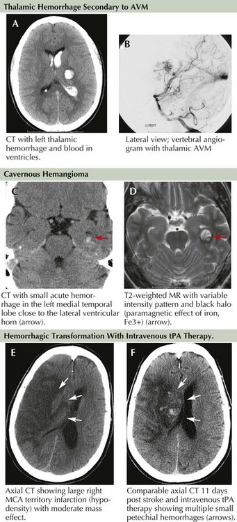
Hemorrhagic brain infarct (HBI), in contrast to primary intracranial hemorrhage, is a secondary phenomenon that occurs as a result of ischemic damage to both the brain parenchyma and the vessel wall distal to the site of occlusion. The vascular wall endothelium and the blood–brain barrier are subsequently damaged and leak with reperfusion as they no longer tolerate normal arterial pressure. Petechial bleeding and, at times, gross hemorrhage through the damaged vessel into the infracted area may be seen (Fig. 58-3E&F). It is estimated that petechial bleeding develops in more than 50% of patients with embolic infarcts. Although it is often implied that cardioembolic strokes are more likely to be associated with development of HBI, some investigators suggest that any large infarct, regardless of mechanism, is predisposed to such bleeding. The use of IV heparin or heparinoid for secondary stroke prevention also promotes the development of HBI. However, the presence of petechial hemorrhage without frank hematoma formation does not seem to worsen neurologic outcome.

Along with typical subarachnoid hemorrhage, aneurysmal rupture can, at times, cause intraparenchymal hematoma with focal neurologic signs (Fig. 58-4A&B). The location of the blood often hints to the site of the aneurysm. Lateral temporal lobe hematomas suggest MCA aneurysmal rupture while medially located bleeds are associated with carotid artery aneurysms. Frontal hematomas indicate anterior communicating artery aneurysm. Posterior communicating artery aneurysms cause thalamic hemorrhages, often with intraventricular extension.

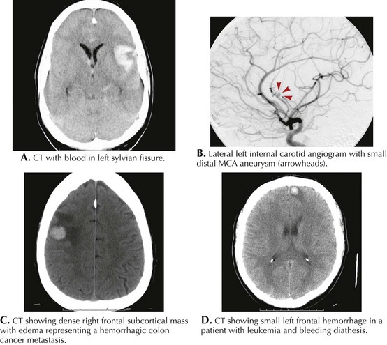
Primary or metastatic brain tumors can lead to ICH (Box 58-3; Fig. 58-4C&D). A single small hemorrhage from a metastatic lesion, as can be seen with melanomas or hypernephromas, may be difficult to distinguish from primary ICH unless evidence of other lesions is identified. Other clues, such as an atypical cortical or subcortical location of the bleed, irregular margins, and unexpected contrast enhancement of the lesion must prompt further investigations to exclude systemic disorders. A detailed dermatologic examination may reveal irregularly pigmented lesions, suggesting melanoma, and ultrasound or body CT scan may uncover a renal tumor.
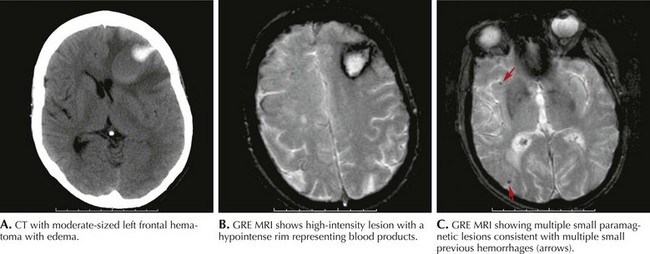
Antithrombotic- and Anticoagulant-Induced ICH

The question of whether aspirin promotes ICH remains unclear, but the Physicians’ Health Study, a randomized, double-blinded, placebo-controlled trial looking at aspirin in cardiovascular disease, suggested a trend toward increased risk. There were 2.1% hemorrhagic strokes in the treatment group and 1.1% in the placebo group. This increase was not seen in many other clinical trials testing the benefits of aspirin for the prevention of stroke. Several trials using warfarin for stroke prevention in patients with atrial fibrillation have demonstrated intracerebral bleeding rates of 0.5–1.8 per year. The highest risk for bleeding was seen in patients older than age 75 years. The combination of warfarin and aspirin suggested similar rates of systemic bleeding, approximately 2.4 per year, and no difference in rates of ICH. The use of intravenous heparin in the setting of acute stroke has not been systematically studied as to benefit or complications. A few studies have suggested no risk of ICH whereas others indicated a risk of approximately 2%, especially when heparin is used in the setting of an acute stroke. The International Stroke Trial used subcutaneous heparin at 12,500 U twice daily versus 5000 U twice daily or a combination of subcutaneous heparin and aspirin. At 14 days, the risk of ICH was 1.8% for the high heparin dose and 0.7% for the low heparin dose. Rates were similar when heparin was combined with aspirin. Heparinoid formulations as well have shown a risk of ICH of 2.4% versus 0.8% for controls.

Endocarditis

The true incidence of endocarditis is unknown. Rheumatic heart disease was formerly the primary cause of bacterial endocarditis, with the most common agent being Streptococcus viridans. More virulent forms of endocarditis have emerged as the use of intravenous drugs has increased. Furthermore, the use of implanted long-term catheters or other similar devices, particularly in hemodialysis or immunocompromised patients, has increased the risk of infection. Native valve acute endocarditis usually has an aggressive course, with Staphylococcus aureus and group B streptococci the typical organisms. Underlying structural valve disease need not be present.

Subacute endocarditis due to alpha-hemolytic streptococci or enterococci usually occurs in the setting of structural valve disease and has a more indolent course. Staph. aureus and fungal infections are surpassing streptococcal bacteria as causes for valvular infection. Mitral and aortic valves are especially vulnerable. The mitral valve is more consistently associated with neurologic complications than is the aortic valve. In one study, more than 28% of those with bacterial endocarditis had neurologic complications. Mortality was 77% with staphylococcal infections and 36% with streptococcal infections. Cerebral infarctions occurred in up to 50% of patients, ICH occurred in 2.1%, and subarachnoid hemorrhage in 0.8%.

Bacterial mycotic aneurysms are often small and located peripherally, unlike berry and fungal mycotic aneurysms found at the bifurcations in the circle of Willis. Corresponding ICH is therefore typically located superficially in the more distal part of the vessels. However, the more peripheral locations are not necessarily less devastating than the more typical deeper hemorrhages. The presumed hemorrhage mechanism is pyogenic arteritis resulting in blood vessel wall erosion and rupture or rupture of mycotic aneurysm (present in only 12% of patients). Patients receiving anticoagulation as treatment of a presumed ischemic stroke were more likely to suffer hemorrhages. ICH occurred in 24% of patients receiving anticoagulation in the setting of ischemic infarcts or TIAs caused by endocarditis. Repeated blood culture and transesophageal echocardiography are the cornerstones of diagnosis in endocarditis and, if both are positive, have a sensitivity higher than 90%.

Varied clinical profiles of TIA, stroke, and subarachnoid hemorrhage typify the presentation of an atrial myxoma. Therefore, a cardiac embolic evaluation, including transesophageal echocardiography and electrocardiography, are needed.

Management and Prognosis

Many ICH patients initially presenting with a modest neurologic deficit may rapidly worsen during the first 24 hours. Serial CT scans demonstrate that ICH can recur or worsen even up to 7 days after the ictus. As in the first vignette of this chapter, recurrent or progressive ICH is often rapid and fatal, particularly if the hemorrhage extends to the ventricular system and produces acute hydrocephalus. Sudden volume increase, and mass effect from enlarging ICH compromises the surrounding microvasculature and leads to both mechanical and ischemic tissue damage to the surrounding brain structures. Tissue shifts compounded by evolving vasogenic cerebral edema over 24 hours may lead to transtentorial herniation. Excessive increases in blood pressure, concomitant infection, fever, hyperglycemia or hypoglycemia, and other medical conditions all worsen outcome. The initial management of ICH, after ensuring adequate ventilation and hemodynamic stability, involves correcting coagulopathies, treating hypertension, and addressing the possibility of increased intracranial pressure. In patients with intraventricular blood and early hydrocephalus, placement of a temporary external drain should be considered. Beyond these basics principles, the best treatment of ICH remains unclear and quite variable from center to center and in different countries. Although some advocate invasive techniques for hematoma evacuation, others rely mostly on medical treatment and supportive care.

At present, there are no strict recommendations for blood pressure control in the setting of primary hypertensive ICH. There is evidence to indicate that elevated BP greater than 210 mm Hg is associated with recurrent or expanding ICH but lower BP measurement may not be. Indeed, far too aggressive lowering of BP may lead to a potential drop in cerebral perfusion pressure (CPP), especially in the presence of elevated ICP, and cause secondary ischemia with worsening outcomes. The present guidelines set by the American Heart Association/American Stroke Association council suggest definite treatment of SBP above 210 mm Hg or of MAP of 150 mm Hg with a continuous infusion of a titratable IV medication such as nicardipine, a beta-blocker, or nitroprusside if needed. For measurements below this level but above 180 mm Hg systolic or MAP of 130 mm Hg and in the presence of suspected ICP, a decrease of BP to keep the CPP above 60 mm Hg is recommended. If ICP is not present, careful monitoring with an attempt to avoid hypertensive episodes is suggested. It is often useful to obtain patients’ prior BP measurements from outpatient records or from primary care providers if available. This may help guide blood pressure control by providing a sense of where each individual patient’s BP range of cerebral autoregulation was before the ICH.

Surgical Trial in Intracerebral Hemorrhage (STICH, 2005) was a multicenter international prospective randomized trial to compare early surgery with initial conservative treatment for patients with spontaneous supratentorial intracerebral hemorrhage. In this study, no overall benefit from early surgery when compared to conservative treatment could be demonstrated. Surgery demonstrated slightly better outcome (26.1% vs. 23.8%), but survival rates appeared to be similar in surgically and medically treated patients. Subgroup analysis suggested that large hemorrhages, older age, and blood in the ventricles predicted poor outcome. Also the subgroup with superficial bleeds and no intraventricular hemorrhage tended to fare better with surgery than with medical treatment alone (49% vs. 37%). The outcome from surgery likely depends on several factors, including the fact that deep-seated basal ganglia or thalamic hemorrhages are difficult to evacuate without disrupting surrounding normal structures and exacerbating brain damage, especially with open craniotomy. A trial to evaluate the role of early surgery in superficial supratentorial lobar hematomas without intraventricular hemorrhage is ongoing. However, when a nondominant hemispheric or cerebellar ICH threatens impending herniation and before the patient’s level of consciousness significantly deteriorates, emergent surgery may be lifesaving and may provide a reasonably good recovery, especially in younger patients.

Patients who have small hematomas (smaller than 30 cm3) seem to do generally well without surgical evacuation. However, larger hematomas (larger than 60 cm3) do poorly, even when evacuated surgically. Hematomas between 30 and 40 cm3 may do best after surgical evacuation. There is no evidence that minimally invasive surgery (microsurgery or endoscopy) hold any advantage over open craniotomy, and the advantage of these technique is yet to be determined. A few neurosurgical studies have investigated the benefits of evacuating deep hematomas using a continuous infusion of thrombolytic agents and suction method. Thrombolytic agents such as tissue plasminogen activator have been infused into the hematoma. Although they produce more rapid hematoma resolution, the long-term clinical outcome seems unchanged. Generally, the therapeutic approach must be individualized.

Rebleeding and hematoma expansion is a common cause of acute deterioration and holds up to a 70% risk of death or unfavorable outcome. Prevention of hemorrhage progression, therefore, has become a central theme in the acute treatment of primary ICH. To that end, pro-coagulants, such as activated Factor VII, have been tested in patients with intracerebral hemorrhage (Factor Seven for Acute Hemorrhagic Stroke [FAST]). Activated Factor VII initiates the clotting cascade by binding to the surface of platelets and generating aX, which, in turn, induces surface thrombin formation. This reaction is specific to the site of bleeding as Factor VII works in the presence of tissue factor released at the site of injured tissue. A safety study in 2005 demonstrated that patients treated within 3 hours of presentation with activated Factor VII had the hematoma increase only by 11–16% compared to a 29% increase in the placebo group. Mortality in this Phase II trial decreased from 29 to 18%. Clotting events such as myocardial infarctions, deep venous thrombosis, pulmonary emboli, and ischemic strokes increased from 2% to 7%. A Phase III trial demonstrated that Factor VIIa did indeed decrease ICH volume, but failed to show a clinical effect with mortality and severe disability rates found comparable in the treatment groups and the placebo group. It was postulated that age might have played a role in diluting out the result and that relatively healthy individuals below the age of 70 years may be the ideal patients to respond to such treatments. At this time, however, no pharmacological treatment is available to limit the expansion of the spontaneous intracerebral hemorrhage in the absence of a coagulopathy.

Expansion of the hematoma or rebleeding in patients taking warfarin is another difficult management issue. Obviously, reversing the effects of warfarin is the first step in trying to limit bleeding. The administration of vitamin K and fresh-frozen plasma is often given acutely, but the benefits are not realized for another 24 hours. Some have advocated the use of prothrombin complex concentrate a (conglomerate of high levels of vitamin K–dependent factors) or activated Factor VII to help reverse the effects of warfarin. However, as mentioned previously, there is a significantly increased risk of thromboembolic events, and there are no studies at this time to help guide such treatment.

Additional Resources

Caplan LR. Caplan’s Stroke: A Clinical Approach, 3rd ed. Woburn, Mass: Butterworth-Heinemann; 2000.

Broderick J, Connolly S, Feldmann E, et al. Guidelines for the management of spontaneous intracerebral hemorrhage in adults: 2007 update: a guideline from the American Heart Association/American Stroke. Stroke. 2007 Jun;38(6):2001-2023. Evidence-based guidelines to help manage intracerebral hemorrhage and its complications

Dennis MS. Outcome after brain haemorrhage. Cerebrovasc Dis. 2003;16(Suppl 1):9-13.

Fisher CM. Pathological observations in hypertensive cerebral hemorrhage. J Neuropathol Exp Neurol. 1971;30:536-550.

Garcia JH, Ho KL. Pathology of hypertensive arteriopathy. Neurosurg Clin North Am. 1992;3:497-507.

Garibi J, Bilbao G, Pomposo I, et al. Prognostic factors in a series of 185 consecutive spontaneous supratentorial intracerebral haematomas. Br J Neurosurg. 2002;16:355-361.

Kaneko M, Tanaka K, Shimada T, et al. Long term evaluation of ultra-early operation for hypertensive intracerebral hemorrhage in 100 cases. J Neurosurg. 1983;58:838-842.

Kase CS. Intracerebral hemorrhage: non-hypertensive causes. Stroke. 1986;17:590-595.

Mayer SA. Intracerebral hemorrhage: natural history and rationale of ultra early hemostatic therapy. Intensive Care Med. 2002;28(Suppl 2):S235-S240.

Mayer SA, Brun NC, Begtrup K, et al. Recombinant activated factor VII for acute intracerebral hemorrhage. N Engl J Med. 2005;352(8):777-785.

Mayer SA, Brun NC, Begtrup K, et al. Efficacy and safety of recombinant activated factor VII for acute intracerebral hemorrhage. N Engl J Med. 2008 May 15;358(20):2127-2137. Treatment with rFVIIa within 4 hours of ICH reduced hematoma volume growth but did not improve survival or functional outcome. Also, there were higher rates of arterial thrombotic events in the higher-dose group

Morgenstern LB, Frankowski RF, Shedden P, et al. Surgical treatment for intracerebral hemorrhage (STICH): a single-center, randomized clinical trial. Neurology. 1998;51(5):1359-1363. This clinical study has influenced our approach to surgical evacuation of ICH with most neurosurgeons and neurologists considering it as generally showing lack of benefit for hematoma evacuation. However, a subset of patients with superficial ICH and no intraventricular blood emerged as potentially benefiting from surgical evacuation and are currently being randomized to STICH II

Skidmore CT, Andrefsky J. Spontaneous intracerebral hemorrhage: epidemiology, pathophysiology, and medical management. Neurosurg Clin North Am. 2002;13:281-288.

Woo D, Broderick JP. Spontaneous intracerebral hemorrhage: epidemiology and clinical presentation. Neurosurg Clin North Am. 2002;13:265-279.