Intestinal Scleroderma

Published on 19/07/2015 by admin

Filed under Radiology

Last modified 22/04/2025

Print this page

rate 1 star rate 2 star rate 3 star rate 4 star rate 5 star
Your rating: none, Average: 0 (0 votes)

This article have been viewed 3080 times

 Smooth muscle is replaced by fibrous tissue

• Gastrointestinal tract: Most common internal organ system involvement (80-90%)

image Esophagus > duodenum > anorectal > small bowel > colon
• Small bowel

image Marked dilatation of small bowel, especially duodenum and jejunum
image Duodenal findings identical to SMA syndrome
image “Hidebound” small bowel: Atonic with closely spaced thin folds, sacculations (pathognomonic of scleroderma)
image Prolonged transit time with barium retention in duodenum and small bowel up to 24 hours
image ± pneumatosis intestinalis and pneumoperitoneum
image ± transient, nonobstructive intussusceptions
• Colon

image Sacculations on border of transverse and descending colon
image Loss of haustrations
image Stercoral ulceration (from retained fecal material in rectosigmoid)

TOP DIFFERENTIAL DIAGNOSES

• SMA syndrome
• Celiac-sprue disease
• Ileus

DIAGNOSTIC CHECKLIST

• Markedly dilated atonic small bowel with thin, crowded circular folds and delayed barium transit time
image
(Left) This 50-year-old man has diffuse scleroderma with progressive dysphagia & abdominal bloating. A film from the upper GI small bowel follow-through (SBFT) shows a dilated, atonic esophagus image 
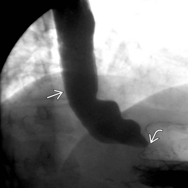
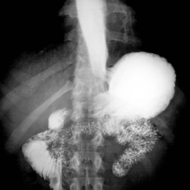
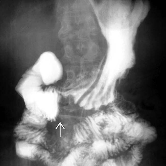
that is slow to empty due to a distal esophageal, peptic stricture image.

image
(Right) A 90-minute film (same case & study) from SBFT shows classic scleroderma of the small bowel with dilated, atonic jejunum & closely spaced, thin transverse folds image with slow transit. “Pseudo-obstruction” is another descriptive term relevant to this case.
image
(Left) Axial CECT in a 40-year-old woman demonstrates closely packed, thin small bowel folds image 
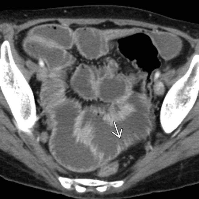
and diffusely dilated lumen, classic features of scleroderma with pseudo-obstruction.

image
(Right) Coronal CECT in the same patient demonstrates the dilated small bowel with a “hidebound” appearance of closely packed, thin folds image 
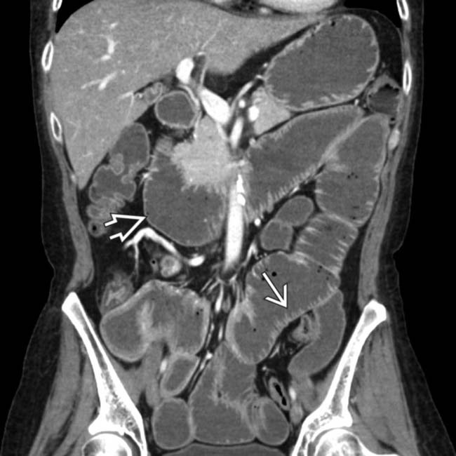
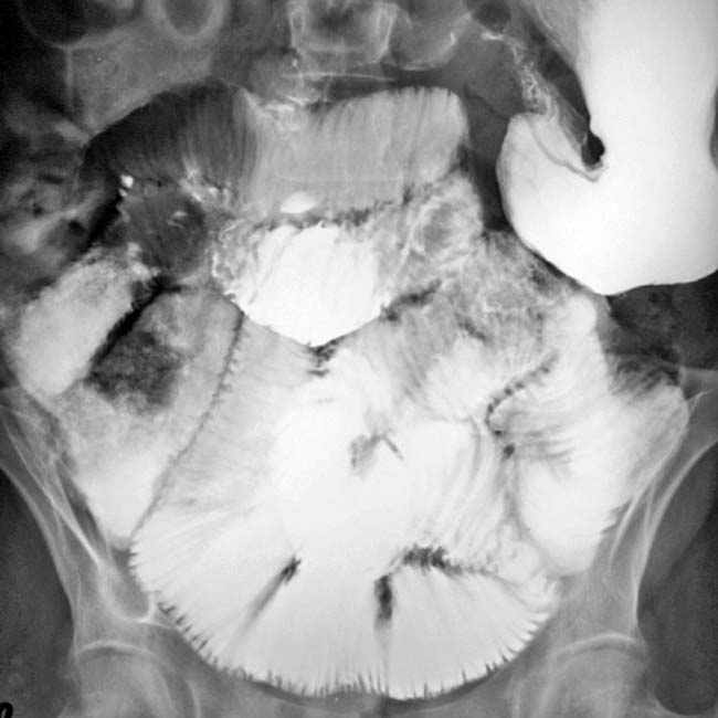
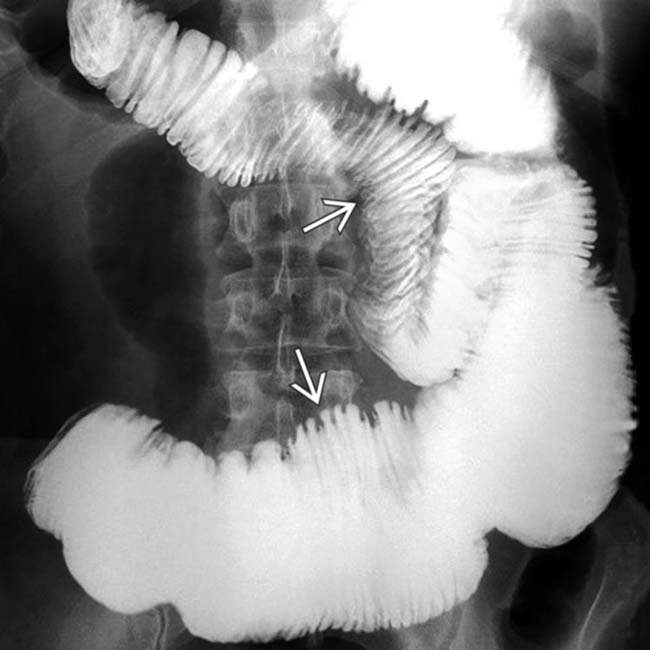
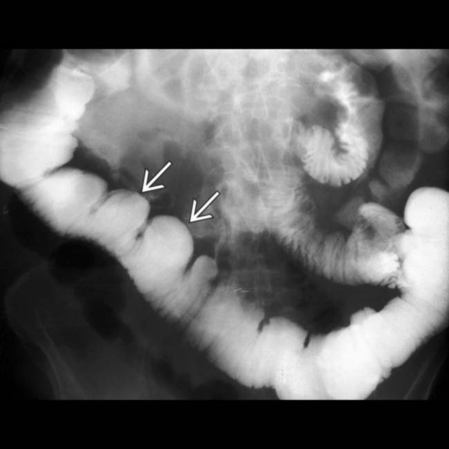
(particularly in the jejunum), a characteristic feature of scleroderma. Also note the disproportionate dilation of the duodenum image, another common feature of scleroderma.

TERMINOLOGY

Synonyms

• Progressive systemic sclerosis

Definitions

• Multisystem disorder of small vessels and connective tissue of unknown etiology

IMAGING

General Features

• Best diagnostic clue

image Dilated, atonic small bowel with crowded folds and wide-mouthed sacculations
• Other general features

image Multisystemic disorder with immunologic and inflammatory changes
image Characterized by atrophy, fibrosis, sclerosis of skin, vessels, and organs
image Involves skin and parenchyma of multiple organs

– GI tract, lungs, heart, kidneys, and nervous system
image Gastrointestinal tract (GI) scleroderma

– 2nd most common manifestation after skin changes (80-90% of patients)
– Most common sites: Esophagus > duodenum > anorectal > small bowel > colon
– Most frequent cause of chronic intestinal pseudo-obstruction
image Scleroderma classified into 2 types

– Diffuse scleroderma
– CREST syndrome (more benign course)
image Diffuse scleroderma: Cutaneous and visceral involvement

– Severe interstitial pulmonary fibrosis
– Organ failure more likely
– Associated with antitopoisomerase I antibody (anti-Scl 70)
image CREST syndrome: Less cutaneous and visceral involvement

– C alcinosis of skin
– R aynaud phenomenon
– E sophageal dysmotility
– S clerodactyly
– T elangiectasia

Radiographic Findings

• Esophagram

image Atony, aperistalsis: Lower 2/3 of esophagus (smooth muscle)
image Mild to moderate dilation of esophagus
image Patulous lower esophageal sphincter: Early finding
image Ulcers, fusiform peptic stricture (reflux esophagitis)

– Reflux predisposes to Barrett metaplasia (present in 40%)
image Hiatal hernia
• Upper GI series

image Stomach: Gastric dilation and delayed emptying
• Small bowel follow-through

image Marked dilatation of small bowel (particularly 2nd and 3rd parts of duodenum and jejunum)

– May have “megaduodenum”: Dilation made worse by compression of 3rd portion as it passes under mesenteric vessels
– Imaging findings identical to SMA syndrome
image Pathognomonic: Hidebound sign of small bowel

– Dilated jejunal lumen with crowded, thin circular folds
– Seen in > 60% of cases of scleroderma-related pseudo-obstruction
– Due to muscle atrophy and uneven replacement by collagen in longitudinal fibers, with intense fibrosis of submucosa
image Wide-mouthed sacculations (true diverticula)
image Prolonged transit time with barium retention in duodenum and small bowel up to 24 hours
image ± pneumatosis intestinalis and pneumoperitoneum

– May result from steroid medications + effect of dilated bowel
image ± transient, nonobstructive intussusceptions
• Barium enema

image Sacculations on border of transverse and descending colon
image Marked dilatation (may simulate Hirschsprung disease)
image Chronic phase: Complete loss of haustrations

– Simulates cathartic colon or chronic ulcerative colitis
image Stercoral ulceration (from retained fecal material in rectosigmoid)

DIFFERENTIAL DIAGNOSIS

Superior Mesenteric Artery (SMA) Syndrome

• Narrowing of angle between SMA and aorta may compress 3rd part of duodenum causing proximal dilatation

image Usually seen in asthenic persons who rapidly lose weight and retroperitoneal fat
image Due to prolonged bed rest or immobilization

– e.g., patients with whole body burns, body casts, spinal injury or surgery
• Upper GI findings

image Marked dilatation of 1st and 2nd parts of duodenum
image Vertical, linear, extrinsic, band-like defect in transverse part of duodenum overlying spine
image Obstruction is partially relieved in prone position
• CT may demonstrate beak-like compression of 3rd part of duodenum between SMA and aorta
• Scleroderma patients are thin; SMA compression may contribute to duodenal dilation

Celiac-Sprue Disease

• Gluten-sensitive enteropathy
• Segmental dilatation of small intestine
• Excess fluid in lumen (flocculation of barium)
• Atrophic duodenal and jejunal folds; relative hypertrophy of ileal folds
• Transient intussusceptions

Ileus

• Diffusely dilated lumen of bowel

image May have inflammatory component (e.g., acute pancreatitis)
• Small bowel folds in ileus usually normal to thick, rather than thin and crowded in scleroderma

PATHOLOGY

General Features

• Etiology

image Unknown; autoimmune with genetic predisposition
image May be initiated by environmental antigens like silica and L-tryptophan
image Immunologic mechanism (delayed hypersensitivity reaction)

– ↑ production of cytokines (TNF-α or IL-1) → increased collagen production
– Vascular damage and activation of fibroblasts
• Genetics

image Diffuse: Antitopoisomerase I antibodies associated with HLA-DR5
image Localized: Anticentromere antibodies associated with HLA-DR 1, 4, and 5
• Associated abnormalities

image May be associated with systemic lupus erythematosus, polymyositis, dermatomyositis
image Small bowel stasis may facilitate bacterial overgrowth

– May exacerbate anemia and malnutrition

Gross Pathologic & Surgical Features

• Rubber-hose inflexibility: Lower 2/3 of esophagus
• Thin and ulcerated mucosa
• Dilated gas and fluid-containing SB loops with sacculations

Microscopic Features

• Perivascular lymphocytic infiltrates
• Early capillary and arteriolar injury
• Atrophy and fragmentation of smooth muscle → collagen deposition and fibrosis

CLINICAL ISSUES

Presentation

• Most common signs/symptoms

image Esophagus

– Dysphagia, regurgitation
– Epigastric fullness and burning pain
image Duodenum

– Nausea, early satiety
image Small bowel

– Bloating, abdominal pain
– Weight loss, diarrhea, anemia
image Colon

– Chronic constipation
image Rectum and anus

– Fecal incontinence
– Fecal impaction; stercoral ulceration
• Laboratory data

image Increased erythrocyte sedimentation rate (ESR)
image Iron, B12, and folic acid deficiency anemias
image Increased antinuclear antibodies (ANA)

Demographics

• Age

image Onset at 30-50 years
• Gender

image M:F = 1:3
• Ethnicity

image African Americans > Caucasians
• Epidemiology

image Incidence: 14.1/1,000,000
image Prevalence: 19-75/100,000 persons

Natural History & Prognosis

• Complications

image Barrett esophagus → adenocarcinoma
image Bowel pseudo-obstruction
• Prognosis

image Limited disease with antinuclear antibodies (ANA): Good prognosis
image Diffuse disease: Poor with involvement of kidneys, heart, and lungs ± GI tract

Treatment

• Small frequent meals; elevation of head of bed
• Avoid tea and coffee
• Cimetidine, ranitidine, omeprazole
• Metoclopramide, laxatives
• Patients with severe malabsorption

image Parenteral hyperalimentation

DIAGNOSTIC CHECKLIST

Consider

• Check for involvement of other organs
• Check for family history of collagen vascular diseases

Image Interpretation Pearls

• Markedly dilated atonic small bowel with thin, crowded circular folds and delayed barium transit time

image
(Left) Film from a SBFT shows dilation of the duodenum with an abrupt narrowing image as it crosses the spine.
image
(Right) Delayed SBFT film from the same patient shows barium within the colon, which has a peculiar appearance of sacculations image 
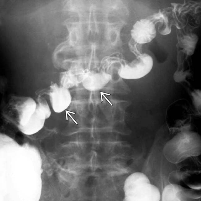
along the mesenteric border. These reflect the muscle atrophy within the bowel wall and its replacement by collagen and fibrosis.
image
(Left) Film from a SMFT in a 25-year-old man with scleroderma shows marked dilation of the duodenum with abrupt narrowing image 
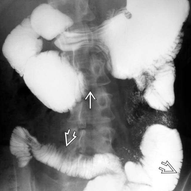
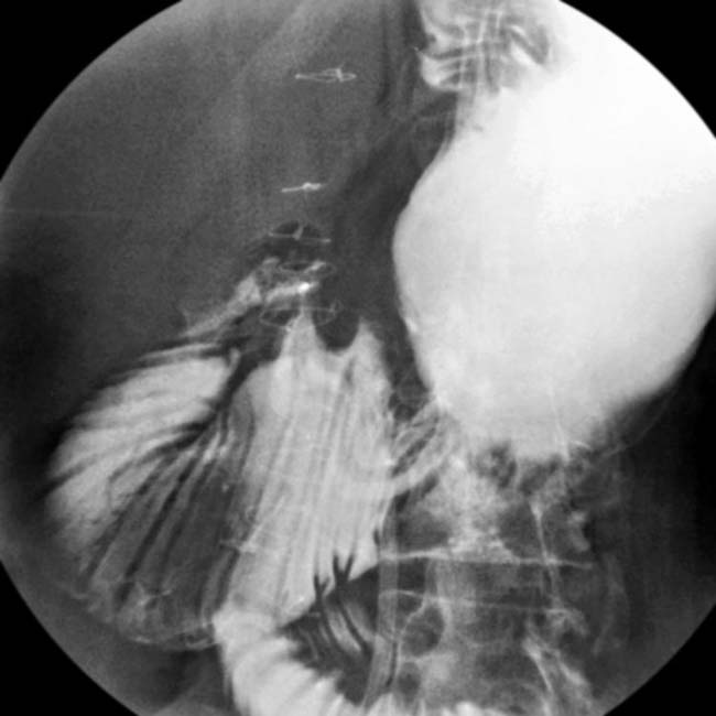
as it crosses the spine. The transverse folds of the jejunum image are thin and abnormally close together.

image
(Right) In the same case, a delayed film from a SBFT shows barium in the colon but persistent dilation of the duodenum.
image
(Left) Spot film from a SBFT shows sacculation image 
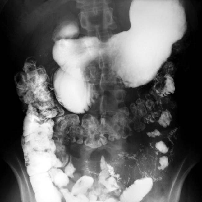
and abnormal folds in the small bowel.

image
(Right) Barium enema shows sacculation image 
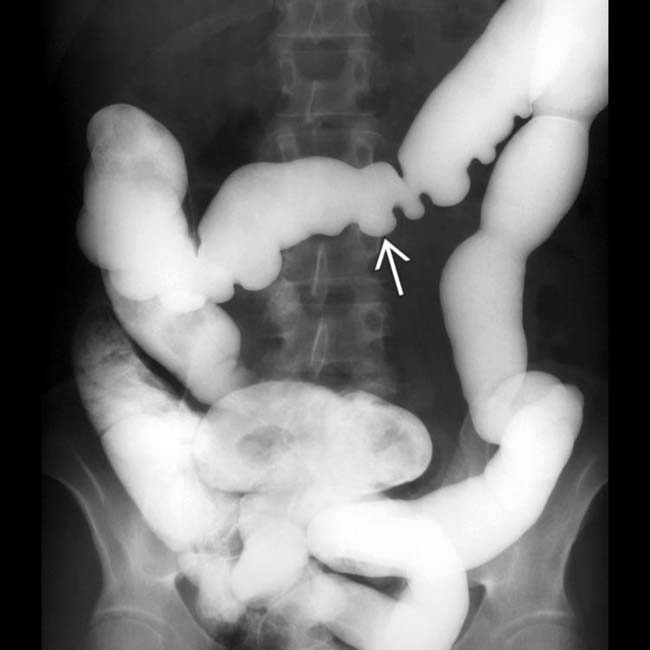
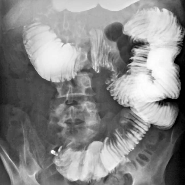
of the transverse colon and loss of a normal haustral pattern throughout most of the colon, all due to scleroderma.
image
SBFT shows dilated duodenum, “pseudo SMA syndrome,” and “hidebound” jejunal folds.

image
SBFT shows dilated, atonic bowel with the “hidebound” fold pattern.
image
SBFT shows a dilated and “hidebound” appearance of the duodenum.
image
UGI series shows a dilated esophagus with patulous GE junction and dilated duodenum with an abrupt “cut-off” as it crosses the midline.

Share this: