Injuries to the Eye

Published on 25/03/2015 by admin

Filed under Pediatrics

Last modified 22/04/2025

Print this page

rate 1 star rate 2 star rate 3 star rate 4 star rate 5 star
Your rating: none, Average: 0 (0 votes)

This article have been viewed 1630 times

Chapter 627 Injuries to the Eye

About 30% of all blindness in children results from trauma. Children and adolescents account for a disproportionate number of episodes of ocular trauma. Boys ages 11-15 yr are the most vulnerable; their injuries outnumber those in girls by a ratio of about 4 : 1. The majority of injuries are related to sports, toy darts, other projectiles, sticks, stones, fireworks, paint balls, and air-powered BB guns. The last causes particularly devastating ocular and orbital injuries. Much of the trauma is avoidable (Chapter 5.1). Any part of the orbit or globe may be affected (Fig. 627-1).

image

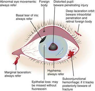
Figure 627-1 The injured eye.

(From Khaw PT, Shah P, Elkington AR: Injury to the eye, BMJ 328:36–38, 2004.)

Foreign Body involving the Ocular Surface

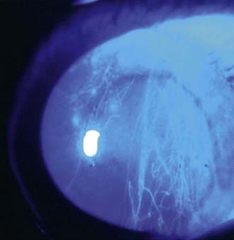
A foreign body usually produces acute discomfort, lacrimation, and inflammation. Most foreign bodies can be detected by examination in good light with the aid of magnification or a direct ophthalmoscope set on a high plus lens (+10 or +12). In many cases, slit-lamp examination is necessary, especially if the particle is deep or metallic. Some conjunctival foreign bodies tend to lodge under the upper eyelid, causing the sensation of corneal foreign body as they come into contact with the globe on eyelid movement; they can also produce vertically oriented linear corneal abrasions (Fig. 627-2). Finding these abrasions should lead to a suspicion of such a foreign body, and eversion of the lid may be necessary (Chapter 611). If a foreign body is suspected but not found, further examination is indicated. If the history suggests injury with a high-velocity particle, radiologic examination of the eye may be needed to explore the possibility of an intraocular foreign body.

Removal of a foreign body can be facilitated by instillation of a drop of topical anesthetic. Many foreign bodies can be removed by irrigating or by gently wiping them away with a moistened cotton-tipped applicator. Embedded foreign bodies or foreign bodies in the central cornea should be treated by an ophthalmologist. Removal of corneal foreign bodies can leave epithelial defects, which are treated as corneal abrasions. Metallic foreign bodies can cause rust to form in the corneal tissues; examination by an ophthalmologist 1 or 2 days after removal of a foreign body is recommended because a rust ring might require further treatment (curettage).

Hyphema

Hyphema is the presence of blood in the anterior chamber of the eye. It can occur with either a blunt or perforating injury and represents a potential vision-threatening situation. Hyphema appears as a bright or dark red fluid level between the cornea and iris or as a diffuse murkiness of the aqueous humor. Children with hyphema present with acute loss of vision and pain.

The treatment of hyphema involves efforts to minimize the vision-threatening sequelae such as rebleeding, glaucoma, and corneal blood staining. The patient is put on bed rest with elevation of the head of the bed to 30 degrees. A shield is placed on the affected eye, and a cycloplegic agent is used to immobilize the iris. Additionally, topical or systemic steroids are used to minimize intraocular inflammation. Antiemetics should be considered if the patient is experiencing nausea. All nonsteroidal anti-inflammatories and aspirin must be avoided. Rarely, hospitalization and sedation are necessary to ensure compliance in some children. If the intraocular pressure is elevated, topical and systemic pressure lowering medications are used. If the pressure is not controllable by such measures, then surgical evacuation of the clot may be required to minimize the risk of permanent vision loss.

Patients with sickle cell disease or trait are at higher risk of acute loss of vision secondary to elevated intraocular pressure or optic nerve infarction and can require more-aggressive intervention. Patients with a history of traumatic hyphema have an increased incidence of glaucoma later in life and should be monitored on a regular basis throughout their life.

Open Globe

A penetrating, perforating, or blunt injury resulting in compromise of the cornea or sclera of the eye is one of the most sight-threatening injuries that can be sustained. This is known as an open globe. An open globe is a true ophthalmologic emergency that requires prompt, careful evaluation and immediate repair to minimize vision loss. Permanent vision loss can result from corneal scarring, loss of intraocular contents, or infection. Evaluation involves careful history including time and mechanism of the injury, as well as tests of visual acuity and inspection of the eye. A full-thickness corneal wound often has prolapse of iris tissue though the wound. If this is not immediately evident, a peaked or irregular pupil may be seen. Scleral compromise may be more difficult to identify because of overlying structures. The thinnest part of the sclera is at the corneoscleral junction (the limbus) and just posterior to the insertion of the rectus muscles. When an open globe is caused by blunt force injury, these are the 2 areas most likely involved. The overlying conjunctiva might not be compromised but a subconjunctival hemorrhage may be present, obscuring the view. In these cases, look for a shallow anterior chamber, low intraocular pressure, or pigment within the involved area. If an open globe is diagnosed, the examination should be stopped, an eye shield should be placed immediately, and the ophthalmologist should be contacted to minimize further ocular compromise.

Chemical Injuries

Chemical burns of the cornea and adnexal tissue are among the most urgent of ocular emergencies. Alkali burns are usually more destructive than acid burns because they react with fats to form soaps, which damage cell membranes, allowing further penetration of the alkali into the eye. Acids generally cause less-severe, more-localized tissue damage. The corneal epithelium offers moderate protection against weak acids, and little damage occurs unless the pH is 2.5 or less. Most stronger acids precipitate tissue proteins, creating a physical barrier against their further penetration.

Mild acid or alkali burns are characterized by conjunctival injection and swelling and mild corneal epithelial erosions. The corneal stroma may be mildly edematous, and the anterior chamber can have mild to moderate cell and flare reactions. With strong acids, the cornea and conjunctiva rapidly become white and opaque. The corneal epithelium can slough, leaving a relatively clear stroma; this appearance can initially mask the severity of the burn. Severe alkali burns are characterized by corneal opacification.

Emergency treatment of a chemical burn begins with copious immediate irrigation with water or saline. Local debridement and removal of foreign particles should be performed while still irrigating. If the nature of the chemical injury is unknown, the use of pH test paper is helpful in determining whether the agent was basic or acidic. Irrigation should continue for at least 30 min or until 2 L of irrigant has been instilled in mild cases and for 2-4 hr or until 10 L of irrigant has been instilled in severe cases. At the end of irrigation, the pH should be within a normal range (7.3-7.7). The pH should be checked again approximately 30 min after irrigation to ensure that it has not changed. The goal of treatment is to minimize vision-threatening sequelae such as conjunctival scarring, corneal scarring and opacification, glaucoma, cataract, vision loss, and phthisis.

Orbital Fractures

The orbit is the bony structure surrounding the eye. Any of these bones can fracture in a traumatic incident. Superior and lateral wall fractures are the least common of the fracture sites, but superior orbital fracture is the most significant because of the potential of intracranial injury. The medial wall of the orbit is very susceptible to fracture because of the thin nature of the lamina papyracea. Perhaps the most common site of fracture from blunt trauma is the orbital floor. This is often referred to as blow-out fracture. At times, the fracture acts as a trapdoor, entrapping orbital contents within the fracture site.

The patient often presents with recent history of periorbital trauma and pain. Diplopia, eyelid swelling, eye movement restriction, and hypesthesia might or might not be present. A complete ophthalmic exam including history of injury, visual acuity, pupils, ocular alignment and motility, anterior segment, and fundus is required because there are often accompanying ocular injuries. Fracture is suspected if the eye is misaligned, movement is restricted, or the eye is sunken. The diagnosis is verified by orbital CT scan.

Medical management includes iced compresses to the orbit and elevation for the head of the bed for the first 24-48 hr. Broad-spectrum antibiotics are sometimes recommended for 14 days because of the exposure of the orbital contents to the sinus cavity. In medial wall fractures, the patient should be instructed not to blow the nose so as to prevent orbital emphysema and subsequent optic nerve compression.

Consider neurosurgical consultation in orbital roof fractures. Indications for surgical repair of orbital fractures are diplopia in primary or downgaze that persists for 2 weeks, enophthalmos, or fracture of the orbital floor involving >50% of the floor. Extraocular muscle entrapment often requires prompt surgical repair because these patients have significant pain, nausea, and vomiting that is difficult to control. Rarely, extraocular muscle entrapment can cause the activation of the oculocardiac reflex, requiring urgent fracture repair.

Child Abuse

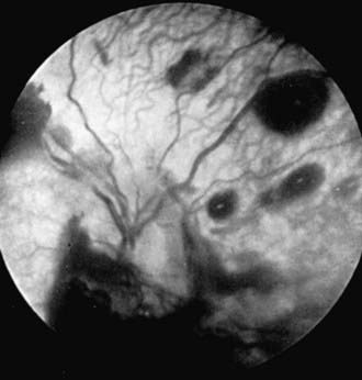
Child abuse (Chapter 37) is a major cause of injuries to the eye and orbital region. The possibility of nonaccidental trauma must be considered in any child with ecchymosis or laceration of the lids, hemorrhage in or about the eye, cataract or dislocated lens, retinal detachment, or fracture of the orbit. Inflicted childhood neurotrauma (shaken baby syndrome) occurs secondary to violent, nonaccidental, repetitive, unrestrained acceleration-deceleration head and neck movements, with or without blunt head trauma, in children typically <3 yr of age. Inflicted childhood neurotrauma accounts for approximately 10% of all cases of child abuse and carries a mortality rate of up to 25%. Detection of abuse is not only important in order to treat the pathology that is discovered but also to prevent further abuse or even death. The ocular manifestations are numerous and can have a prominent role in recognition of this syndrome. Retinal hemorrhage is the most common ophthalmic finding and occurs at all levels of the retina. The pattern of hemorrhage helps to distinguish this disorder from other causes of retinal hemorrhage or from accidental injuries (Fig. 627-3). Retinal hemorrhages can occur without associated intracranial pathology.

Sports-Related Ocular Injuries and Their Prevention

Although sports injuries occur in all age groups, far more children and adolescents participate in high-risk sports than do adults. The greater number of participating children, their athletic immaturity, and the increased likelihood of their using inadequate or improper eye protection account for their disproportionate share of sports-related eye injuries (Chapters 680 and 684).

The sports with the highest risk of eye injury are those in which no eye protection can be worn, including boxing, wrestling, and martial arts. High-risk sports include those that use a rapidly moving ball or puck, bat, stick, racquet, or arrow (baseball, hockey, lacrosse, racquet sports, and archery) or involve aggressive body contact (football and basketball). Related to both risk and frequency of participation, the highest percentage of eye injuries are in basketball and baseball.

Protective eyewear, designed for a specific activity, is available for most sports. For basketball, racquet sports, and other recreational activities that do not require a helmet or face mask, molded polycarbonate sports goggles that are secured to the head by an elastic strap are suggested. For hockey, football, lacrosse, and baseball (batter), specific helmets with polycarbonate face shields and guards are available. Children should also wear sports goggles under the helmets. For baseball, goggles and helmets should be worn for batting, catching, and base running; goggles alone are usually sufficient for other positions.

Bibliography

American Academy of Pediatrics Committee on Sports Medicine and Fitness, American Academy of Ophthalmology Committee on Eye Safety and Sports Ophthalmology. Protective eyewear for young athletes. Pediatrics. 1996;98:311-313.

Buys YM, Levin AV, Enzenauer RW, et al. Retinal findings after head trauma in infants and young children. Ophthalmology. 1992;99:1718-1723.

Centers for Disease Control and Prevention. Serious eye injuries associated with fireworks—United States, 1990–1994. MMWR Morb Mortal Wkly Rep. 1995;44:449-452.

Crouch ERJr, Crouch ER. Management of traumatic hyphema: therapeutic options. J Pediatr Ophthalmol Strabismus. 1999;36:238-250.

Egbert JE, May K, Kersten RC, et al. Pediatric orbital floor fracture: direct extraocular muscle involvement. Ophthalmology. 2000;107:1875-1879.

Forbes BJ, Christian CW, Judkins AR, et al. Inflicted childhood neurotrauma (shaken baby syndrome): ophthalmic findings. J Pediatr Ophthalmol Strabismus. 2004;41:80-88.

Goldenberg DT, Wu D, Capone AJr, et al. Nonaccidental trauma and peripheral retinal nonperfusion. Ophthalmology.. 2010;117:561-566.

Khaw PT, Shah P, Elkington AR. Injury to the eye. BMJ. 2004;328:36-38.

Kivlin JD, Simons KB, Lazoritz S, et al. Shaken baby syndrome. Ophthalmology. 2000;107:1246-1254.

Lane K, Penne RB, Bilyk JR. Evaluation and management of pediatric orbital fractures in primary care setting. Orbit. 2007;26:183-191.

Michael JG, Hug D, Dowd MD. Management of corneal abrasion in children: a randomized clinical trial. Ann Emerg Med. 2002;40:67-72.

Morad Y, Avni I, Benton SA, et al. Normal computerized tomography of brain in children with shaken baby syndrome. J AAPOS. 2004;8:445-450.

Prevent Blindness America. Most eye injuries in school-age children are sports-related. Prevent Blindness America urges public to make eye safety while playing sports a priority. Insight. 2009;34:26.

Smith GA, Knapp JF, Barnett TM, et al. The rocket’s red glare, the bombs bursting in air: fireworks-related injuries to children. Pediatrics. 1996;98:1-9.