Inflammatory Bowel Disease

Published on 27/03/2015 by admin

Filed under Pediatrics

Last modified 22/04/2025

Print this page

rate 1 star rate 2 star rate 3 star rate 4 star rate 5 star
Your rating: none, Average: 0 (0 votes)

This article have been viewed 1907 times

Chapter 328 Inflammatory Bowel Disease

The term inflammatory bowel disease (IBD) is used to represent 2 distinctive disorders of idiopathic chronic intestinal inflammation: Crohn disease and ulcerative colitis. Their respective etiologies are poorly understood, and both disorders are characterized by unpredictable exacerbations and remissions. The most common time of onset of IBD is during the preadolescent/adolescent era and young adulthood. A bimodal distribution has been shown with an early onset at 10-20 yr of age and a 2nd, smaller peak at 50-80 yr of age. About 25% of patients present before 20 yr of age. IBD may begin as early as the 1st yr of life, and an increased incidence among young children has been observed since the turn of the century. Children with early-onset IBD are more likely to have colonic involvement. In developed countries, these disorders are the major causes of chronic intestinal inflammation in children beyond the 1st few yr of life. A 3rd, less-common category, indeterminate colitis, represents ∼10% of pediatric patients.

Genetic and environmental influences are involved in the pathogenesis of IBD. The prevalence of Crohn disease in the United States is much lower for Hispanics and Asians than for whites and blacks. The risk of IBD in family members of an affected person has been reported in the range of 7-30%; a child whose parents both have IBD has a >35% chance of acquiring the disorder. Relatives of a patient with ulcerative colitis have a greater risk of acquiring ulcerative colitis than Crohn disease, whereas relatives of a patient with Crohn disease have a greater risk of acquiring this disorder; the 2 diseases can occur in the same family. The risk of occurrence of IBD among relatives of patients with Crohn disease is somewhat greater than for patients with ulcerative colitis.

The importance of genetic factors in the development of IBD is noted by a higher chance that both twins will be affected if they are monozygotic rather than dizygotic. The concordance rate in twins is higher in Crohn disease (36%) than in ulcerative colitis (16%). Genetic disorders that have been associated with IBD include Turner syndrome, the Hermansky-Pudlak syndrome, glycogen storage disease type Ib, and various immunodeficiency disorders. In 2001 the first IBD gene, NOD2, was identified through association mapping. A few months later, the IBD 5 risk haplotype was identified. These early successes were followed by a long period without notable risk factor discovery. Since 2006, the year of the first published genome wide array study on IBD, there has been an exponential growth in the set of validated genetic risk factors for IBD.

A perinuclear antineutrophil antibody (pANCA) is found in ∼70% of patients with ulcerative colitis compared with <20% of those with Crohn disease and is believed to represent a marker of genetically controlled immunoregulatory disturbance. About 55% of those with Crohn disease are positive for anti-Saccharomyces cerevisiae (ASCA) antibody. Additional markers including antibody to Escherichia coli outer membrane porin (anti-OmpC) and anti-flagellin (anti-CBir1) antibodies are associated with Crohn disease.

Environmental factors are also important and presumably explain discordance between twins and changes in risk among the same race in different geographic regions; the precise factors remain unknown. Persons migrating to developed countries often appear to acquire the higher rates of IBD associated with these regions. Cigarette smoking is a risk factor for Crohn disease but paradoxically protects against ulcerative colitis. No specific infectious agent has been reproducibly associated with IBD, although this is an active area of investigation.

An abnormality in intestinal mucosal immunoregulation may be of primary importance in the pathogenesis of IBD. The gut is under constant immunologic stimulation from microbial agents and dietary antigens. In response, the mucosa normally displays “physiologic” inflammation. In IBD, the mechanisms that keep “physiologic” inflammation in check fail and pathologic inflammation ensues. It is not clear if this represents an abnormal response to customary enteric antigens or a normal response to an as-yet-unidentified microbe. Mediators of inflammation (cytokines, arachidonic acid metabolites, reactive oxygen metabolites, growth factors) are involved, leading to tissue destruction and remodeling with fibrosis. Most therapies are aimed at interfering with these mediators.

It is usually possible to distinguish between ulcerative colitis and Crohn disease by the clinical presentation and radiologic, endoscopic, and histopathologic findings (Table 328-1). It is not possible to make a definitive diagnosis in ∼10% of patients with chronic colitis; this disorder is called indeterminate colitis. Occasionally, a child initially believed to have ulcerative colitis on the basis of clinical findings is subsequently found to have Crohn colitis. This is particularly true for the youngest patients, because Crohn disease in this patient population can more often manifest as exclusively colonic inflammation, mimicking ulcerative colitis. The medical treatments of Crohn disease and ulcerative colitis overlap.

Table 328-1 COMPARISON OF CROHN DISEASE AND ULCERATIVE COLITIS

FEATURE CROHN DISEASE ULCERATIVE COLITIS
Rectal bleeding Sometimes Common
Diarrhea, mucus, pus Variable Common
Abdominal pain Common Variable
Abdominal mass Common Not present
Growth failure Common Variable
Perianal disease Common Rare
Rectal involvement Occasional Universal
Pyoderma gangrenosum Rare Present
Erythema nodosum Common Less common
Mouth ulceration Common Rare
Thrombosis Less common Present
Colonic disease 50-75% 100%
Ileal disease Common None except backwash ileitis
Stomach-esophageal disease More common Chronic gastritis can be seen
Strictures Common Rare
Fissures Common Rare
Fistulas Common Rare
Toxic megacolon None Present
Sclerosing cholangitis Less common Present
Risk for cancer Increased Greatly increased
Discontinuous (skip) lesions Common Not present
Transmural involvement Common Unusual
Crypt abscesses Less common Common
Granulomas Common None
Linear ulcerations Uncommon Common

Extraintestinal manifestations occur slightly more commonly with Crohn disease than with ulcerative colitis (Table 328-2). Growth retardation is seen in 15-40% of children with Crohn disease at diagnosis. Of the extraintestinal manifestations that occur with IBD, joint, skin, eye, mouth, and hepatobiliary involvement tend to be associated with colitis, whether ulcerative or Crohn. The presence of some manifestations, such as peripheral arthritis, erythema nodosum, and anemia, correlates with activity of the bowel disease. Activity of pyoderma gangrenosum correlates less well with activity of the bowel disease, whereas sclerosing cholangitis, ankylosing spondylitis, and sacroiliitis do not correlate with intestinal disease. Arthritis occurs in 3 patterns: migratory peripheral arthritis involving primarily large joints, ankylosing spondylitis, and sacroiliitis. The peripheral arthritis of IBD tends to be nondestructive. Ankylosing spondylitis begins in the 3rd decade and occurs most commonly in patients with ulcerative colitis who have the human leukocyte antigen B27 phenotype. Symptoms include low back pain and morning stiffness; back, hips, shoulders, and sacroiliac joints are typically affected. Isolated sacroiliitis is usually asymptomatic but is common when a careful search is performed. Among the skin manifestations, erythema nodosum is most common. Patients with erythema nodosum or pyoderma gangrenosum have a high likelihood of having arthritis as well. Glomerulonephritis, uveitis, and a hypercoagulable state are other rare manifestations that occur in childhood. Cerebral thromboembolic disease has been described in children with IBD.

Table 328-2 EXTRAINTESTINAL COMPLICATIONS OF INFLAMMATORY BOWEL DISEASE

MUSCULOSKELETAL

SKIN AND MUCOUS MEMBRANES

DERMATOLOGIC

OCULAR

BRONCHOPULMONARY

CARDIAC

MALNUTRITION

HEMATOLOGIC

RENAL AND GENITOURINARY

PANCREATITIS

HEPATOBILIARY

ENDOCRINE AND METABOLIC

NEUROLOGIC

G6PD, glucose-6-phosphate dehydrogenase; IBD, inflammatory bowel disease; PSC, primary sclerosing cholangitis.

Modified from Kugathasan S: Diarrhea. In Kliegman RM, Greenbaum LA, Lye PS, editors: Practical strategies in pediatric diagnosis and therapy, ed 2, Philadelphia, 2004, Saunders, p 285.

328.1 Chronic Ulcerative Colitis

Ulcerative colitis, an idiopathic chronic inflammatory disorder, is localized to the colon and spares the upper gastrointestinal (GI) tract. Disease usually begins in the rectum and extends proximally for a variable distance. When it is localized to the rectum, the disease is ulcerative proctitis, whereas disease involving the entire colon is pancolitis. Approximately 50-80% of pediatric patients have extensive colitis, and adults more commonly have distal disease. Ulcerative proctitis is less likely to be associated with systemic manifestations, although it may be less responsive to treatment than more-diffuse disease. About 30% of children who present with ulcerative proctitis experience proximal spread of the disease. Ulcerative colitis has rarely been noted to present in infancy. Dietary protein intolerance can easily be misdiagnosed as ulcerative colitis in this age group. Dietary protein intolerance (cow’s milk protein) is a transient disorder; symptoms are directly associated with the intake of the offending antigen.

The incidence of ulcerative colitis has remained relatively constant, in contrast to an increase in Crohn disease, but varies with country of origin. Incidence rates are highest in northern European countries and the United States (15/100,000) and lowest in Japan and South Africa (1/100,000). The incidence of ulcerative colitis in Israel varies with the country of origin; those born in Asia or Africa have the lowest risk. The prevalence of ulcerative colitis in northern European countries and the United States varies from 100 to 200/100,000 population. Men are slightly more likely to acquire ulcerative colitis than are women; the reverse is true for Crohn disease.

Clinical Manifestations

Blood, mucus, and pus in the stool as well as diarrhea are the typical presentation of ulcerative colitis. Constipation may be observed in those with proctitis. Symptoms such as tenesmus, urgency, cramping abdominal pain (especially with bowel movements), and nocturnal bowel movements are common. The mode of onset ranges from insidious with gradual progression of symptoms to acute and fulminant. Fever, severe anemia, hypoalbuminemia, leukocytosis, and more than 5 bloody stools per day for 5 days define fulminant colitis. Chronicity is an important part of the diagnosis; it is difficult to know if a patient has a subacute, transient infectious colitis or ulcerative colitis when a child has had 1-2 weeks of symptoms. Symptoms beyond this duration often prove to be secondary to IBD. Anorexia, weight loss, and growth failure may be present, although these complications are more typical of Crohn disease.

Extraintestinal manifestations that tend to occur more commonly with ulcerative colitis than with Crohn disease include pyoderma gangrenosum, sclerosing cholangitis, chronic active hepatitis, and ankylosing spondylitis. Iron deficiency can result from chronic blood loss as well as decreased intake. Folate deficiency is unusual but may be accentuated in children treated with sulfasalazine, which interferes with folate absorption. Chronic inflammation and the elaboration of a variety of inflammatory cytokines can interfere with erythropoiesis and result in the anemia of chronic disease. Secondary amenorrhea is common during periods of active disease.

The clinical course of ulcerative colitis is marked by remission and relapse, often without apparent explanation. After treatment of initial symptoms, ∼5% of children with ulcerative colitis have a prolonged remission (>3 yr). About 25% of children presenting with severe ulcerative colitis require colectomy within 5 yr of diagnosis, compared with only 5% of those presenting with mild disease. It is important to consider the possibility of enteric infection with recurrent symptoms; these infections can mimic a flare-up or actually provoke a recurrence. The use of nonsteroidal anti-inflammatory drugs is considered by some to predispose to exacerbation.

It is generally believed that the risk of colon cancer begins to increase after 8-10 yr of disease and can then increase by 0.5-1% per yr. The risk is delayed by ∼10 yr in patients with colitis limited to the descending colon. Proctitis alone is associated with virtually no increase in risk over the general population. Because colon cancer is usually preceded by changes of mucosal dysplasia, it is recommended that patients who have had ulcerative colitis for >10 yr be screened with colonoscopy and biopsies every 1-2 yr. Although this is the current standard of practice, it is not clear if morbidity and mortality are changed by this approach. Two competing concerns about this plan of management remain unresolved: The original studies may have overestimated the risk of colon cancer and, therefore, the need for surveillance has been overemphasized; and screening for dysplasia might not be adequate for preventing colon cancer in ulcerative colitis if some cancers are not preceded by dysplasia.

Differential Diagnosis

The major conditions to exclude are infectious colitis, allergic colitis, and Crohn colitis. Every child with a new diagnosis of ulcerative colitis should have stool cultured for enteric pathogens, stool evaluation for ova and parasites, and perhaps serologic studies for amebae (Table 328-3). In the setting of antibiotic use, pseudomembranous colitis secondary to Clostridium difficile should be considered. Cytomegalovirus infection can mimic ulcerative colitis or be associated with an exacerbation of existing disease. The most difficult distinction is from Crohn disease because the colitis of Crohn disease can initially appear identical to that of ulcerative colitis, particularly in younger children. The gross appearance of the colitis or development of small bowel disease eventually leads to the correct diagnosis; this can occur years after the initial presentation.

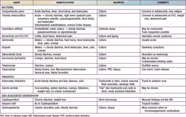
At the onset, the colitis of hemolytic-uremic syndrome may be identical to that of early ulcerative colitis. Ultimately, signs of microangiopathic hemolysis (the presence of schistocytes on blood smear), thrombocytopenia, and subsequent renal failure should confirm the diagnosis of hemolytic-uremic syndrome. Although Henoch-Schönlein purpura can manifest as abdominal pain and bloody stools, it is not usually associated with colitis. Behçet disease can be distinguished by its typical features (Chapter 155). Other considerations are radiation proctitis, viral colitis in immunocompromised patients, and ischemic colitis (Table 328-4). In infancy, dietary protein intolerance can be confused with ulcerative colitis, although the former is a transient problem that resolves on removal of the offending protein, and ulcerative colitis is extremely rare in this age group. Hirschsprung disease can produce an enterocolitis before or within months after surgical correction; this is unlikely to be confused with ulcerative colitis.

Diagnosis

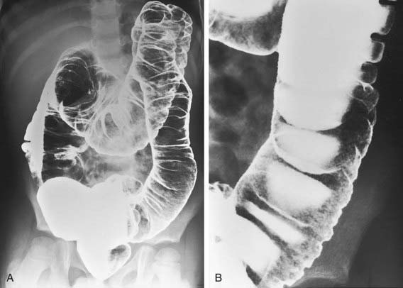
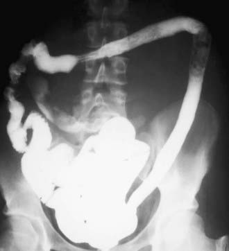
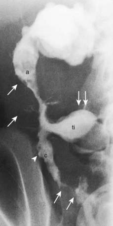
The diagnosis of ulcerative colitis or ulcerative proctitis requires a typical presentation in the absence of an identifiable specific cause (see Tables 328-3 and 328-4) and typical endoscopic and histologic findings (see Table 328-1). One should be hesitant to make a diagnosis of ulcerative colitis in a child who has experienced symptoms for <2-3 wk until infection has been excluded. When the diagnosis is suspected in a child with subacute symptoms, the physician should make a firm diagnosis only when there is evidence of chronicity on colonic biopsy. Laboratory studies can demonstrate evidence of anemia (either iron deficiency or the anemia of chronic disease) or hypoalbuminemia. Although the sedimentation rate and C-reactive protein are often elevated, they may be normal even with fulminant colitis. An elevated white blood cell count is usually seen only with more-severe colitis. Fecal calprotectin levels are usually elevated. Barium enema is suggestive but not diagnostic of acute (Fig. 328-1) or chronic burned-out disease (Fig. 328-2).

The diagnosis of ulcerative colitis must be confirmed by endoscopic and histologic examination of the colon. Classically, disease starts in the rectum with a gross appearance characterized by erythema, edema, loss of vascular pattern, granularity, and friability. There may be a “cutoff” demarcating the margin between inflammation and normal colon, or the entire colon may be involved. There may be some variability in the intensity of inflammation even in those areas involved. Flexible sigmoidoscopy can confirm the diagnosis; colonoscopy can evaluate the extent of disease and rule out Crohn colitis. A colonoscopy should not be performed when fulminant colitis is suspected because of the risk of provoking toxic megacolon or causing a perforation during the procedure. The degree of colitis can be evaluated by the gross appearance of the mucosa. One does not generally see discrete ulcers, which would be more suggestive of Crohn colitis. The endoscopic findings of ulcerative colitis result from microulcers, which give the appearance of a diffuse abnormality. With very severe chronic colitis, pseudopolyps may be seen. Biopsy of involved bowel demonstrates evidence of acute and chronic mucosal inflammation. Typical histologic findings are cryptitis, crypt abscesses, separation of crypts by inflammatory cells, foci of acute inflammatory cells, edema, mucus depletion, and branching of crypts. The last finding is not seen in infectious colitis. Granulomas, fissures, or full-thickness involvement of the bowel wall (usually on surgical rather than endoscopic biopsy) suggest Crohn disease.

Perianal disease, with the exception of mild local irritation or anal fissures associated with diarrhea, should make the clinician think of Crohn disease. Plain radiographs of the abdomen might demonstrate loss of haustral markings in an air-filled colon or marked dilation with toxic megacolon. With severe colitis, the colon may become dilated; a diameter of >6 cm, determined radiographically, in an adult suggests toxic megacolon. If it is necessary to examine the colon radiologically in a child with severe colitis (to evaluate the extent of involvement or to try to rule out Crohn disease), it is sometimes helpful to perform an upper GI contrast series with small bowel follow-through and then look at delayed films of the colon. A barium enema is contraindicated in the setting of a potential toxic megacolon.

Treatment

Medical

A medical cure for ulcerative colitis is not available; treatment is aimed at controlling symptoms and reducing the risk of recurrence, with a secondary goal of minimizing steroid exposure. The intensity of treatment varies with the severity of the symptoms. About 20-30% of patients with ulcerative colitis have spontaneous improvement in symptoms.

The first drug class to be used with mild colitis is an aminosalicylate. Sulfasalazine is composed of a sulfur moiety linked to the active ingredient 5-aminosalicylate (5-ASA). This linkage prevents the premature absorption of the medication in the upper GI tract, allowing it to reach the colon, where the 2 components are separated by bacterial cleavage. The dose of sulfasalazine is 50-75 mg/kg/24 hr (divided into 2-4 doses). Generally, the dose is not more than 2-4 g/24 hr. Hypersensitivity to the sulfa component is the major side effect of sulfasalazine and occurs in 10-20% of patients. Because of poor tolerance, sulfasalazine is used less commonly than other, better tolerated 5-ASA preparations (mesalamine, 50-100 mg/kg/day; balsalazide 110-175 mg/kg/day). Sulfasalazine and the 5-ASA preparations have been shown to effectively treat active ulcerative colitis and to prevent recurrence. It is recommended that the medication be continued even when the disorder is in remission. These medications might also decrease the lifetime risk of colon cancer.

Approximately 5% of patients have an allergic reaction to 5-ASA, manifesting as rash, fever, and bloody diarrhea, which can be difficult to distinguish from symptoms of a flare of ulcerative colitis. 5-ASA can also be given in enema or suppository form and is especially useful for proctitis. Hydrocortisone enemas are used to treat proctitis as well, but they are probably not as effective. Oral and rectal 5-ASA has been shown to be more effective than just oral 5-ASA for distal colitis.

Probiotics have been shown to be effective in adults for maintenance of remission for ulcerative colitis, although they have not been shown to induce remission during an active flare. The most promising role for probiotics has been to prevent pouchitis, a common complication following surgery.

Children with moderate to severe pancolitis or colitis that is unresponsive to 5-ASA therapy should be treated with oral corticosteroids, most commonly, prednisone. The usual starting dose of prednisone is 1-2 mg/kg/24 hr (40-60 mg maximum dose). This medication can be given once daily. With severe colitis, the dose can be divided twice daily and can be given intravenously. Steroids are considered an effective medication for acute flares, but they are not appropriate maintenance medications due to loss of effect and side effects, including growth retardation, adrenal suppression, cataracts, osteopenia, aseptic necrosis of the head of the femur, glucose intolerance, risk of infection, and cosmetic effects. For a hospitalized patient with persistence of symptoms despite intravenous steroid treatment for 5-7 days, escalation of therapy or surgical options should be considered.

With medical management, most children are in remission within 3 mo; however, 5-10% continue to have symptoms unresponsive to treatment beyond 6 mo. Many children with disease requiring frequent corticosteroid therapy are started on immunomodulators such as azathioprine (2.0-2.5 mg/kg/day) or 6-mercaptopurine (1-1.5 mg/kg/day). Uncontrolled data suggest a corticosteroid-sparing effect in many treated patients. Lymphoproliferative disorders are associated with thiopurine use. Cyclosporine, which has been associated with improvement in some children with severe or fulminant colitis, is rarely used owing to high side-effect profile, the inability of this medication to change the natural history of disease, and increasing use of infliximab, a chimeric monoclonal antibody to tumor necrosis factor (TNF)-α, also effective in cases of fulminant colitis. Infliximab has also been shown to be effective for induction and maintenance therapy in adults with moderate to severe disease. TNF blocking agents are associated with an increased risk of infection (particularly tuberculosis) and malignancies (lymphoma, leukemia).

Surgical

Colectomy is performed for intractable disease, complications of therapy, and fulminant disease that is unresponsive to medical management. No clear benefit of the use of total parenteral nutrition or a continuous enteral elemental diet in the treatment of severe ulcerative colitis has been noted. Nevertheless, parenteral nutrition is used if oral intake is insufficient so that the patient will be nutritionally ready for surgery if medical management fails. With any medical treatment for ulcerative colitis, the clinician should always weigh the risk of the medication or therapy against the fact that colitis can be successfully treated surgically.

Surgical treatment for intractable or fulminant colitis is total colectomy. The optimal approach is to combine colectomy with an endorectal pull-through, where a segment of distal rectum is retained and the mucosa is stripped from this region. The distal ileum is pulled down and sutured at the internal anus with a J pouch created from ileum immediately above the rectal cuff. This procedure allows the child to maintain continence. Commonly, a temporary ileostomy is created to protect the delicate anastomosis between the sleeve of the pouch and the rectum. The ileostomy is usually closed within several months, restoring bowel continuity. At that time, stool frequency is often increased but may be improved with loperamide. The major complication of this operation is pouchitis, which is a chronic inflammatory reaction in the pouch, leading to bloody diarrhea, abdominal pain, and, occasionally, low-grade fever. The cause of this complication is unknown, although it is more common when the ileal pouch has been constructed for ulcerative colitis than for other indications (e.g., familial polyposis coli). Pouchitis is seen in 30-40% of patients who had ulcerative colitis. It commonly responds to treatment with oral metronidazole or ciprofloxacin. Probiotics have also been shown to decrease the rate of pouchitis as well as the recurrence of pouchitis following antibiotic therapy.

Bibliography

AGA Institute Medical Position Panel. AGA medical position statement on the diagnosis and management of colorectal neoplasia in inflammatory bowel disease. Gastroenterology. 2010;138:738-745.

Anderson CA, Boucher G, Lees CW, et al. Meta-analysis identifies 29 additional ulcerative colitis risk loci, increasing the number of confirmed associations to 47. Nat Genet. 2011;43:246-252.

Baumgart DC, Carding SR. Inflammatory bowel disease: cause and immunobiology. Lancet. 2007;369:1627-1640.

Beattie RM, Croft NM, Fell JM, et al. Inflammatory bowel disease. Arch Dis Child. 2006;91:426-432.

Brest P, Lapaquette P, Souidi M, et al. A synonymous variant in IRGM alters a binding site for miR-196 and causes deregulation of IRGM-dependent xerophagy in Crohn’s disease. Nat Genet. 2011;43(3):242-245.

Colombel JF, Sandborn WJ, Reinisch W, et al. Infliximab, azathioprine, or combination therapy for Crohn’s disease. N Engl J Med. 2010;362(15):1383-1394.

Dolgin SE, Shlasko E, Gorfine S, et al. Restorative proctocolectomy in children with ulcerative colitis utilizing rectal mucosectomy with or without diverting ileostomy. J Pediatr Surg. 1999;34:837-839.

Dubois PC, van Heel DA. New susceptibility genes for ulcerative colitis. Nat Genet. 2008;40:686-688.

Franke A, Balschun T, Karlsen TH, et al. Sequence variants in IL10, ARPC2 and multiple other loci contribute to ulcerative colitis susceptibility. Nat Genet. 2008;40:1319-1323.

Gionchetti P, Rizello F, Helwig U, et al. Prophylaxis of pouchitis onset with probiotic therapy: a double-blind, placebo-controlled trial. Gastroenterology. 2003;124:1202-1209.

Heyman M, Kirschner BS, Gold BD, et al. Children with early onset inflammatory bowel disease (IBD): analysis of a pediatric IBD consortium registry. J Pediatr. 2004;146:35-40.

Ho GT, Mowat C, Goddard CJ, et al. Predicting the outcome of severe ulcerative colitis: development of a novel risk score to aid early selection of patients for second-line medical therapy or surgery. Aliment Pharmacol Ther. 2004;19:1079-1087.

IBD in EPIC Study Investigators. Linoleic acid, a dietary n-6 polyunsaturated fatty acid, and the aetiology of ulcerative colitis: a nested case control study within a European prospective cohort study. Gut. 2009;58:1606-1611.

Jose FA, Heyman MB. Extraintestinal manifestations of inflammatory bowel disease. J Pediatr Gastroenterol Nutr. 2008;46:124-133.

Kruis W, Fric P, Pokrotnieks J, et al. Maintaining remission of ulcerative colitis with the probiotic Escherichia coli Nissle 1917 is as effective as with standard mesalazine. Gut. 2004;53:1617-1623.

Kugathasan S, Judd RH, Hoffmann RG, et al. Epidemiologic and clinical characteristics of children with newly diagnosed inflammatory bowel disease in Wisconsin: a statewide population-based study. J Pediatr. 2003;143:525-531.

Leggieri N, Marques-Vidal P, Cerwenka H, et al. Migrated foreign body liver abscess. Medicine. 2010;89(2):85-95.

Malaty HM, Fan X, Opekun AR, et al. Rising incidence of inflammatory bowel disease among children: a 12-year study. J Pediatr Gastroenterol Nutr. 2010;50(1):27-31.

Mamula P, Markowitz JE, Baldassano RN. Inflammatory bowel disease in early childhood and adolescence: special considerations. Gastroenterol Clin North Am. 2003;32:967-995.

Mamula P, Markowitz JE, Cohen LJ, et al. Infliximab in pediatric ulcerative colitis: two-year follow-up. J Pediatr Gastroenterol Nutr. 2004;38:298-301.

Medical Letter. Encapsulated mesalamine granules (Apriso) for ulcerative colitis. Med Lett. 2009;51:38-40.

Momozawa Y, Mni M, Nakamura K, et al. Resequencing of positional candidates identifies low frequency IL23R coding variants protecting against inflammatory bowel disease. Nat Genet. 2011;43(1):43-47.

Quail MA, Russell RK, Van Limbergen JE, et al. Fecal calprotectin complements routine laboratory investigations in diagnosing childhood inflammatory bowel disease. Inflamm Bowel Dis. 2009;15:756-759.

Romano C, Famiani A, Gallizzi R, et al. Indeterminate colitis: a distinctive clinical pattern of inflammatory bowel disease in children. Pediatrics. 2008;122:e1278-e1281.

Rutgeerts P, Sandborn WJ, Feagan BG, et al. Infliximab for induction and maintenance therapy for ulcerative colitis. N Engl J Med. 2005;353:2462-2476.

Sanderson JD, Parkes GC. Timing of surgery for inflammatory bowel disease. BMJ. 2007;335:1006-1007.

Schwiertz A, Jacobi M, Frick JS, et al. Microbiota in pediatric inflammatory bowel disease. J Pediatr. 2010;157:240-244.

Timmer A, Behrens R, Buderus S, et al. Childhood onset inflammatory bowel disease: predictors of delayed diagnosis from the CEDATA German-language pediatric inflammatory bowel disease registry. J Pediatr. 2011;158:467-473.

Timmer A, Obermeier F. Reduced risk of ulcerative colitis after appendectomy. BMJ. 2009;338:781-782.

Turner D, Walsh CM, Benchimol EI, et al. Severe paediatric ulcerative colitis: incidence, outcomes, and optimal timing for second-line therapy. Gut. 2008;57:331-338.

Turner D, Walsh CM, Steinhart H, et al. Response to corticosteroids in severe ulcerative colitis: a systematic review of the literature and a meta-regression. Clin Gastroenterol Hepatol. 2007;5:103-110.

UK IBD Genetics Consortium and the Wellcome Trust Case Control Consortium. Genome-wide association study of ulcerative colitis identifies three new susceptibility loci, including the HNF4A region. Nat Genet. 2009;41:1330-1334.

328.2 Crohn Disease (Regional Enteritis, Regional Ileitis, Granulomatous Colitis)

Crohn disease, an idiopathic, chronic inflammatory disorder of the bowel, involves any region of the alimentary tract from the mouth to the anus. Although there are many similarities between ulcerative colitis and Crohn disease, there are also major differences in the clinical course and distribution of the disease in the GI tract (see Table 328-1). The inflammatory process tends to be eccentric and segmental, often with skip areas (normal regions of bowel between inflamed areas). Although inflammation in ulcerative colitis is limited to the mucosa (except in toxic megacolon), GI involvement in Crohn disease is often transmural.

Compared to adult-onset disease, pediatric Crohn disease is more likely to have extensive anatomic involvement. At initial presentation, >50% of patients have disease that involves ileum and colon (ileocolitis), 20% have exclusively colonic disease, and upper GI involvement (esophagus, stomach, duodenum) is seen in up to 30% of children. Isolated small bowel disease is much less common in the pediatric population compared to adults. Isolated colonic disease is common in children <8 yr of age and may be indistinguishable from ulcerative colitis. Anatomic location of disease tends to extend over time in children.

Crohn disease tends to have a bimodal age distribution, with the 1st peak beginning in the teenage years. The incidence of Crohn disease has been increasing, whereas that of ulcerative colitis has been stable. In the United States, the reported incidence of pediatric Crohn disease is 4.56/100,000 and the pediatric prevalence is 43/100,000.

Clinical Manifestations

Crohn disease can be characterized as inflammatory, stricturing, or penetrating. Patients with small bowel disease are more likely to have an obstructive pattern (most commonly with right lower quadrant pain) characterized by fibrostenosis, and those with colonic disease are more likely to have symptoms resulting from inflammation (diarrhea, bleeding, cramping). Disease phenotypes often change as duration of disease lengthens (inflammatory becomes structuring or penetrating).

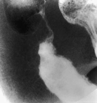
Systemic signs and symptoms are more common in Crohn disease than in ulcerative colitis. Fever, malaise, and easy fatigability are common. Growth failure with delayed bone maturation and delayed sexual development can precede other symptoms by 1 or 2 yr and is at least twice as likely to occur with Crohn disease as with ulcerative colitis. Children can present with growth failure as the only manifestation of Crohn disease. Causes of growth failure include inadequate caloric intake, suboptimal absorption or excessive loss of nutrients, the effects of chronic inflammation on bone metabolism and appetite, and the use of corticosteroids during treatment. Primary or secondary amenorrhea and pubertal delay are common. In contrast to ulcerative colitis, perianal disease is common (tags, fistula, abscess). Gastric or duodenal involvement may be associated with recurrent vomiting and epigastric pain. Partial small bowel obstruction, usually secondary to narrowing of the bowel lumen from inflammation or stricture, can cause symptoms of cramping abdominal pain (especially with meals), borborygmus, and intermittent abdominal distention (Fig. 328-3). Stricture should be suspected if the child notes relief of symptoms in association with a sudden sensation of gurgling of intestinal contents through a localized region of the abdomen.

image

Figure 328-3 Stenotic Crohn disease. Severe stenosis of the terminal ileum is present in this 16 yr old boy. Inflammatory effacement of the mucosal folds and small ulcerations characterize the proximal nonstenotic segment.

(From The child with diarrhea. In Hoffman AD, Hilton SW, Edwards DK, editors: Practical pediatric radiology, ed 2, Philadelphia, 1994, WB Saunders, p 267.)

Penetrating disease is demonstrated by fistula formation. Enteroenteric or enterocolonic fistulas (between segments of bowel) are often asymptomatic but can contribute to malabsorption if they have high output or result in bacterial overgrowth (Fig. 328-4). Enterovesical fistulas (between bowel and urinary bladder) originate from ileum or sigmoid colon and appear as signs of urinary infection, pneumaturia, or fecaluria. Enterovaginal fistulas originate from the rectum, cause feculent vaginal drainage, and are difficult to manage. Enterocutaneous fistulas (between bowel and abdominal skin) often are caused by prior surgical anastomoses with leakage. Intra-abdominal abscess may be associated with fever and pain but might have relatively few symptoms. Hepatic or splenic abscess can occur with or without a local fistula. Anorectal abscesses often originate immediately above the anus at the crypts of Morgagni. The patterns of perianal fistulas are complex because of the different tissue planes. Perianal abscess is usually painful, but perianal fistulas tend to produce fewer symptoms than anticipated. Purulent drainage is commonly associated with perianal fistulas. Psoas abscess secondary to intestinal fistula can present as hip pain, decreased hip extension (psoas sign), and fever.

Extraintestinal manifestations occur more commonly with Crohn disease than with ulcerative colitis; those that are especially associated with Crohn disease include oral aphthous ulcers, peripheral arthritis, erythema nodosum, digital clubbing, episcleritis, renal stones (uric acid, oxalate), and gallstones. Any of the extraintestinal disorders described in the section on IBD can occur with Crohn disease (see Table 328-2). The peripheral arthritis is nondeforming. The occurrence of extraintestinal manifestations usually correlates with the presence of colitis.

Extensive involvement of small bowel, especially in association with surgical resection, can lead to short bowel syndrome, which is rare in children. Complications of terminal ileal dysfunction or resection include bile acid malabsorption with secondary diarrhea and vitamin B12 malabsorption. Chronic steatorrhea can lead to oxaluria with secondary renal stones. Increasing calcium intake can actually decrease the risk renal stones secondary to ileal inflammation. The risk of cholelithiasis is also increased secondary to bile acid depletion.

A disorder with this diversity of manifestations can have a major impact on an affected child’s lifestyle. Fortunately, the majority of children with Crohn disease are able to continue with their normal activities, having to limit activity only during periods of increased symptoms.

Differential Diagnosis

The most common diagnoses to be distinguished from Crohn disease are the infectious enteropathies (in the case of Crohn disease: acute terminal ileitis, infectious colitis, enteric parasites, and periappendiceal abscess) (see Tables 328-3, 328-4, and 328-5). Yersinia can cause many of the radiologic and endoscopic findings in the distal small bowel that are seen in Crohn disease. The symptoms of bacterial dysentery are more likely to be mistaken for ulcerative colitis than for Crohn disease. Celiac disease and Giardia infection have been noted to produce a Crohn-like presentation including diarrhea, weight loss, and protein-losing enteropathy. GI tuberculosis is rare but can mimic Crohn disease. Foreign-body perforation of the bowel (toothpick) can mimic a localized region with Crohn disease. Small bowel lymphoma can mimic Crohn disease but tends to be associated with nodular filling defects of the bowel without ulceration or narrowing of the lumen. Bowel lymphoma is much less common in children than is Crohn disease. Recurrent functional abdominal pain can mimic the pain of small bowel Crohn disease. Lymphoid nodular hyperplasia of the terminal ileum (a normal finding) may be mistaken for Crohn ileitis. Right lower quadrant pain or mass with fever can be the result of periappendiceal abscess. This entity is occasionally associated with diarrhea as well.

Table 328-5 DIFFERENTIAL DIAGNOSIS OF PRESENTING SYMPTOMS OF CROHN DISEASE

PRIMARY PRESENTING SYMPTOM DIAGNOSTIC CONSIDERATIONS
Right lower quadrant abdominal pain, with or without mass Appendicitis, infection (e.g., Campylobacter, Yersinia spp.), lymphoma, intussusception, mesenteric adenitis, Meckel diverticulum, ovarian cyst
Chronic periumbilical or epigastric abdominal pain Irritable bowel syndrome, constipation, lactose intolerance, peptic disease
Rectal bleeding, no diarrhea Fissure, polyp, Meckel diverticulum, rectal ulcer syndrome
Bloody diarrhea Infection, hemolytic-uremic syndrome, Henoch-Schönlein purpura, ischemic bowel, radiation colitis
Watery diarrhea Irritable bowel syndrome, lactose intolerance, giardiasis, Cryptosporidium infection, sorbitol, laxatives
Perirectal disease Fissure, hemorrhoid (rare), streptococcal infection, condyloma (rare)
Growth delay Endocrinopathy
Anorexia, weight loss Anorexia nervosa
Arthritis Collagen vascular disease, infection
Liver abnormalities Chronic hepatitis

From Kugathasan S: Diarrhea. In Kliegman RM, Greenbaum LA, Lye PS, editors: Practical strategies in pediatric diagnosis and therapy, ed 2, Philadelphia, 2004, Saunders, p 287.

Growth failure may be the only manifestation of Crohn disease; other disorders such as growth hormone deficiency, gluten-sensitive enteropathy (celiac disease), Turner syndrome, or anorexia nervosa must be considered. If arthritis precedes the bowel manifestations, an initial diagnosis of juvenile idiopathic arthritis may be made. Refractory anemia may be the presenting feature and may be mistaken for a primary hematologic disorder. Leukemia can manifest with abdominal pain in association with an abnormal blood cell count and might initially be mistaken for Crohn disease. Chronic granulomatous disease of childhood can cause inflammatory changes in the bowel as well as perianal disease. Antral narrowing in this disorder may be mistaken for a stricture secondary to Crohn disease.

Diagnosis

Crohn disease can manifest as a variety of symptom combinations. At the onset, symptoms may be subtle (growth retardation, abdominal pain alone); this explains why the diagnosis might not be made until 1 or 2 years after the start of symptoms. The diagnosis of Crohn disease depends on finding typical clinical features of the disorder (history, physical examination, laboratory studies, and endoscopic or radiologic findings), ruling out specific entities that mimic Crohn disease, and demonstrating chronicity. The history can include any combination of abdominal pain (especially right lower quadrant), diarrhea, vomiting, anorexia, weight loss, growth retardation, and extraintestinal manifestations. Only 25% initially have the triad of diarrhea, weight loss, and abdominal pain. Most do not have diarrhea, and only 25% have GI bleeding.

Children with Crohn disease often appear chronically ill. They commonly have weight loss and growth failure, and they are often malnourished. The earliest of sign of growth failure is decreased height velocity, which can be present in up to 88% of prepubertal patients with Crohn disease and typically precedes symptoms. Children with Crohn disease often appear pale, with decreased energy level and poor appetite; the latter finding sometimes results from an association between meals and abdominal pain or diarrhea. There may be abdominal tenderness that is either diffuse or localized to the right lower quadrant. A tender mass or fullness may be palpable in the right lower quadrant. Perianal disease, when present, may be characteristic. Large anal skin tags (1-3 cm diameter) or perianal fistulas with purulent drainage suggest Crohn disease. Digital clubbing, findings of arthritis, and skin manifestations may be present.

A complete blood cell count commonly demonstrates anemia, often with a component of iron deficiency. Although the erythrocyte sedimentation rate is often elevated, it may be normal; an elevated platelet count is common. The white blood cell count may be normal or mildly elevated. The serum albumin level may be low, indicating small bowel inflammation or protein-losing enteropathy. Fecal calprotectin or lactoferrin is often elevated. Anti–Saccharomyces cerevisiae antibodies, antibody to Escherichia coli outer membrane porin (anti-OmpC), and anti-flagellin (anti-CBir1) antibodies are associated with Crohn’s disease.

The small and large bowel and the upper GI tract should be examined by both endoscopic and radiologic studies in the child with suspected Crohn disease. Esophagogastroduodenoscopy and ileocolonoscopy should be performed to properly assess the upper GI tract, terminal ileum, and entire colon. Findings on colonoscopy can include patchy, nonspecific inflammatory changes (erythema, friability, loss of vascular pattern), aphthous ulcers, linear ulcers, nodularity, and strictures. Findings on biopsy may be only nonspecific chronic inflammatory changes. Noncaseating granulomas, similar to those of sarcoidosis, are the most characteristic histologic findings, although often they are not present. Transmural inflammation is also characteristic but can be identified only in surgical specimens.

Radiologic studies are necessary to assess the entire small bowel and investigate for evidence of structuring or penetrating disease. A variety of findings may be apparent on radiologic studies. Plain films of the abdomen may be normal or might demonstrate findings of partial small bowel obstruction or thumbprinting of the colon wall. An upper GI contrast study with small bowel follow-through might show aphthous ulceration and thickened, nodular folds as well as narrowing or stricturing of the lumen. Linear ulcers can give a cobblestone appearance to the mucosal surface. Bowel loops are often separated as a result of thickening of bowel wall and mesentery. Other manifestations on radiographic studies that suggest more-severe Crohn disease are fistulas between bowel (enteroenteric or enterocolonic), sinus tracts, and strictures (see Figs. 328-3 and 328-4).

An upper GI contrast examination with small bowel follow-through has typically been the study of choice for imaging of the small bowel, but CT and MR enterography and small bowel ultrasound are increasingly being used to assess for intestinal wall thickening and extraluminal findings such as abscesses or fistulas. MR of the pelvis is also useful for delineating the extent of perianal involvement. Video capsule endoscopy has revealed evidence of small bowel mucosal lesions in some patients with normal radiologic evaluation.

Treatment

Crohn disease cannot be cured by medical or surgical therapy. The aim of treatment is to relieve symptoms and prevent complications of chronic inflammation (anemia, growth failure), prevent relapse, minimize corticosteroid exposure, and, if possible, effect mucosal healing.

Medical

The specific therapeutic modalities used depend on geographic localization of disease, severity of inflammation, age of the patient, and the presence of complications (abscess). For mild terminal ileal disease or mild Crohn disease of the colon, an initial trial of mesalamine (50-100 mg/kg/day, maximum 3-4 g) may be attempted. Specific pharmaceutical preparations have been formulated to release the active 5-ASA compound throughout the small bowel, in the ileum and colon, or exclusively in the colon. Rectal preparations are used for distal colonic inflammation. Antibiotics such as metronidazole (10-20 mg/kg/day) are used for infectious complications, are first-line therapy for perianal disease (although perianal disease usually recurs when antibiotic is discontinued), and they may be effective for treatment of mild to moderate Crohn disease.

Corticosteroids continue to be a mainstay of therapy for acute exacerbations of pediatric Crohn disease because they effectively suppress acute inflammation, rapidly relieving symptoms. For more extensive or severe small bowel or colonic disease, most clinicians initiate therapy with corticosteroids (prednisone, 1-2 mg/kg/day, maximum 40-60 mg). The goal is to taper dosing as soon as the disease becomes quiescent. Clinicians vary in their tapering schedules, and the disease can flare during this process. There is no role for continuing corticosteroids as maintenance therapy because, in addition to their side effects, tolerance develops and steroids have not been shown to change disease course or promote healing of mucosa. A special controlled ileal-release formulation of budesonide, a corticosteroid with local anti-inflammatory activity on the bowel mucosa and with high hepatic first-pass metabolism, is also used for mild to moderate ileal or ileocecal disease (adult dose, 9 mg daily). Budesonide appears to be more effective than mesalamine in the treatment of active ileocolonic disease but is less effective than prednisone. Although less effective than traditional corticosteroids, budesonide does cause less steroid-related side effects.

Unfortunately, up to 50% of children with Crohn disease either become refractory to corticosteroid therapy or become dependent on daily dosing and quickly experience flare of the disease when the dose is decreased. Immunomodulators such as azathioprine (2.0-2.5 mg/kg/day) or 6-mercaptopurine (1.0-1.5 mg/kg/day) may be effective in some children who have a poor response to prednisone or who are steroid dependent. Because a beneficial effect of these drugs can be delayed for 3-6 mo after starting therapy, they are not helpful acutely. The early use of these agents can decrease cumulative prednisone dosages over the 1st 1-2 years of therapy. Genetic variations in the enzyme systems responsible for metabolism of these agents (thiopurine S-methyltransferase [TPMT]) can affect response rates and potential toxicity. Lymphoproliferative disorders have developed from thiopurine use in patients with IBD. Methotrexate is another immunomodulator that is effective in the treatment of active Crohn’s disease and has been shown to improve height velocity in the 1st year of administration. The advantages of this medication include once-weekly dosing by either subcutaneous or oral route (10 mg weekly for 20-29 kg, 15 mg weekly for 30-39 kg, 20 mg weekly for 40-49 kg, 25 mg weekly for >50 kg) and a more-rapid onset of action (6-8 wk) than azathioprine or 6-mercaptopurine. Folic acid is usually administered concomitantly to decrease medication side effects. The immunomodulators are effective for the treatment of perianal fistulas.

Therapy with antibodies directed against mediators of inflammation is used for patients with Crohn disease. Infliximab (5 mg/kg IV), a chimeric monoclonal antibody to TNF-α, has been shown to be effective for the induction and maintenance of remission and mucosal healing in chronically active moderate to severe Crohn disease, healing of perianal fistulas, and steroid sparing. Pediatric data have additionally supported improved growth with the administration of this medication. The onset of action of infliximab is quite rapid and it is initially given as 3 infusions over a 6 wk period (0, 2, and 6 wk). The durability of response to infliximab is variable and can be as short as 4-8 wk, making maintenance therapy necessary. Side effects include infusion reactions, increased incidence of infections (especially reactivation of latent tuberculosis), increased risk of lymphoma, and the development of autoantibodies and autoimmune disorders (leukocytoclastic vasculitis). The development of antibodies to infliximab (ATI) is associated with an increased incidence of infusion reactions and decreased durability of response. Regularly scheduled dosing of infliximab, as opposed to episodic dosing on an as-needed basis, is associated with decreased levels of ATI. A purified protein derivative (PPD) test for tuberculosis should be done before starting infliximab. Active or latent intra-abdominal infection (abscess) is a contraindication to infliximab therapy. Adalimumab, a subcutaneously administered, fully humanized monocloncal antibody against TNF-α, has also been shown to be effective for the treatment of chronically active moderate to severe Crohn’s disease in adults, and it has been used extensively in children for this purpose. Antibodies against interleukin 6, 10, 11, 12, 23, and 23/p40 and intracellular adhesion molecules are being tested.

Exclusive enteral nutritional therapy is an effective primary as well as adjunctive treatment. The enteral nutritional approach (elemental or polymeric diets) is as rapid in onset of response and as effective as the other treatments. Pediatric studies have suggested similar efficacy to prednisone for improvement in clinical symptoms, but enteral nutritional therapy is superior to steroids for actual healing of mucosa. Because elemental diets are relatively unpalatable, they are administered via a nasogastric or gastrostomy infusion, usually overnight. Most children are hesitant to use nasogastric infusion, but once it is begun, most find it is not difficult. The advantages are that it is relatively free of side effects, avoids the problems associated with corticosteroid therapy, and simultaneously addresses the nutritional rehabilitation. Children can participate in normal daytime activities. A major disadvantage of this approach is that patients are not able to eat a regular diet because they are receiving all of their calories from formula. In addition, perianal and colon disease does not respond well. For children with growth failure, this approach may be ideal, however.

High-calorie oral supplements, although effective, are often not tolerated because of early satiety or exacerbation of symptoms (abdominal pain, vomiting, or diarrhea). Nonetheless, they should be offered to children whose weight gain is suboptimal even if they are not candidates for exclusive enteral nutritional therapy. The continuous administration of nocturnal nasogastric feedings for chronic malnutrition and growth failure has been effective with a much lower risk of complications than parenteral hyperalimentation. The efficacy of probiotics and omega-3 fatty acids in the treatment of Crohn disease is controversial.

Surgery

Surgical therapy should be reserved for very specific indications. Recurrence rate after bowel resection is high (>50% by 5 yr); the risk of requiring additional surgery increases with each operation. Potential complications of surgery include development of fistula or stricture, anastomotic leak, postoperative partial small bowel obstruction secondary to adhesions, and short bowel syndrome. Surgery is the treatment of choice for localized disease of small bowel or colon that is unresponsive to medical treatment, bowel perforation, fibrosed stricture with symptomatic partial small bowel obstruction, and intractable bleeding. Intra-abdominal or liver abscess sometimes is successfully treated by ultrasonographic or CT-guided catheter drainage and concomitant intravenous antibiotic treatment. Open surgical drainage is necessary if this approach is not successful. Growth retardation was once considered an indication for resection; without other indications, this approach has not been shown to be beneficial, and medical or nutritional therapy, or both, is preferred.

Perianal abscess often requires drainage unless it drains spontaneously. In general, perianal fistulas should be managed by a combined medical and surgical approach. Often, the surgeon places a seton through the fistula to keep the tract open and actively draining while medical therapy is administered, to help prevent the formation of a perianal abscess. A severely symptomatic perianal fistula can require fistulotomy, but this procedure should be considered only if the location allows the sphincter to remain undamaged.

The surgical approach for Crohn disease is to remove as limited a length of bowel as possible. There is no evidence that removing bowel up to margins that are free of histologic disease has a better outcome than removing only grossly involved areas. The latter approach reduces the risk of short bowel syndrome. Laparoscopic approach is increasingly being used, with decreased postoperative recovery time. One approach to symptomatic small bowel stricture has been to perform a strictureplasty rather than resection. The surgeon makes a longitudinal incision across the stricture but then closes the incision with sutures in a transverse fashion. This is ideal for short strictures without active disease. The reoperation rate is no higher with this approach than with resection, whereas bowel length is preserved. Postoperative medical therapy with agents such as mesalamine, metronidazole, azathioprine, and, more recently, infliximab, is often given to decrease the likelihood of postoperative recurrence.

Severe perianal disease can be incapacitating and difficult to treat if unresponsive to medical management. Colon diversion can allow the area to be less active, but on reconnection of the colon, disease activity usually recurs. Therefore, surgical treatment of severe perianal disease can require colectomy. Procedures that create a continent ileostomy or endorectal pull-through are generally discouraged in Crohn disease because of the risk of recurrence of the disease in remaining bowel. With colectomy, a conventional ileostomy is performed.

Prognosis

Crohn disease is a chronic disorder that is associated with high morbidity but low mortality. Symptoms tend to recur despite treatment and often without apparent explanation. Weight loss and growth failure can usually be improved with treatment and attention to nutritional needs. Up to 15% of patients with early growth retardation secondary to Crohn disease have a permanent decrease in linear growth. Osteopenia is particularly common in those with chronic poor nutrition and frequent exposure to high doses of corticosteroids. Some of the extraintestinal manifestations can, in themselves, be major causes of morbidity, including sclerosing cholangitis, chronic active hepatitis, pyoderma gangrenosum, and ankylosing spondylitis.

The region of bowel involved often increases with time, although rapid progression typically occurs early and is subsequently slow. Complications of the inflammatory process tend to increase with time and include bowel strictures, fistulas, perianal disease, and intra-abdominal or retroperitoneal abscess. A majority of patients with Crohn disease eventually require surgery for one of its many complications; the rate of reoperation is high. The time between the onset of symptoms and the need for surgery appears to be shorter in children than in adults. Surgery is unlikely to be curative and should be avoided except for the specific indications noted previously. Repeated small bowel resection, which may be unavoidable, can lead to malabsorption secondary to short bowel syndrome (Chapter 330.7). Resection of terminal ileum can result in bile acid malabsorption with diarrhea and vitamin B12 malabsorption. The risk of colon cancer in patients with long-standing Crohn colitis approaches that associated with ulcerative colitis, and screening colonoscopy after 10 years of colonic disease is indicated.

Despite these complications, most children with Crohn disease lead active, full lives with intermittent flare-up in symptoms.

Bibliography

Abraham C, Cho JH. Inflammatory bowel disease. N Engl J Med. 2009;361:2066-2076.

Ardizzone S, Maconi G, Sampietro GM, et al. Azathioprine and mesalamine for prevention of relapse after conservative surgery for Crohn disease. Gastroenterology. 2004;127:730-740.

Akobeng AK. Crohn’s disease: current treatment options. Arch Dis Child. 2008;93:787-792.

Barrett JC, Hansoul S, Nicolae DI, et al. Genome-wide association defines more than 30 distinct susceptibility loci for Crohn’s disease. Nat Genet. 2008;40:955-962.

Baumgart DC, Sandborn WJ. Inflammatory bowel disease: clinical aspects and established and evolving therapies. Lancet. 2007;369:1641-1657.

Beaugerie L, Brousse N, Bouvier AM, et al. Lymphoproliferative disorders in patients receiving thiopurines for inflammatory bowel disease: a prospective observational cohort study. Lancet. 2009;374:1617-1624.

Borelli O, Cordischi L, Cirulli M, et al. Polymeric diet alone versus corticosteroids in the treatment of active pediatric Crohn’s disease: a randomized controlled open-label trial. Clin Gastroenterol Hepatol. 2006;4:744-753.

Canani RB, de Horatio LT, Terrin G, et al. Combined use of noninvasive tests is useful in the initial diagnostic approach to a child with suspected inflammatory bowel disease. J Pediatr Gastro Nutr. 2006;42:9-15.

Cooney R, Baker J, Brain O, et al. NOD2 stimulation induces autophagy in dendritic cells influencing bacterial handling and antigen presentation. Nat Med. 2010;16:90-97.

Coulombe F, Behr MA. Crohn’s disease as an immune deficiency? Lancet. 2009;374:769-770.

D’Haens G, Baert F, van Assche G, et al. Early combined immunosuppression or conventional management in patients with newly diagnosed Crohn’s disease: an open randomized trial. Lancet. 2008;371:660-667.

Escher JC. Budesonide vs. prednisolone for the treatment of of active Crohn’s disease in children: a randomized, double-blind, controlled, multicentre trial. Eur J Gastroenterol Hepatol. 2004;16:47-54.

Farhi D, Cosnes J, Zizi N, et al. Significance of erythema nodosum and pyoderma gangrenosum in inflammatory bowel diseases. Medicine. 2008;87:281-293.

Glocker EO, Kotlarz D, Boztug K, et al. Inflammatory bowel disease and mutations affecting the interleukin-10 receptor. N Engl J Med. 2009;361:2033-2044.

Grainge MJ, West J, Card TR. Venous thromboembolism during active disease and remission in inflammatory bowel disease: a cohort study. Lancet. 2010;375:657-662.

Gupta N, Bostrom AG, Kirschner BS, et al. Gender differences in presentation and course of disease in pediatric patients with Crohn disease. Pediatrics. 2007;120:e1418-e1425.

Hanauer SB, Korelitz BI, Rutgeerts P, et al. Postoperative maintenance of Crohn’s disease remission with 6-mercaptopurine, mesalamine, or placebo: a 2 year trial. Gastroenterology. 2004;127:723-729.

Heyman MB, Garnett EA, Wojcicki J, et al. Growth hormone treatment for growth failure in pediatric patients with Crohn’s disease. J Pediatr. 2008;153:651-658.

Hyams J, Crandall W, Kugathasan S, et al. Induction and maintenance infliximab therapy for the treatment of moderate-to-severe Crohn’s disease in children. Gastroenterology. 2007;132:863-873.

Kappelman MD, Rifas-Shiman SL, Kleinman K, et al. The prevalence of geographic distribution of Crohn’s disease and ulcerative colitis in the United States. Clin Gastroenterol Hepatol. 2007;5:1424-1429.

Kugathasan S, Judd RH, Hoffmann RG, et al. Epidemiologic and clinical characteristics of children with newly diagnosed inflammatory bowel disease in Wisconsin: a statewide population-based study. J Pediatrics. 2003;143:525-531.

McCarroll SA, Huett A, Kuballa P, et al. Deletion polymorphism upstream of IRGM associated with altered IRGM expression and Crohn’s disease. Nat Genet. 2008;40:1107-1112.

The Medical Letter. Certolizumab (Cimzia) for Crohn’s disease. Med Lett. 2008;50:81-82.

The Medical Letter. Natalizumab (Tysabri) for Crohn’s disease. Med Lett. 2008;50:34-36.

Otley A, Steinhart AH: Budesonide for induction of remission in Crohn’s disease, Cochrane Database Syst Rev (4)CD000296, 2005.

Peyrin-Biroulet L, Desreumaux P, Sandborn WJ, et al. Crohn’s disease: beyond antagonists of tumour necrosis factor. Lancet. 2008;372:67-81.

Ramos-Casals M, Brito-Zerón P, Muñoz S, et al. Autoimmune diseases induced by TNF-targeted therapies. Medicine. 2007;86:242-251.

Sandborn WJ, Feagan BG, Stoinov S, et al. Certolizumab pegol for the treatment of Crohn’s disease. N Engl J Med. 2007;357:228-238.

Sandborn WJ, Rutgeerts P, Enns R, et al. Adalimumab induction therapy for Crohn disease previously treated with infliximab. Ann Intern Med. 2007;146:829-838.

Sands BE, Anderson FH, Bernstein CN, et al. Infliximab maintenance therapy for fistulizing Crohn’s disease. N Engl J Med. 2004;350:876-885.

Schreiber S, Khaliq-Kareemi M, Lawrence IC, et al. Maintenance therapy with certolizumab pegol for Crohn’s disease. N Engl J Med. 2007;357:239-250.

Sentongo TA, Semeao EJ, Piccoli DA, et al. Growth, body composition, and nutritional status in children and adolescents with Crohn’s disease. J Pediatr Gastroenterol Nutr. 2002;31:33-40.

Sinha R, Nwokolo C, Murphy PD. Magnetic resonance imaging in Crohn’s disease. BMJ. 2008;336:273-276.

Turner D, Grossman AB, Rosh J, et al. Methotrexate following unsuccessful thiopurine therapy in pediatric Crohn’s disease. Am J Gastroenterol. 2007;102:2804-2812.

Van Limbergen JV, Russell RK, Drummond HE, et al. Definition of phenotypic characteristics of childhood-onset inflammatory bowel disease. Gastroenterology. 2008;135:1114-1122.

von Roon AC, Karamountzos L, Purkayastha S, et al. Diagnostic precision of fecal calprotectin for inflammatory bowel disease and colorectal malignancy. Am J Gastroenterol. 2007;102:803-813.