Inflammatory and infectious disorders of the brain

Published on 09/04/2015 by admin

Filed under Neurology

Last modified 22/04/2025

Print this page

rate 1 star rate 2 star rate 3 star rate 4 star rate 5 star
Your rating: none, Average: 0 (0 votes)

This article have been viewed 2362 times

Inflammatory and infectious disorders of the brain

JUDITH A. DEWANE, PT, DSc, NCS

The diversity of neurological sequelae that may occur after an inflammatory disorder in the brain (brain abscess, encephalitis, or meningitis) provides a range of challenges to the rehabilitation team. The therapist must identify the problems underlying the individual’s movement dysfunctions without the template of the cluster of “typical” problems available with some other neurological diagnoses. Each client presents a combination of problems that is unique to that client and that requires the creative design of an intervention program. The following discussion of the therapeutic management of individuals recovering from an inflammatory disorder in the brain focuses on the process of designing an intervention plan to address the specific dysfunctions of the individual client. Because the management of the clinical problems is built on an understanding of the underlying pathological condition and because therapists may not be as familiar with these disease processes, an overview of the inflammatory disorders of the brain is presented.

Overview of inflammatory disorders in the brain

Categorization of inflammatory disorders

Inflammatory disorders of the brain can be categorized based on the anatomical location of the inflammatory process and the cause of the infection, as follows:

In most individuals, the defense mechanisms of the central nervous system (CNS) provide protection from infecting organisms. Compromises of the protective barriers can result in CNS infections as complications of common infections. The response of the CNS to the infection depends on several factors, including the type of organism, its route of entry, the CNS location of the infection, and the immunological competence of the individual. CNS infections occur with greater frequency and severity in individuals who are very young or elderly, immunodeficient, or antibody deficient.

The inflammatory process may be a localized, circumscribed collection of pus; may involve primarily the leptomeninges; may involve the brain substance; or may involve both the meninges and the brain substance. The infecting agents may be bacteria, viruses, prions, fungi, protozoa, or parasites. The most common agents producing meningitis are bacterial; the most common agents producing encephalitis are viral. However, bacterial encephalitis and viral meningitis also are disease entities. The following overview of the inflammatory processes within the brain is organized based on the anatomical location of the infection. More comprehensive discussions based on specific infecting organisms can be found in the references at the end of the chapter.1 The site of the infection will determine the signs and symptoms of the CNS infections, whereas the infecting organism determines the prognosis, including the time course and severity of the problems.2

Brain abscess

Brain abscesses occur when microorganisms reach brain tissue from a penetrating wound to the brain, by extension of local infection such as sinusitis or otitis, or by hematogenous spread from a distant site of infection. The route of infection influences the CNS region involved. The extension of a local infection tends to produce a solitary brain abscess in an adjacent lobe. Multiple abscesses may originate from the spread of microorganisms through the blood. The introduction of microorganisms by a penetrating trauma may result in an abscess soon after the trauma or several years later. As with the disorders presented in the subsequent discussions, circumstances that result in a compromised immune system (chronic corticosteroid or other immunosuppressive drug administration, administration of cytotoxic chemotherapeutic agents, or human immunodeficiency virus [HIV] infection) may predispose the individual to develop opportunistic infections.

The site and size of the abscess influence the initial symptoms. Evidence of increased intracranial pressure, a focal neurological deficit, and fever are described as the classic presenting triad.3 However, the classic triad occurs in less than 50% of patients.4 Most individuals experience an alteration of consciousness. In 47% of the cases, the frontal, parietal, or temporal lobe is involved.4,5 Medical management of the abscess typically consists of antibiotic therapy (depending on the infecting agent and size and site of the abscess) and, often, surgical aspiration or excision. Bharucha and colleagues3 describe neurological sequelae in 25% to 50% of the survivors, with 30% to 50% having persistent seizures, 15% to 30% having hemiparesis, and 10% to 20% having disorders of speech or language.

Meningitis

Meningitis (synonymous with leptomeningitis) denotes an infection spread through the cerebrospinal fluid (CSF) with the inflammatory process involving the pia and arachnoid maters, the subarachnoid space, and the adjacent superficial tissues of the brain and spinal cord. Pachymeningitis denotes an inflammatory process involving the dura mater. Meningitis can be caused by a wide variety of organisms, some of which cross the blood-brain barrier and the blood-CSF barrier. The CSF also can become contaminated by a wound that penetrates the meninges as a result of trauma or a medical procedure, such as implantation of a ventriculoperitoneal (VP) shunt. Once the organism compromises the blood-brain and blood-CSF barriers, the CSF provides an ideal medium for growth. All the body’s typical major defense systems are essentially absent in the normal CSF. The blood-brain barrier may impede the clearance of infecting organisms by leukocytes and interfere with the entry of pharmacological agents from the blood. The infecting organism is disseminated throughout the subarachnoid space as the contaminated CSF bathes the brain. Entry into the ventricles occurs either from the choroid plexuses or by reflux through the exit foramen of the fourth ventricle. The spread of the organism through the CSF circulation accounts for the differences in the variety and extent of the neurological sequelae that can result from meningitis.

Bacterial meningitis

Clinical problems.

The diagnostic categorization of meningitis depends on the infecting agent (e.g., Haemophilus influenzae meningitis, Streptococcus pneumoniae meningitis, and viral meningitis) and on the acute or chronic nature of the meningitis (acute, subacute, or chronic meningitis). The term acute bacterial meningitis denotes infections caused by aerobic bacteria (both gram-positive and gram-negative).5,6 The most common infecting organism producing acute bacterial meningitis varies according to the age of the population. During the neonatal period and in the older adult, infections by gram-negative enterobacilli, especially Escherichia coli, and group B streptococci occur most frequently. Typical causative agents in children include H. influenzae, Neisseria meningitidis, and S. pneumoniae.7 S. pneumoniae, N. meningitidis, and H. influenzae are the most common causes of community-acquired meningitis.5,7,8 Individuals with a condition such as sickle cell anemia, alcoholism, or diabetes mellitus and individuals who are immunosuppressed are at increased risk.1,9 Meningococci have been implicated in meningitis that strikes young children most often but can also infect adolescents and young adults. Freshman who live in dormitories are almost four times more likely to get meningitis than other college students.

An example of an organism that uses a typical systemic route of bacterial infection is the H. influenzae organism—a member of the normal flora of the nose and throat. During an upper respiratory tract infection, the organism may gain entry to the blood. The route of transmission of the organism from the blood to the CSF is not well established.

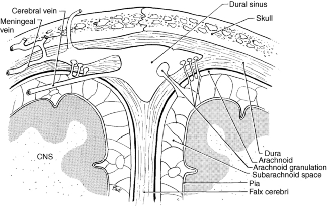
The circulation of CSF spreads the infecting organism through the ventricular system and the subarachnoid spaces (Figure 26-1). The pia and arachnoid maters become acutely inflamed, and as part of the inflammatory response, a purulent exudate forms in the subarachnoid space. The exudate may undergo organization, resulting in an obstruction of the foramen of Monro, the aqueduct of Sylvius, or the exit foramen of the fourth ventricle. The supracortical subarachnoid spaces proximal to the arachnoid villi may be obliterated, resulting in a noncommunicating or obstructive hydrocephalus caused by the accumulation of CSF. As the CSF accumulates, the intracranial pressure rises. The increased intracranial pressure produces venous obstruction, precipitating a further increase in the intracranial pressure. The rise in the CSF pressure compromises the cerebral blood flow, which activates reflex mechanisms to counteract the decreased cerebral blood flow by raising the systemic blood pressure. An increased systemic blood pressure accompanies increased CSF pressure.

The mechanism producing the headaches that accompany increased intracranial pressure may be the stretching of the meninges and pain fibers associated with blood vessels. Vomiting may occur as a result of stimulation of the medullary emetic centers. Papilledema may occur as intracranial pressure increases.

Other routes of bacterial infection may involve a local spread as the result of an infection of the middle ear or mastoid air cells. Meningitis may occur as a complication of a skull fracture, which exposes CNS tissue to the external environment or to the nasal cavity. Fractures of the cribriform plate of the ethmoid bone producing CSF rhinorrhea provide another route for infection. Meningitis may be a further complication to the clinical problems of a traumatic head injury.

Clinical features of acute bacterial meningitis include fever, severe headache, altered consciousness, convulsions (particularly in children), and nuchal rigidity. Nuchal rigidity is indicative of an irritative lesion of the subarachnoid space. Signs of meningeal irritation are painful cervical flexion, the Kernig sign, the Brudzinski sign, and the jolt sign.10 Cervical flexion is painful because it stretches the inflamed meninges, nerve roots, and spinal cord. The pain triggers a reflex spasm of the neck extensors to splint the area against further cervical flexion; however, cervical rotation and extension movements remain relatively free.

The Kernig sign refers to a test performed with the client supine in which the thigh is flexed on the abdomen and the knee extended (Figure 26-2, A). Complaints of lumbar or posterior thigh pain indicate a positive test result.10 This movement pulls on the sciatic nerve, which pulls on the covering of the spinal cord, causing pain in the presence of meningeal irritation. The same results are achieved with passive hip flexion with the knee remaining in extension. This is the same procedure described by Hoppenfeld11 as the straight leg raising test for determining pathology of the sciatic nerve or tightness of the hamstrings. Passive hip flexion with knee extension can be painful because of meningeal irritation, spinal root impingement, sciatic nerve irritation, or hamstring tightness. The Brudzinski sign refers to the flexion of the hips and knees elicited when cervical flexion is performed10 (see Figure 26-2, B). These signs will not be present in the deeply comatose client who has decreased muscle tone and absence of muscle reflexes. The signs may also be absent in the infant or elderly patient. Finally, the jolt test, which has the patient turn his or her head from side to side quickly (two to three rotations per second), has a positive result if the maneuver worsens the patient’s headache.10
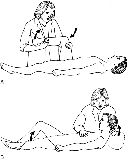
The diagnosis of bacterial meningitis can be established based on blood cultures and a sample of CSF obtained by a lumbar puncture. CSF pressure is consistently elevated. The CSF sample in bacterial meningitis typically reveals an increased protein count and a decreased glucose level.

The type and severity of the sequelae of acute bacterial meningitis relate directly to the area affected, the extent of CNS infection, the age and general health of the individual, the level of consciousness at the initiation of pharmacological therapy, and the pathological agent involved. Some of the common CNS complications include subdural effusions, altered levels of consciousness, seizures, involvement of the cranial nerves, and increased intracranial pressure.

Potential neurological sequelae.

Even with optimal antimicrobial therapy, bacterial meningitis continues to have a finite mortality rate, which varies with the infecting organism, age of the individual, and time lapse to initiation of treatment, and has the potential for marked neurological morbidity. Neurological sequelae occur in 20% to 50% of the cases.1,9 Bacterial meningitis is considered a medical emergency; delays in initiation of antibacterial therapy increase the risk of complications and permanent neurological dysfunction.1,9

Reports of the long-term outcome of individuals with bacterial meningitis indicate that up to 20% have long-term neurological sequelae.1,5 The sequelae may be the result of the acute infectious pathological condition or subacute or chronic pathological changes. The acute infectious pathological condition could result in sequelae such as inflammatory or vascular involvement of the cranial nerves or thrombosis of the meningeal veins. Cranial nerve palsies, especially sensorineural hearing loss, are common complications. The risk of an acute ischemic stroke is greatest during the first 5 days.9 Weeks to months after treatment, subacute or chronic pathological changes may develop, such as communicating hydrocephalus, which manifests as difficulties with gait, mental status changes, and incontinence.10,12 Approximately 5% of the survivors will have weakness and spasticity.6 Focal cerebral signs that may occur either early or late in the course of bacterial meningitis include hemiparesis, ataxia, seizures, cranial nerve palsies, and gaze preference.1,13 Cognitive slowness has been found in 27% of clients after pneumococcal meningitis, even with good recovery as documented by a Glasgow Outcome Scale score of 5.14

Damage to the cerebral cortex can result in numerous expressions of dysfunction. Motor system dysfunction may be the observable expression of the damage within the CNS, but the location of the damage may include sensory and processing areas, as well as those areas typically categorized as belonging to the motor system. Perceptual deficits or regression in cognitive skills may present residual problems. Cranial nerve involvement is most frequently expressed as dysfunction of the eighth cranial nerve complex and produces auditory and vestibular deficits.

Aseptic meningitis.

Aseptic meningitis refers to a nonpurulent inflammatory process confined to the meninges and choroid plexus, usually caused by contamination of the CSF with a viral agent, although other agents can trigger the reactions. The symptoms are similar to those of acute bacterial meningitis but typically are less severe. The individual may be irritable and lethargic and complain of a headache, but cerebral function remains normal unless unusual complications occur.5,15 Aseptic meningitis of a viral origin usually causes a benign and relatively short course of illness.16,17

A variety of neurotropic viruses can produce aseptic (viral) meningitis. The enteroviruses (echoviruses and the Coxsackie viruses), herpesviruses, and HIV are the most common causes.5,7,18 The primary nonviral causes of aseptic meningitis are Lyme Borrelia and Leptospira.19 The diagnosis of this type of aseptic meningitis may be established by isolation of the infecting agent within the CSF or by other techniques. The glucose level of the CSF in bacterial meningitis is usually depressed; however, the glucose level in viral meningitis is normal.2

Treatment of aseptic meningitis consists of management of symptoms. The condition does not typically produce residual neurological sequelae, and full recovery is anticipated within a few days to a few weeks.

Encephalitis

Clinical problems.

Encephalitis refers to a group of diseases characterized by inflammation of the parenchyma of the brain and its surrounding meninges. Although a variety of agents can produce encephalitis, the term usually denotes a viral invasion of the cells of the brain and spinal cord.

Different cell populations within the CNS vary in their susceptibility to infection by a specific virus. (For example, the viruses responsible for poliomyelitis have a selective affinity for the motor neurons of the brain stem and spinal cord. Viruses such as Coxsackie viruses and echoviruses typically infect meningeal cells to cause the benign viral meningitis discussed in the previous section.) In acute encephalitis, neurons that are vulnerable to the specific virus are invaded and undergo lysis. Viral encephalitis causes a syndrome of elevated temperature, headache, nuchal rigidity, vomiting, and general malaise (symptoms of aseptic or viral meningitis), with the addition of evidence of more extensive cerebral damage such as coma, cranial nerve palsy, hemiplegia, involuntary movements, or ataxia. The difficulty in differentiating between acute viral meningitis and acute viral encephalitis is reflected in the use of the term meningoencephalitis in some cases.

The pathological condition includes destruction or damage to neurons and glial cells resulting from invasion of the cells by the virus, the presence of intranuclear inclusion bodies, edema, and inflammation of the brain and spinal cord. Perivascular cuffing by polymorphonuclear leukocytes and lymphocytes may occur, as well as angiitis of small blood vessels. Widespread destruction of the white matter by the inflammatory process and by thrombosis of the perforating vessels can occur. Increased intracranial pressure, which can result from the cerebral edema and vascular damage, presents the potential for a transtentorial herniation. The likelihood of residual impairment of neurological functions depends on the infecting viral agent. Patients with mumps meningoencephalitis have an excellent prognosis, whereas 55% of the individuals with herpes simplex encephalitis treated with acyclovir have some neurological sequelae.6 Because of the slow recovery of injured brain tissue, even in patients who recover completely, return to normal function may take months.20

Plum and Posner21 discuss viral encephalitis in terms of five pathological syndromes. Acute viral encephalitis is a primary or exclusively CNS infection. An example would be herpes simplex encephalitis, in which the virus shows a partiality for the gray matter of the temporal lobe, insula, cingulate gyrus, and inferior frontal lobe. Other examples are the mosquito-borne viruses, such as the St. Louis encephalitis, California virus encephalitis, and most recently the West Nile virus (WNV). From 1999 to 2005 in the United States, the incidence of infection from the WNV has increased from 62 cases to 3000 cases.22 Currently all states have some level of WNV activity, and only four states are without human cases (Figure 26-3). The majority (80%) of people infected by the WNV will be asymptomatic; of the remaining people infected, less than 1% will develop severe illness.23 Risk of neuroinvasive disease from WNV is 40%; neuroinvasive disease involves meningitis, encephalitis, or poliomyelitis, with wide variety in clinical presentation (Figure 26-4).24 Parainfectious encephalomyelitis is associated with viral infections such as measles, mumps, or varicella. Acute toxic encephalopathy denotes encephalitis that occurs during the course of a systemic infection with a common virus. The clinical symptoms are produced by the cerebral edema in acute toxic encephalopathy, which results in increased intracranial pressure and the risk of transtentorial herniation. Reye syndrome is an example. Global neurological signs, such as hemiplegia and aphasia, are usually present, rather than focal signs. The clinical symptoms of the previous three syndromes may be similar. Specific diagnosis may be established only by biopsy or autopsy.
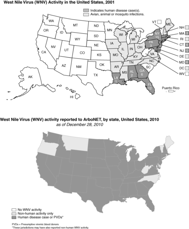
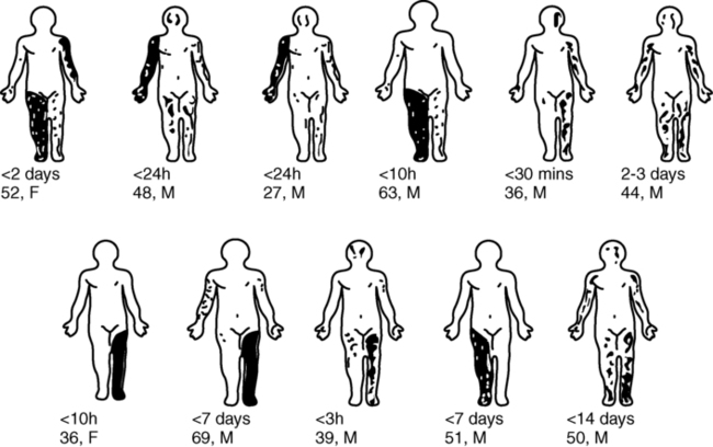
Progressive viral infections occur from common viruses invading susceptible individuals, such as those who are immunosuppressed or during the perinatal to early childhood period. Slow, progressive destruction of the CNS occurs, as in subacute sclerosing panencephalitis. The final category of encephalitis syndromes consists of “slow virus” infections by unconventional agents (the prion diseases) that produce progressive dementing diseases such as Creutzfeldt-Jakob disease and kuru.25

Clinical picture of the individual with inflammatory disorders of the brain

An individual within the acute phase of meningitis or encephalitis or with residual neurologic dysfunction from these disorders may demonstrate signs and symptoms similar to those of generalized brain trauma, tumor disorder, or other identified abnormal neurological state. The variability in the clinical picture is reflected in the inclusion of the category “infectious diseases that affect the central nervous system” in the Guide to Physical Therapist Practice.27 Practice patterns for physical therapists that apply to this population include the following:

Although these patterns have been identified within the physical therapy practice patterns, the concepts and selection of evaluation and intervention procedures are just as applicable for occupational therapists and other individuals working on movement dysfunction. In the acute phase the inflammatory process may result in impairments in arousal and attention that range from nonresponsiveness to agitation. The degree of agitation may range from mild to severe, depending on both the client’s unique CNS characteristics and the degree of inflammation. The agitated state may be the result of alterations in the processing of sensory input, with the consequence of inappropriate or augmented responses to sensory input. The client may respond to a normal level of sound as though it were an unbearably loud noise. Low levels of artificial light may be perceived as extremely bright.

Perceptual and cognitive impairments may be present, resulting in a variety of functional limitations and disabilities. Clients may have distortions in their perception of events as well as memory problems. As their memory returns, accuracy of time and events may be distorted, leading to frustration and anxiety for both the client and those family members and friends who are interacting within the environment.

In addition to alterations in mentation, the individual may demonstrate impaired affect, such as a hypersensitivity or exaggerated emotional responses to seemingly normal interactions. For example, when upset about dropping a spoon on the floor, a client may throw the tray across the table. When another individual was told his girlfriend would be a little late for her afternoon visit, the client became extremely upset and stated his intent to kill himself because his girlfriend did not love him anymore.

Because of the variety of pathological problems after acute inflammation, the client may have residual problems manifested as generalized or focal brain damage. The specifics of these impairments cannot be described as a typical clinical picture because they are extremely dependent on the individual client. These variations require the therapist to conduct a thorough examination and evaluation process to develop an appropriate individualized intervention program. Although content from the Guide to Physical Therapist Practice has been incorporated into the discussion of the examination, evaluation, and intervention processes, the model presented provides a structure that can accommodate the specific disciplinary expertise of both occupational and physical therapists.

Examination and evaluation process

Just as the medical intervention with clients who have an inflammatory disorder of the CNS is, to a large extent, symptomatic, so is the intervention by therapists. Designing an individualized intervention program based on the client’s problems requires a comprehensive initial and ongoing evaluation to define the impairments, functional limitations, and disabilities and to note changes in them. Although the discussion of examination procedures is separated from the discussion of intervention strategies, it must be recognized that the separation is artificial and does not reflect the image of practice. The evaluation process should be considered in relationship to both the long-term assessment of the individual’s changes and the short-term within-session and between-session variations. For example, documentation of the level of consciousness of a client on day one of intervention will provide a starting point for calculation of the distance spanned at the time of discharge. Perhaps more critical to the final outcome is determination of the level of consciousness before, during, and after a particular intervention technique to determine its impact on the individual’s level of arousal and ability to interact with the environment. The evaluation process is a constant activity intertwined with intervention. The observations and data from the process are periodically recorded to establish the course of the disease process and the success of the therapeutic management of the client.

Observation of current functional status

The examination process should be conceptualized as a decision-making tree that requires the therapist to determine actively which components are to be included in a detailed examination and which can be eliminated or deferred. The first step in this process is the observation of the client’s current functional status. If the client is comatose and nonmobile, the focus of the initial session might be an assessment of the stability of physiological functions, level of consciousness, responses to sensory input, and joint mobility. If the client is an outpatient with motor control deficits, the initial session might focus on defining motor abilities and components contributing to movement dysfunctions with a more superficial assessment of physiological functions and level of consciousness. The therapist must be alert to indications of the need for a more detailed evaluation of perceptual and cognitive function (e.g., the client cannot follow two-step commands, indicating the need to assess cognitive skills).

Some of the components discussed in this process may be examination skills that are more typically possessed by other professions (e.g., assessment of emotional or psychological status). The inclusion of these items is not meant to suggest that the therapist must complete the formal testing. The items are included to indicate factors that will affect goal setting for the client and that will have an impact on the intervention strategy. Although the therapist may not be the health care team member who has primary responsibility for evaluation of these areas, he or she should recognize these areas as potential contributors to movement dysfunctions.

Observation of the current functional status of the client provides the therapist with an initial overview of his assets and deficits. This provides the framework into which the pieces of information from the evaluation of specific aspects of function can be fit. The therapist must not allow assumptions made during the initial observation to bias later observations. The therapist might note that the client is able to roll from the supine to the side-lying position to interact with visitors in the room. When the same activity is not repeated on the mat table in the treatment area, the therapist, knowing the client has the motor skill to roll, might conclude that he is uncooperative or apraxic or has perceptual deficits. The therapist may have failed to consider that the difference between the two situations is the type of support surface or the presence or absence of side rails, which may have enabled the client to roll in bed by pulling over to the side-lying position. It is characteristic of human observation skills that we tend to “see” what we expect to see. The therapist must attempt to observe behaviors and note potential explanations for deviations from normal without biasing the results of the subsequent observations.

The following discussion of the specific considerations within the examination process does not necessarily represent the temporal sequence to be used during the data collection process. As different items are discussed, suggestions for potential combinations of items will be made. The sequence of the process is best determined by the interaction of therapist and client. Figure 26-5 outlines the components that should be considered during the evaluation process and provides a synopsis of the following discussion.
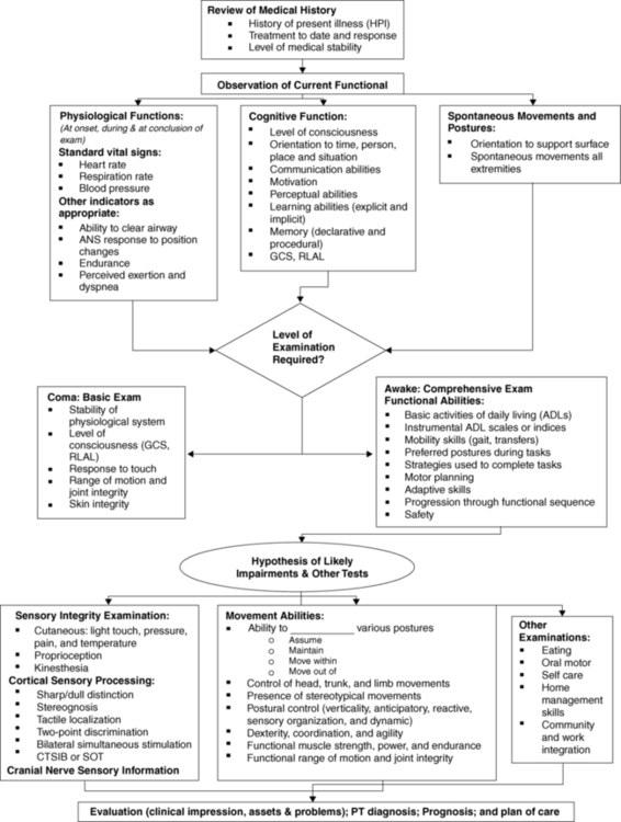
The general philosophy in the evaluation of the client with neurological deficits as a result of brain inflammation is a whole-part-whole approach. General observations of the client’s performance provide an overall description of the client’s abilities while indicating deficits in his or her performance. The cause(s) of the deficits (impairments) are explored to provide the pieces of data defining her or his performance. These pieces of data then are arranged within the framework provided by the general observation to define the whole of the client’s assets and deficits. As the whole picture is established (with the realization that it will be constantly adjusted), the process of goal setting is initiated. These goals need to consider the patient’s and family’s desires. The process presented for refining evaluation data into an intervention plan is applicable whether the client’s neurological dysfunction is the result of a bacterial or viral infection, cerebrovascular accident, trauma, or other factors.

Evaluation of physiological responses to therapeutic activities

It is assumed that the therapist enters the initial interaction with a client after reviewing the available background information. This may provide the therapist with information on the baseline status of the client’s vital physiological functions. Any control problems in these areas should be particularly noted. Until the therapist determines that the vital functions, such as rate of respiration, heart rate, and blood pressure, vary appropriately with the demands of the intervention process, these factors should be monitored. The monitoring process should include consideration of the baseline rate, rate during exercise, and time to return to baseline. The pattern of respiration and changes in that pattern also should be noted.

Other tests and measures of the status of ventilation, respiration, and circulation may be indicated in specific individuals. Individuals with limited mobility or motor control of the trunk or those with cranial nerve dysfunctions may demonstrate difficulty with functions such as moving secretions out of the airways. Inactivity during a prolonged recovery period may result in cardiovascular adaptations that compromise endurance and contribute to increases in the perceived exertion during activities.

Autonomic nervous system dysfunctions may be expressed as inappropriate accommodations to positional changes, such as orthostatic hypotension. Clients with depressed levels of consciousness may display temperature regulation dysfunctions. One mechanism for assessing the client’s ability to maintain a homeostatic temperature is to review the nursing notes. The events surrounding any periods of diaphoresis should be examined. If no causative factors have been identified, then interventions that involve thermal agents as discussed elsewhere in this text should be used judiciously.

Evaluation of cognitive status

Because the evaluation process encompasses the stages of recovery from the critical acute phase through discharge from therapy, a range of aspects are included under the evaluation of cognitive status. As indicated previously, the observation of current functional status will direct the therapist toward the appropriate component tests and measures.

Acute bacterial meningitis and various forms of viral encephalitis may result in changes in the client’s level of consciousness. Consciousness is a state of awareness of one’s self and one’s environment.21 Coma can be defined as a state in which one does not open the eyes, obey commands, or utter recognizable words.28 The individual does not respond to external stimuli or to internal needs. The term vegetative state is sometimes used to indicate the status of individuals who open their eyes and display a sleep-wake cycle but who do not obey commands or utter recognizable words. DeMeyer29 presents a succinct description of the neuroanatomy of consciousness and the neurological examination of the unconscious patient. Plum and Posner21 also provide extensive information in this area.

Several scales have been developed to provide objective guidelines to assess alterations in the state of consciousness. The Glasgow Coma Scale28 assesses three independent items: eye opening, motor performance, and verbal performance. The scale yields a figure from 3 (lowest) to 15 (highest) that can be used to indicate changes in the individual’s state of consciousness. The evaluation format is simple, and the scale demonstrates both interrater and intrarater reliability. Refer to Box 24-4 for the specific scale. The Rancho Los Amigos scale assesses level of consciousness and behavior.28 The therapist can use assessment tools such as the Glasgow Coma Scale and the companion Glasgow Outcome Scale to determine if the intervention program has resulted in any recordable changes in the client’s level of consciousness. Ideally, the client with decreased levels of consciousness will be monitored at consistent intervals to determine changes in status. Any carryover or delayed effects of the intervention could then be noted. The record of the client’s level of consciousness might also display a pattern of peak awareness at a particular point in the day. Scheduling an intervention session during the client’s peak awareness time may maximize the benefit of the therapy.

Performed in conjunction with the assessment of the client’s state of consciousness is the determination of the individual’s orientation to person, time, place, and situation. Because the individual’s level of orientation (documented as oriented times 4) is frequently recorded by multiple members of the rehabilitation team, the information in the medical chart may provide insights into fluctuations over the course of a day or a week.

Gross assessment of the individual’s ability to communicate, both the expressive and receptive aspects of the process, is an important component of the examination. If a dysfunction is present in the client’s ability to communicate, the client should be evaluated by an individual with expertise in this area so that strategies for dealing with the communication deficit can be developed. Evaluation of the movement abilities of the client with communication deficits requires creative planning on the part of the therapist but usually can be accomplished if generalized movement tasks are used. With the client who cannot comprehend a verbal command to roll, the therapist should use an alternate form of communication, such as manual cueing or guidance. The therapist could structure the situation to elicit the desired behavior by activities such as placing the client in an uncomfortable position or positioning a desired object so that it can be reached only by rolling.

As the therapist progresses through the examination and intervention process, ongoing data collection should be occurring on factors that influence the motivation of the individual. Individuals with damage to certain areas within the frontal lobe will have difficulty with committing to long-term projects and may not be motivated to work during a therapy session by an explanation detailing the relationship of the current activity to the larger goal of returning home. In these situations, the therapist must create appropriate immediate rewards, such as a 2-minute rest break after completion of a specific movement task.

Deficits in cognition may be evident as problems in the area of explicit (declarative) or implicit (procedural) learning. Explicit learning is used in the acquisition of knowledge that is consciously recalled. This is information that can be verbalized in declarative sentences, such as the sequential listing of the steps in a movement sequence. Implicit or procedural learning is used in the process of acquiring movement sequences that are performed automatically without conscious attention to the performance. Procedural learning occurs through repetitions of the movement task (refer to Chapter 4). Because explicit and implicit learning use different neuroanatomical circuits, implicit learning can occur in individuals with deficits in the components underlying explicit learning (awareness, attention, higher-order cognitive processes) (refer to Chapter 5 for additional information regarding implicit and explicit learning).

The emotional and psychological aspects of the client and the higher-order cognitive and retention skills of the client should be evaluated informally by the therapist, with referral to appropriate professionals if dysfunction in these areas is suspected. A coordinated team approach is necessary for clients with emotional and psychological, cognitive, perceptual, or communication problems or a combination of these problems. A consistent strategy used by all team members eliminates the necessity on the part of the client of trying to cope with different approaches by different people in an area in which she or he already has a deficit. The impact of cognitive deficits on the process of learning motor skills is further discussed in the next section on movement assessment. The assessment of the impact of perceptual dysfunctions is incorporated within the evaluation of sensory channels.

Examination of functional abilities

As indicated in the introduction to the evaluation of clients with inflammatory and infectious disorders of the brain, the examination process is not compartmentalized. As the therapist is examining the movement abilities of the individual through the format described in the previous section, he or she is also collecting information on the functional abilities of the individual. The components underlying the movement abilities of the client can be examined within the framework of the basic or instrumental activities of daily living (ADLs), depending on the functional level of the person. The treatment setting and documentation requirements within that setting will determine whether the data on basic ADL and instrumental ADL skills are recorded with use of a formal scale or index or are gathered through an individualized process.

The introduction of specific tasks provides the therapist with the opportunity to observe the preferred posture used to accomplish the different tasks. The therapist should construct situations that require the individual to respond to unexpected occurrences to provide some insight into the person’s ability to adapt to the unexpected. Throughout the process of examining a patient’s movement abilities and functional abilities, the therapist is assessing the individual’s awareness of safety considerations and judgment in attempting tasks.

The presence of motor planning dysfunctions can be noted as the client attempts a movement sequence or a functional task. The therapist may have to cue the client physically to initiate the sequence, which then flows smoothly. The therapist may observe that the client has the correct components to a movement sequence, but that the sequence of the components is incorrect. Or the client may demonstrate the ability to produce a movement sequence under one set of conditions but not another. Indications of these types of motor planning problems can be observed during the initial interactions with the client. Similarly, the therapist also should be aware of indications of problems with dexterity, coordination, and agility, as well as with signs of cerebellar dysfunctions.

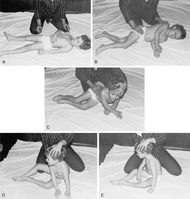
Another aspect of the evaluation process that can be integrated in the observations of movement abilities is identification of perceptual deficits. Aspects of the client’s motor performance can provide indications for detailed perceptual testing to classify the deficits. This testing should be conducted by the health care team member qualified in the area of perceptual testing. During the general evaluation procedures, the therapist can screen the client for signs of perceptual deficits. Clients’ abilities to cross their midlines with their upper extremities can be demonstrated in movement sequences, such as moving from the supine to the side-sitting to the sitting position (Figure 26-6). The quality of the integration of information from the two sides of the body can be indicated by the symmetry or asymmetry of posture in positions that should be symmetrical. The therapist may suspect that the client has a deficit in body awareness or body image by the poor quality of movement patterns that are within the motor capability of the individual. Spontaneous comments by the client as to how he or she feels when moving (“my leg feels so heavy”) also add to the therapist’s assessment of the client’s body image. Problems with verticality can be seen with the client who lists to one side when in an upright posture. When the therapist corrects the list to a vertical posture, clients may express that they now feel that they are leaning to one side. Individuals who cannot appropriately relate their positions to the position of objects in their environments may have a figure-ground deficit or a problem with the concept of their position in space. When approaching stairs, these clients may fail to step up or may attempt to step up too soon. These examples should provide an indication of the observations that can indicate the need for detailed perceptual testing.

The preceding aspects of evaluation of movement abilities focused on facets of motor performance. Within this process the therapist should intertwine an appraisal of the individual’s ability to learn motor tasks (or elements of the task). The therapist attempts to determine whether the client can maintain a change in the ability to perform a movement throughout a therapy session and into the next session. The client’s ability to capture and integrate changes into the movement repertoire is fundamental to the success of the intervention program. The program can focus on the learning of movement sequences and the generalization of these sequences to movements within other contexts. Individuals with lowered levels of consciousness (typically Rancho Los Amigos Levels I to III) will be unable to learn or have difficulty learning and generalizing new motor skills. Therapy sessions may be more successful if the focus remains on the performance of motor tasks that were previously “overlearned” and automatic. Although the therapist may be able to manually guide the individual in coming to sit on the edge of the bed, until the individual demonstrates a higher level of processing, it may be unrealistic to expect that he or she will consistently reposition the legs without cueing before attempting the movement sequence. From looking at the “whole” of function, the therapist needs to determine the impairments that require further examination.

Evaluation of sensory channel integrity and processing

The examination process must include an assessment of the channels for sensory input. Knowledge gained in the assessment of the sensory systems will be used in the program-planning process to select the intervention strategies that have the highest probability of success. Although movements can be performed (and in some cases even learned) in the absence of typical sensory feedback, the presence of altered sensory function creates more challenges for both the client/learner and the therapist/teacher. The therapist assesses both the client’s ability to perceive the sensory stimulus and the appropriateness of the response to the stimulus. Therefore it is important to determine if the sensory modality is intact, impaired, or absent, and if it is impaired, whether it hyperresponsive, hyporesponsive, or inconsistent. In addition, variations in the interpretation of sensory input may occur in some clients. Gentle tactile contact may be perceived by the person as a noxious input. Some individuals will have difficulty processing and discriminating information with high levels of one type of sensory input (e.g., the noisy clinic area) or with multiple simultaneous inputs (e.g., talking to the therapist while walking down a hallway with people moving toward the individual).

The therapist should develop a systematic approach to the initial cursory screen of the sensory systems. Deficits identified in the initial examination will provide structure for scheduling more comprehensive evaluation of deficits in specific systems. The therapist must also monitor changes in the status of physiological vital functions during sensory input, especially if the client has a history of instability of heart rate, blood pressure, or rate of respiration.

Based on the information from the screen, the therapist will organize the components of the more detailed examination. Components to be considered include the integrity of the peripheral sensory circuits, the cortical level processing of the sensory information, the integrity of the cranial nerve sensory circuits, and the processing of multichannel input.

Cutaneous input has several aspects that must be assessed. Some of the inflammatory diseases of the brain may result in cutaneous distributions in which sensation is absent or diminished. These areas should be routinely evaluated for changes in distribution of level of sensation. Tests of light touch, pressure, and pain can be used if the client can communicate reliably. In most cases, inclusion of assessment of differentiation of hot and cold will not add appreciably to the information needed for treatment planning unless thermal modalities are a consideration.

A gross assessment of the intactness of the touch system can be made in the noncommunicative client by introducing a mildly aversive (not painful) stimulus, such as a light scratch, while monitoring the client for changes in facial expression, posture, or tonus. The possibility of a spinal-level reflex response should be kept in mind when interpreting the results of such a gross assessment.

Assessment of the client’s response to proprioceptive input is incorporated within the assessment of the client’s movement abilities and is intertwined with the intervention process because a variety of intervention techniques are based on proprioceptive input. Evaluation of the proprioceptive channels can be conducted through assessment of the client’s static position sense and dynamic kinesthesis. These tests allow the therapist to make inferences concerning the client’s cognitive abilities to interpret proprioceptive information. Inherent in the successful completion of these tests is the necessity for the client to be able to understand directions and to be able to communicate data to the therapist. Because information input, processing, and output are involved in these tests, failure to comply with the test instructions cannot be definitively attributed to dysfunction of the proprioceptive system. The therapist also should consider information obtained from watching the client move before drawing a conclusion concerning the intactness of the proprioceptive channels. Some of the factors to consider include disregard of an extremity and variations in quality of performance between visually directed and non–visually directed movements. Although tests of position sense and kinesthetics provide one aspect of the evaluation of the proprioceptive system, the therapist also must be involved constantly in assessing the client’s response to the intervention techniques that are part of the treatment plan. This again illustrates the intermingling of assessment and intervention. Intervention places a demand for movement on the client. As the movement occurs, the therapist assesses the quality of the movement. If the quality is not appropriate, the therapist initiates intervention to improve the quality. If the technique does not produce the desired result, a second technique can be tried and the cyclical process continues.

In addition to determining the integrity of the peripheral sensory pathways and recognition of the input, it is important to assess the individual’s ability to process more complex presentations of cutaneous input. Difficulties in the cortical-level processing of cutaneous stimuli are identified through tests of sharp and dull discrimination, stereognosis, tactile localization, texture recognition, two-point discrimination, and bilateral simultaneous stimulation.

Central processing and integration of sensory information as it affects postural control can be examined using the Clinical Test of Sensory Integration and Balance (CTSIB)30 or with computerized dynamic posturography using the Sensory Organization Test (SOT). With both tests, the effectiveness of using vision or somatosensory or vestibular sensation at the appropriate time (sensory weighting) and changing from one sensory system to the other is examined.

Assessment of the integrity of the cranial nerve sensory channels is typically incorporated within the standard cranial nerve examination. Review of the physician’s notes may provide sufficient information; however, the therapist may need to complete more specific tests before considering certain intervention techniques. Of specific note are vision and vestibular screening, both of which are discussed in detail elsewhere in this text. Simple visual system tests, such as identification of field deficits, assessment of tracking abilities, and a gross evaluation of visual acuity, can be performed quickly.

The complex functions of the vestibular system can be assessed through a variety of avenues such as the Ayres Post-Rotatory Nystagmus Test,31,32 the SOT, and tests of the vestibular ocular reflex (head-thrust test, head-shaking test, and test of dynamic visual acuity [DVA]), and are discussed in detail elsewhere in this text. It is important to examine the vestibular influence in both postural control and gaze stability. The integrity of the connections underlying a vestibularly induced nystagmus response is assessed by physicians through the caloric test (warm and cold water or air introduced into the ear channel to induce nystagmus). The effect of rapid linear accelerations and decelerations can be evaluated as potential activating mechanisms increasing the level of consciousness or level of muscle activity. An example is the Dynamic Gait Index, a functional test that incorporates changes in speed and head movements during walking.33 Slow, rhythmical reversals of linear movements may have a calming effect on the client’s behavior or level of muscle activation. Linear movements in all planes and diagonals should be explored.

During the evaluation of the client as well as during intervention with the client, the therapist must be aware of the potential to bombard him or her with sensory input and overload his or her ability to respond discriminatively to it. If the therapist detects that the client has difficulty in responding appropriately to sensory input, as with a client in a lowered state of consciousness or an agitated state, or demonstrating tactile defensiveness, sensory input should be used selectively during the initial examination or intervention sessions. If multiple sensory inputs are used, the positive or negative effects cannot be attributed to a specific input or necessarily to the series of inputs. Evaluation as well as intervention with sensory inputs should proceed in a controlled fashion. Inclusion of additional sensory modalities in the intervention plan should occur systematically.

The individual’s response to multichannel sensory conflict input is typically assessed as a component of higher-level balance assessment and locomotor abilities. A more thorough discussion of sensory assessment is discussed elsewhere in the text. The therapist should apply these concepts during the evaluation of all motor tasks. Consider the following example: a client who relies on visual input to supplement vestibular and somatosensory information is performing the task of sitting on the edge of the mat table. She remains relatively steady until someone walks directly toward her from across the clinic. This change in the environmental context of the performance requires her to assess whether she is moving toward the individual or the individual is moving toward her. Without reliable vestibular and somatosensory check points, the client may activate a postural response to the incorrect assessment. As this example demonstrates, the evaluation of the sensory channels is intertwined with the evaluation of the person’s movement abilities.

Examination of movement abilities

The initial assessment of the individual’s movement abilities is conducted by observing as she or he moves through a sequence of functional postures. The therapist determines the functional postures to be examined for a specific client, ranging from bed mobility activities (assessment of movement in prone and supine positions) through upright ambulation. The medical status of the individual, the extent of involvement, the intervention setting, and the age of the individual are considerations in determining the appropriate functional postures to be examined. The therapist gathers information on the movement abilities of the client as he or she moves into, within, and out of the position.

The assessment focuses on both the quantity and quality of motor performance. The quantitative aspect of the movement assessment involves the number of different functional postures the individual can use. The quality of the movement abilities is assessed within the posture as well as in the process of moving between postures. For example, the therapist should assess the quality of the head, trunk, and extremity control demonstrated throughout the movement sequences. The use of stereotypical movement patterns should be noted because their presence may limit the adaptability of movements required to accomplish functional tasks. Other items relating to the client’s movement abilities are assessed during this process.

Indications of abnormal ranges of movement of all joints can be obtained. The range may show a limitation of movement or an indication of joint instability. Once the gross deviations are identified, these joints can be examined to determine the source of the problem: joint capsular, ligamentous, bony, skin, or muscular and fascial dysfunction. Conducting the gross assessment of range while the client is moving eliminates the time spent in performing a joint-by-joint goniometric evaluation of articulations with normal excursions.

As the individual is moving (either independently or with the therapist assisting), an assessment of the distribution and fluctuations in muscle activity can be made and will provide information on functional muscle strength, power, and endurance. The timing, accuracy, and sequencing of muscle activation within the movement should be noted. The therapist can identify the postures that will be the most conducive to optimal motor performances and those that should be avoided. As the client is moving through various postures, the function of specific musculature can be examined. Muscle groups should be examined with regard to their ability to function in both stability (distal segment fixed) and mobility (distal segment free) situations. Because numerous demands are being placed on each muscle group, therapists can assess the ability to perform isometric and isotonic (concentric and eccentric) contractions. Each different posture introduces a new set of variables; therefore the performance of a muscle group must be reexamined as each new movement pattern is performed.

The therapist can identify postural control in a variety of functional positions. Within each posture, the therapist must examine the control the client displays over the posture. Because the assessment takes place as part of a dynamic sequence, the therapist can assess the client’s ability to assume the posture. If the posture cannot be achieved independently, the therapist assesses the factors interfering with achieving the position, the type of assistance necessary to facilitate assumption of the posture, and the effect of the various intervention techniques used to assist the client in achieving the position. Once the client is in the posture, her or his ability to maintain the posture is examined. Factors that interfere with the performance are noted. The client’s ability to move within the posture is identified. Movement demands placed on the client should include aspects of both static and dynamic equilibrium. Static balance in the sitting position (such as on the side of the bed) could be demonstrated by the individual matching the strength of a force attempting to displace him backward and maintaining the position when the force is suddenly released.

The presence of dynamic balance of the upper torso in the sitting position could be demonstrated by the individual reacting to a quick sideways displacement force administered to the shoulder by activating the trunk lateral flexors to compensate for the displacement. Equally important is the individual’s ability to demonstrate appropriate equilibrium responses to self-imposed perturbations. The absence of anticipatory control in standing could be demonstrated by having the client do the rapid arm raise test with a 5-lb weight and noticing reactive stepping instead of doing a posterior weight shift in anticipation of the destabilizing force.

The final stage in examining the individual’s movement abilities explores the individual’s ability to move out of the posture. The client should have the ability to move out of the posture to a lower-level posture and to a higher-level posture before mastery of the posture is considered to have been achieved.

Many aspects of the client’s performance are analyzed simultaneously. When the therapist assists the client in moving to a new posture, an analysis of the influence of facilitation and inhibition techniques is being conducted. The individual’s response to these handling techniques cues the therapist in projecting the client’s response to an intervention program. The therapist is constantly monitoring the client for changes in physiological functions or changes in the level of consciousness. Anything that results in expressions of pain by the client should be noted. Intervention programs should be a learning experience for clients. If they are attending to pain, they cannot attend to learning. The factor(s) producing the pain should be identified and measures instituted to eliminate the factor(s). If the factors producing the pain cannot be resolved, the intervention program should be designed to avoid triggering the pain.

In addition to looking at postural control in a variety of postures, it is also important to determine the type of postural control dysfunction, such as vertical orientation to the surface or to gravity as the situation dictates, anticipatory postural control, reactive postural control, sensory organization for postural control, and dynamic balance for gait.

Prognosing and goal setting

Ideally, the process of establishing the prognosis and setting the goals for a client is a coordinated effort that involves all members of the health care team, including the client (if feasible) and family. If the therapist is not functioning in a setting where involvement of many disciplines is viable, the therapist can progress through the goal-setting process in the context of his or her role in the client’s care.

Having collected data from the examination process, the first steps are to establish two lists: one dealing with specific problems (impairments, functional limitations, and disabilities) the client is encountering and the second dealing with her or his assets. Formulating an asset list focuses on the positive data elicited from the evaluation process and is critical for prognosing outcomes. Items on the asset list could be observations, such as the client being able to assume the position of sitting on the side of the bed with setup assistance only, improved head control in this posture being facilitated by approximation, and controlled weight shifting being elicited by alternated tapping. The asset list provides a reference defining the postures and intervention techniques that are effective. This reference is used to develop the intervention goals and plan. Formulating and recording a problem list and an asset list can be completed relatively quickly as one gains familiarity with the process. Whereas novice therapists will benefit from generating a written asset list, experienced clinicians may formulate a mental asset list while completing the written evaluation format required by the facility. Just as the evaluation process is ongoing, so are the steps involved in goal setting. The asset and problem lists are redefined as the client’s status changes.

After assets and problems have been identified, the next step is to establish the expected outcomes from this episode of care. This is considered the prognosis. These outcome statements represent the general objectives toward which the intervention process is oriented. They identify the end point of the intervention process and are the exit criteria for terminating the episode of care.

The Guide to Physical Therapist Practice views outcomes in relationship to “minimization of functional limitations, optimization of health status, prevention of disability, and optimization of patient/client satisfaction,” whereas goals “relate to the remediation (to the extent possible) of impairments.”27 The breadth of acceptance of these definitions with the neurorehabilitation professions remains to be determined. These definitions at least give professionals a place to start communicating with consistency. The International Classification of Functioning, Disability and Health (ICF) model of the World Health Organization provides similar definitions, with the focus being client centered.

Measurable, interim objectives should be established in relation to the outcome statements. To determine if the objective has been achieved, the objective should be measurable—either in terms of producing a numerical indicator of performance, such as time span, number of repetitions, distance covered, or accuracy of performance, or in terms of a precise description of the target motor behavior. The appropriate objective indicator must be carefully selected. Performing a movement more quickly may indicate that the individual is performing it with more normal control and therefore greater ease of movement, or it may indicate that the individual has become more skilled in using an abnormal pattern based on inappropriate muscle activation. If it is not appropriate to formulate the objective in terms of a numerical indicator, the objective can be formulated in terms of an observable behavior. The therapist can precisely describe body segment movements based on the component method of movement analysis presented by VanSant.34 For example, the task of coming to standing from supine can be described in terms of the upper-extremity component, axial component, and lower-extremity component.35 Formulation of an appropriate short-term objective could specify use of the upper extremities in a push-and-reach pattern during the task of coming to standing from supine. The interim objectives should be constructed so that observing the client’s behavior will allow the therapist to state whether the criteria of the short-term objective were achieved. Table 26-1 gives an example of some components of short-term objectives leading to mastery of functional activities in sitting.

The client will maintain the posture For ________ seconds     The client will make postural adjustments of the head and trunk Appropriate to the degree of displacement   The client will bring the right foot to the left knee (as if to put on a shoe)  

image

*Outcome: The client will master functional activities in sitting. Short-term objective: Select one phrase from each column.

Therapist needs to consider all aspects of each variable (i.e., 1—a, b, c, d, e; 2—a, b, c, d, e; 3—a, b, c, d, e).

The outcome statements define the client’s destination. The interim objectives define the mileposts. The therapist then uses the asset list to design the intervention program, which is the vehicle to get the client to his or her destination. From the asset list, the therapist knows the intervention techniques that have the highest probability of success. Adopting this process simplifies the task of outlining the strategy for intervention.

As the therapist considers the appropriate outcomes and goals for the client, a decision must be made as to whether the format of the intervention will focus on a “training” approach or a “motor learning” approach. During the assessment process, if the therapist concludes that the individual’s level of cognitive function precludes the development of insight into movement errors (both the detection and correction of an incorrect performance) or the ability to retain the insight over time, then the therapist should delineate the outcomes and intervention plan to accommodate this limitation. The “training” approach requires more structure and repetition of activities within that structure. If it is more appropriate to design the intervention plan according to motor learning considerations, the therapist must consider the appropriate schedule and environmental context for the practice, the type and schedule for the feedback provided, and techniques to promote the generalization of the learning beyond the specific practice session.

General goals for the intervention process

Whereas the goal-setting process described earlier results in specification of the outcomes, goals, and objectives for a specific client, the general goals for the intervention process can be delineated to guide the process. As described in the overview of inflammatory disorders at the beginning of this chapter, the extent of the neurological sequelae may range from a single discrete problem to a devastating clinical picture composed of compromised functions in multiple areas. The goals for the intervention process address the problem areas that (1) jeopardize the efficiency and effectiveness of functional activities and (2) are the primary or secondary results of compromised neurological function. The listing of goals does not directly include consideration of secondary problems (such as decreases in joint range of motion [ROM], cardiovascular fitness, and endurance). The therapist should integrate these considerations in the overall assessment of the components of the movement problems.

The following goals are written as outcomes of the intervention process and not as goals for a specific client. Because of the broad nature of the goals, other professions also will contribute to the attainment of the goals. The goals of the therapeutic intervention program for clients with inflammatory CNS disorders are as follows:

Each of these goals is discussed in conjunction with the general therapeutic intervention procedures that can be used to achieve the goal.

General therapeutic intervention procedures in relation to intervention goals

Because it is assumed that functional abilities are built on the base of the ability to control postures, the intervention goal of promoting optimal postural control underlies the ability to make selective, voluntary movement patterns (goal 2) and the performance of functional activities (goal 3). Optimization of a postural set includes the concepts of decreasing muscle activity that is too high to allow performance of movement sequences, augmenting activation that is too low to support the accomplishment of a movement sequence, and fostering proper timing of the postural responses. Intervention techniques to achieve this goal demand that the therapist constantly monitor the client’s performance so that appropriate interventions are added when needed and continued only as long as they are needed.

Optimal postural control is defined by two elements. The client should have the ability to maintain a vertical orientation with regard to gravity and should be able to maintain balance in the presence of both internal and external perturbations. Automatic adjustments in the postural set should occur in anticipation of and continuously during movements (internal perturbations). Both elements should be performed with minimal physical or cognitive effort on the part of the client. Horak describes five components of normal postural control, including vertical orientation; anticipatory, reactive, sensory organization; and dynamic postural control for gait.36 By looking at the subsets of postural control, interventions can be designed to specifically match the impairment (Table 26-2).

TABLE 26-2 image

INTERVENTIONS FOR POSTURAL CONTROL PROBLEMS

POSTURAL CONTROL PROBLEM POSSIBLE INTERVENTIONS
Malalignment and verticality problems  Augment sensory feedback:

Limits of stability perception problems Anticipatory control problems  

Hold on and slow down (lessen the need for anticipatory control—substitution)

Mental rehearsal (weight shift, then move)

Practice limb movements where balance must be controlled (start slow and get faster)

Practice rapid limb movements where balance must be controlled (opening a door, opening a drawer, lifting a briefcase, lifting a bag of groceries, lifting a suitcase, wearing a backpack). Practice order:

Example: After the patient is doing better on a lifting task involving one object, he or she can work to be successful with several objects of different weights; first cognitively solve what must change for achievement of success in lifting one object versus another; then much repetition of alternating one object versus another; and eventually, work with a variety of objects in a varied pattern.

Reactive postural control problems  

Sensory integration problems  

Dynamic balance problems in gait  

image

Verticality, or maintaining an upright posture, first requires the client to recognize the desired alignment. Augmenting internal feedback mechanisms with the use of mirrors, force plates, or scales or even using a flashlight attached to the client that shines on a target when he or she is vertical can be effective. Manual skills such as positioning the client and using approximation to reinforce the position can be added to the treatment. Progression of intervention strategies can be done by having the client maintain the posture and then begin to manipulate objects with the extremities. Research suggests that the CNS is organized around tasks and not movement patterns. So, as the client is learning to maintain vertical and move in and out of the position, designing a task will likely give a better outcome. For example, Paul developed encephalitis, which left him with residual deficits in verticality. The simple task of keeping a book balanced on his head while sitting or walking gave him the type of feedback he needed without constant cues to “stand up straight” being the focus. Further progression involves teaching the client to move to his limits of stability and find vertical again.

Anticipatory postural control involves the postural preset, which positions the trunk to allow skilled use of the extremities without loss of balance. It requires the client to recognize the situation and the likely destabilizing force that will result, and posturally preset so destabilizing will not occur. The process requires memory and the ability to recognize the critical environmental and task cues. Interventions focus on practicing both the postural adjustment and the focal action before the two components are combined. Table 26-2 also has suggested interventions for reactive, sensory integrative, and dynamic balance in gait problems. Postural control is affected as well by biomechanical constraints, such as tonal abnormalities.

The client’s ability to demonstrate optimal postural control may be restricted by the presence of hypertonicity or hypotonicity in various muscle groups. These states may be relatively static or may fluctuate with the demands of a particular situation. Inappropriately high levels of muscle activity may be present in a stereotypical muscle distribution in the extremities, whereas the activity of the trunk musculature may be too low to support an antigravity posture. The therapist must design the interventions creatively to meet the shifting responses of the demands of a particular activity.

Being cognizant that spasticity is a reaction to initial peripheral instability, the therapist must select treatment that deals with the fact that as spasticity is modified, weakness or hypotonicity may be present. Inappropriately high levels of activity in a muscle group or groups may limit the client’s ability to demonstrate optimal postural control (and optimal selective movements as addressed in the second goal). The therapist can select intervention techniques that are mediated through any of the sensory channels functional for that client. The choice of which channel or combination of channels to use for the input is based on the therapist’s initial and continuing evaluation of the client’s response to specific types of sensory input.

The therapist must address the hypertonicity influencing postural control as a generalized problem before demanding selective voluntary activation of specific muscle groups. Vestibular input that is slow and rhythmical may promote a generalized relaxation of skeletal muscle activity. In some clients, the trunk remains “stiff” in movement sequences in which a segmental response between the upper and lower trunk should occur. Repetitions of rhythmical movements in side lying in which the therapist gently and progressively stretches the client’s pelvis in one direction around the body axis while moving the shoulder girdle in the opposite direction and then reverses the movement may effectively alter the biomechanical and neurological contributions to the stiffness (Figure 26-7).

For some clients, changing the dynamics of a spastic extremity may permit the emergence of more optimal levels of postural control. The appropriately designed ankle-foot orthosis (AFO) may alter the individual’s need to rigidly control the position of the pelvis to remain upright. Use of a soft webbing thumb loop to alter the resting position of the first metacarpal may change the overactivity of musculature throughout the upper extremity and allow appropriate adjustment of the shoulder girdle as part of postural responses.

If the client is sufficiently alert that attending to and understanding directions is a possibility, the therapist should direct the person to focus on the effects of the movement responses rather than focusing attention on the movement of the body.37 As the person begins to appreciate the consequences of what is transpiring, he or she should be asked to assist in maintaining the changes that promote the more skillful movement response. Unless otherwise indicated by the client’s status, interventions must actively involve the individual in the process of planning, initiating, completing, and evaluating the movement. Although the therapist may manipulate the environment (internal and external) in which the response is made, the client must be an active participant for learning to occur.

Although some clients will demonstrate a pattern of generalized overactivity of the postural muscles of the trunk, many will have difficulty generating sufficient activity in the appropriate groups to sustain a posture or to permit movement in the posture. With generalized hypotonia, temporary improvement in postural responses may occur by providing vestibular input that is characterized by rapid and irregular changes. The labyrinths should be stimulated by quick stops and starts with changes in direction. The program should include the introduction of movements in all planes. Approximation can be effective in developing appropriate postural activity from a state of either hypertonicity or hypotonicity. Empirically, it seems that more force is applied to increase than to decrease the postural response. Approximation appears to elicit a response in all the muscles surrounding a joint in preparation for responding to the demands on the erect posture or the demands of weight bearing. Approximation lends itself to combination with other proprioceptive techniques, such as quick stretch or tapping. Although the changes evoked by these techniques may be of short duration, the alterations can evoke movement components that would not otherwise occur and thereby provide the opportunity for the individual to learn from the movement. As the therapist applies various techniques in an attempt to elicit a specific response, the therapist must evaluate the desired response in relationship to the environmental context. If the client is sitting on the edge of a mat table, the activity of the trunk musculature will vary depending on whether the feet are flat on the floor, the client is engaged in an activity, the client is leaning on one arm for support, or the client is resting between activities. The client who slouches in sitting when fatigued, bored, or overwhelmed by the sensory input may present a different clinical picture when the appropriate factors are altered.

The concept of the influence of the environment on the quality of a movement response, discussed in relation to the first goal of the intervention process, is also incorporated in the second goal. High-quality, selective, voluntary movement patterns are sought within the framework of functional activities, rather than as isolated and abstract movements. Optimization of the selective movement patterns may require a decrease in the stereotypical linkages of certain muscle groups, an increase in the ability to selectively activate certain muscle groups, the development of the ability to execute the movement in different postures, or a number of other variations.

Performance of functional activities requires that the individual have the capability of performing both mobility and stability patterns with the extremities. Mobility patterns are open kinetic chain movements in which the distal segment is free. These patterns are necessary for placing the extremities (e.g., swing phase of gait or reaching for a doorknob).

Clients who exhibit stereotypical posturing of the upper extremity with a restricted repertoire of available movement patterns require intervention to change the initial position of the extremity before movements are attempted. The influence of the spasticity that interferes with the repositioning of the extremity can be reduced by applying approximation through the long axis of the extremity. Preferably, the therapist’s manual contacts for the application of the approximation force are on the weight-bearing surfaces of the hand. If the flexed position of the wrist prohibits application of the force to the heel of the palm, the approximation can be applied gradually through the fisted hand. As the resistance to passive movement diminishes, the wrist can be moved toward the neutral position so that the therapist can apply the approximation through the heel of the palm (Figure 26-8). The therapist is moving the extremity toward an alternative resting position so that a new movement sequence can be attempted. It is important to use an intervention technique, such as approximation, to reduce the level of spasticity before passive movement is attempted so that a more appropriate position can be assumed without inappropriately stretching the spastic muscles.

The client is asked to assist the therapist with the movement, with the person being cued to do so with a minimum of effort. Too often, clients attempt to make a selective movement through a massive effort and overactivation of the muscle groups, which compounds the underlying spasticity. Clients should be encouraged to make easy, effortless movements—those they are instructed to perform with reduced effort so that they can relearn selective activation of motor units rather than mass firing patterns. Working “harder” often creates additional impairments versus increasing normal functional movement responses. It is critical that the movements requested relate to a functional activity or skill. Research suggests that skilled movements require attention and therefore motivation. Shaping the activity to both interest the client and afford some level of success is important.33

Electrical stimulation can be used as an adjunct to facilitate performance of a particular component of a mobility pattern. The wrist extension component of the proprioceptive neuromuscular facilitation (PNF) pattern of flexion, abduction, and external rotation can be reinforced by using a portable electrical stimulation unit with adjustable surge duration. The electrical stimulation elicits the correct movement so that the client could learn from the feel of the correct pattern. Adjusting the practice schedule so that the pattern is performed with and without the electrical stimulation support of the movement avoids the potential problem of reliance on the device to produce the movement. Electromyographic biofeedback can be a useful adjunct to achieve activation of specific muscle groups or to guide the client’s attempts to reduce the level of activity of a muscle group.

Mobility patterns in the upper extremity have as their foundation the freedom of the scapula to adjust appropriately to the position of the humerus. The mobility of the scapula can be addressed through techniques that result in a general decrease in muscle activity and diagonal movement patterns of the scapula. The scapular stabilizers, such as the rhomboids, trapezius, and serratus anterior, must be capable of allowing appropriate adjustment of the scapula, as well as providing the fixation base on which humeral elevation can occur.

In stability patterns, the distal segment of the extremity is fixed (closed kinetic chain). These patterns are used in the weight-bearing components of the functional activities, such as the stance phase of gait or creeping. The components of the stability patterns are enhanced by proprioceptive input, such as approximation. During the performance of both stability and mobility patterns, the therapist should control the situation so that the client learns the appropriate movement patterns and not those imposed on top of inappropriate muscle activation.

Performance of movement patterns should progress toward an ability to easily reverse the direction of the movement. This can be promoted by incorporating rhythmical movements within a posture or between postures as early as possible in the intervention sequence. The end point at which the reversal is required should vary. In preparation for mastering the movements required to move from supine to sitting on the edge of the mat table, the client might be asked to move from the supine position to side sitting and back to supine; then the client could move from the supine position to side lying propped on one elbow (the halfway point in the overall movement), then reverse to supine. Incorporating reversal of movement patterns within the intervention program prepares the client to deal with situations that mandate unexpected adjustments in the movement sequence.

Clients who demonstrate problems with the sequencing of movements, such as those with motor dyspraxia, frequently perform better if the movement is performed at a speed that is close to normal. Clients who had normal movement sequences before the brain infection seem to be able to trigger better movement responses at normal speeds than at slower speeds. The slower movement speeds appear to disrupt the typical flow of the movement. In working with clients with sequencing problems, all team members should provide the same, consistent sensory cues to elicit a movement pattern. For example, the therapist may establish a coupling of the verbal cue “roll” with a quick stretch to the ankle dorsiflexors to elicit a rolling pattern. These same cues can be used by other team members to assist the client in changing positions in bed or in performing dressing activities. The consistency of cues may elicit a consistent response from the client. Once the pattern is well established, the intervention program can be designed to reduce the cues progressing toward the ability of the client to perform the activity in response to the demands of the situation rather than to externally imposed cues.

The flow of a movement pattern may be disrupted by problems categorized as incoordination. The origin of the coordination problems could be dysfunction of the visual-perceptual system (see Chapter 28) or vestibular system (see Chapter 22), dyspraxia (see Chapter 14), or dysfunction caused by cerebellar damage (see Chapter 21). If possible, the factors involved in producing a lack of coordination should be identified.

As the client develops more appropriate postural control and the ability to perform selective movement patterns within functional activities, she or he is developing the basis to perform increasingly challenging functional activities. The movement patterns (and the postural control that underlies them) provide the building blocks for mastering an expanding variety of activities.

As the therapist designs the expansion of activities within the intervention program, the demands of each new functional activity and posture must be scrutinized. The client’s ability to meet these demands was examined in the evaluation process. The intervention strategy must focus on the quality of the client’s ability to assume a posture, maintain the posture, move within the posture (static and dynamic equilibrium responses to both self-generated and external perturbations), and move out of the posture. The therapist will change the sequence of this progression of activities to meet the needs of the client. The client may achieve independence in maintaining a posture while still requiring assistance in assuming the posture.

This progression should be grounded within the context of functionally relevant activities. Unless the individual has difficulty tolerating change, activities should be practiced within different environments to enhance generalization of learning. The creative therapist can design a variety of functionally relevant activities that require similar movement components.

With infants, the therapist may choose to use the developmental sequence as a general model for the functional activities progression. Progression through the developmental sequence should be viewed as a dynamic process so that the intervention incorporates movement both within and between postures. For individuals through the remainder of the life span, the focus should be on the age-appropriate functional activities essential to the individual’s daily life, such as bed mobility, sitting to standing, standing to sitting, ambulation, reaching, and manipulation.

Examples of handling techniques that can be adapted to enhance the individual’s progression through the sequence of functional activities can be found in the works of Bobath,38 Carr and Shepherd,39,40 Duncan and Badke,41 Levitt,42 Ryerson and Levit,43 Sullivan and Markos,44 and Voss and colleagues,45 as well as throughout this book. These authors can provide the therapist with ideas for ways to enhance the client’s performance within a specific activity.

At the same time that the therapist addresses the previous intervention goals, the goal of fostering integration of sensory input must be considered. Unless the therapist has advanced knowledge of sensory integration theories, this goal may be secondary rather than primary; nevertheless, it cannot be ignored.

The potential for an exaggerated and inappropriate response to sensory input was discussed as part of the clinical picture. Before the therapist expects the client to exhibit adaptive behavior to the potential bombardment of input from combinations of cutaneous, proprioceptive, auditory, and visual input, the therapist must assess the client’s ability to respond to multisensory inputs. The ability to respond adaptively progresses from a response to a single sensory system input, to a response to the input in the presence of multiple system input, and then to an adaptive response based on inputs from two or more sources. The therapist must be sure that adding more sensory inputs augments an adaptive response rather than detracting from it. The client may respond to handling techniques that provide proprioceptive and cutaneous cues but may demonstrate a deterioration of performance when auditory input is added. When verbal cues are added, the therapist should follow the philosophy that verbal commands should be concise, sparse, and appropriately timed.45

All sensory inputs should evoke the correct response on the part of the client, rather than cause her or him to sift through the jumble of inputs to recognize the appropriate inputs to which a response should be made. At the highest level the client will demonstrate cross-modal learning in which input from one sensory system will evoke a response based on input previously obtained through a different system. Recognition of a comb by touch is based on the precept of “combness” usually obtained initially by visual input. If the therapist recognizes the hierarchy in the process of integrating sensory input, intervention situations that require too high a level of performance from the client can be avoided. The client who can respond adaptively to input from only one source would not be expected to perform in a crowded treatment area that presents extraneous visual and auditory input. The therapist will also recognize the need to include in the intervention plan situations that involve the controlled introduction of sensory inputs so that the client progresses toward the ability to deal with multiple inputs. Carr and Shepherd40 discuss some general principles that can be used during the training of motor tasks in the presence of somatosensory and perceptual-cognitive impairments.

Dysfunctions in perceptual integration are addressed as the client moves through functional sequence activities. Although these movement activities would not provide the total program for an individual with a specific perceptual integration dysfunction, goals in this area can be addressed if the therapist is aware of indications of dysfunctions. The therapist must critically observe the performance of a movement sequence to identify substitute actions to compensate for problems such as inability to cross the midline. The therapist must then attempt to redesign the demands of the situation to elicit the desired behavior. The client who moves from the supine to the side sitting to the long sitting positions without the upper extremities crossing the midline could be required to side sit to the left and transfer objects with the right hand from the left side of the body to the right side (Figure 26-9). The therapist must determine whether the client is truly crossing the midline or rotating the midline of the body to continue to avoid crossing it.

Therapists may be most aware of disturbances in the client’s ability to integrate sensory information into an appropriate response when this dysfunction disrupts balance. The ability to maintain and move in upright postures requires successful processing of information from the sensory triad of postural control: the visual, vestibular, and somatosensory systems. When one component is missing, unreliable, or discrepant with the other two, the person is at risk for loss of balance. During the ongoing evaluation process, the therapist gathers information on the integrity of each system and any evidence of central processing difficulties. Incorporated within the practice of activities to develop postural control, to promote selective movements with functional activities, and to develop mastery of increasingly difficult functional activities is the simultaneous practice of integrating sensory information so that a successful response can be generated.

Clients who are performing at higher levels can be challenged to maintain balance when one element of the sensory triad is missing (e.g., vision occluded) or altered (e.g., sitting, standing, or walking on a soft, compliant surface). Successful maintenance of balance outside the protective environment of the therapy clinic requires the ability to switch the primary information source to any one of the three systems. Walking in the dark requires the person to rely on vestibular and somatosensory input. Standing on a moving bus looking out a window requires resolution of the conflict between visual input (the external world is moving), vestibular input (you are moving), and somatosensory input (you are stationary). Movement experiences within the therapy program should foster practice of this sensory integration process (see Table 26-2).

In addition to attending to the factors directly related to motor performance, the therapist also must attend to the client’s psychosocial and cognitive responses. Although the therapist does not have primary responsibility in this area, a goal of the intervention process should be to enhance the individual’s psychosocial and cognitive responses.

Particularly in the agitated state that may be a component of the response to the inflammatory process, the client may demonstrate exaggerated and inappropriate emotional responses to events. Dealing with these emotional fluctuations can become a major determinant in goal attainment in the other areas. Maintaining a positive, nonthreatening interaction allows the client to use the therapist as a reference for judging the appropriateness of emotional responses.

If the client’s state of agitation is interfering with the intervention program, the therapist may alter the program to include techniques that have a calming effect. For example, the individual can be wrapped in a cotton sheet blanket and rocked in a slow, rhythmical, repetitive manner to decrease agitation. Auditory and visual input should be controlled to avoid overloading sensory processing mechanisms.

Earlier in this text the psychosocial adjustment that occurs in the process of recovering from a neurological disability was discussed. The therapist must be aware of how the client’s regression in affective and cognitive domains affects the intervention process. The therapist should seek assistance from the health care team members responsible for intervention in these areas to deal with the client constructively. The therapist must remember that both the family members and the client are in the process of adjusting to the client’s changed and, it is hoped, changing status. Family members may be an asset or a liability to the client’s recovery process. During the therapist’s interactions with the family members in activities such as instructions in the client’s home program, the therapist should be prepared to deal with expressions of the individual’s difficulty in adjusting to the situation. The therapist also should be prepared to assist family members in identifying appropriate sources to help them deal with their problems.

Changes in mentation, perception of events, and memory losses present challenges to both the client and the therapist. Repetition in the recounting of past events may help reorder past knowledge. Use of brief verbal or visual cues may assist the client in recalling safety instructions or the components of the exercise program. The therapist should try to generate a nonstressful environment when working on these deficits so that attention and recall are not overshadowed by emotional pressure. As the therapist works with the client on an intervention program, situations arise that require problem solving to determine a way to accomplish a task. If the task is to accomplish an independent transfer from a wheelchair into a bathtub, decisions must be made concerning the sequence of movements. Therapists can approach this situation in two ways. They can instruct clients step by step in what to do, or they can involve clients to the extent possible in the process of deciding what to do. If the therapist instructs the client step by step, the client may master the task but may not be able to perform it under different conditions. If the therapist involves the client in the decision-making process, the client may be learning not only how to accomplish the specific task, but also how to accomplish the task under varied conditions. The intervention process should lead to the ability to respond to the demands of a situation, and involvement of clients in the problem-solving process helps prepare them for independence. The therapist must structure the client’s role in decision making to the level of the client’s ability to participate so that the experience is not frustrating. Although the client’s participation may initially increase the time required to complete a task, it promotes skills that may lead more quickly to independence of function.

Summary

This chapter has presented a brief discussion of the pathology and medical management of various inflammatory processes that affect the brain. The process of assessment, the role of assessment in designing an intervention program, the goals of the intervention process, and the means to meet those goals were presented to assist the reader in more effective management of clients with these diagnoses.

Although the problem-solving process presented in this chapter for assessment, prognosis, goal identification, and treatment planning is not limited to clients with inflammatory supraspinal disorders, its application in the presence of typical neurological sequelae has been described. When dealing with inflammatory disorders of the brain, the variability of neurological sequelae is examined based on the anatomical location of the inflammatory process and the cause of the infection.

Although the neurological disorders discussed in this chapter are life-threatening, many clients recover and return to their previous lifestyles. Clients will vary within the spectrum of minimal to severe involvement and from specific to generalized CNS dysfunction and will demonstrate little to full recovery after the acute distress. Prognosis for recovery depends on the type of infecting organism and the extent of involvement. The therapist must remain flexible and willing to adjust every aspect of therapeutic intervention to meet the specific needs of each client. Yet the therapist must also remember that learning requires active participation on the part of the client.

CASE STUDY 26-1

A 74-year-old client with cryptococcal (Cryptococcus gattii) meningitis was seen in the outpatient setting 1½ years after onset with residual complaints of constant dizziness, imbalance, and need for assistance with mobility tasks.

Summary of key findings

The client was alert and oriented to person, place, time, and reason for referral. He had some short-term memory loss. He lived with his wife in a single-story home. He was walking with a two-wheeled walker with standby assistance as his primary mode of mobility. He had difficulty with steadiness during transfers and when turning. He complained of constant dizziness, which he described as a sense of movement and at times spins. All movements worsened the symptoms, and being still decreased the symptoms.

Vision screen was normal, but he complained of increased dizziness with eye movement. Vestibular ocular testing findings were abnormal; client had a positive head-thrust test result bilaterally, and abnormal DVA with head movements at 2 Hz. Light touch and proprioception were intact for both legs. The accompanying table presents a summary of objective findings.

  FTSST Strength Berg SOT Ambulation DVA (2 Hz)
Initial 18.73 s LE 5/5 except hip extensor 4/5 and PF 2/5 bilaterally 38/56 28% (fell 9/18 trials—all sway referenced support) Two-wheeled walker with SBA 20/200
1 month 15.12 s Not tested 55/56 Not tested Two-wheeled walker independent 20/125
2 months Not tested LE 5/5 except PF 4/5 Not tested 39% (fell 6/18 trials—conditions 5 and 6) Single-point cane 20/80 horz
        Use of vision significantly improved from 0% to 55%    
3 months 15 s Not tested 55/56 Not tested Single-point cane 20/63−2* at 2 Hz (horz); 20/80 (vert)
4 months 12.1 LE 5/5 with PF 4/5 Not tested 56% (fell 4/18 trials – first 2 attempts conditions 5 and 6) No device on level surfaces, cane on uneven surfaces 20/50
5 months 51 s Hip abductor 4/5 and ankle PF 2+/5 34/56 Fell conditions 3 and 4 of M-CTSIB in <5 s Two-wheeled walker 20/100
10 months 16.9 s LE 5/5 except PF only 2+/5 52/56 Only falls condition 4 of M-CTSIB Single-point cane all surfaces 20/125

image

DVA, Dynamic visual acuity test; FTSST, five-times-sit-to-stand test; LE, lower extremity; horz, horizontal; M-CTSIB, Modified Clinical Test of Sensory Interaction and Balance; PF, plantarflexors; SBA, standby assist; SOT, Sensory Organization Test; vert, vertical.

*The patient read the line with two errors.

Interventions

The client lived 2 hours from the clinic, so many of the interventions were through progressive home exercises. Client was seen one or two times each month, and the program consisted of balance retraining, gaze stability exercises (vestibuloocular reflex [VOR] retraining beginning in sitting, plain background), lower-extremity strengthening, endurance activities, and gait training. Given the patient’s hearing loss and memory deficits, teaching included demonstration, written instructions, and instruction of the client’s wife. The client progressed steadily the first 4 months, then he began not feeling well. He complained of increased nausea and trouble eating and had a slow decline in mentation. At the 5-month visit, the client’s condition had declined significantly, and after discussion with the client’s physician, he underwent a series of tests which concluded that the patient was having intermittent shunt malfunction. After the shunt revision, the client did improve as noted at the 10-month follow-up visit.