Infections of the Urinary Tract

Published on 06/06/2015 by admin

Filed under Pediatrics

Last modified 22/04/2025

Print this page

rate 1 star rate 2 star rate 3 star rate 4 star rate 5 star
Your rating: none, Average: 0 (0 votes)

This article have been viewed 1488 times

93 Infections of the Urinary Tract

For patients from infancy to adolescence, infections of the urinary tract are an important topic for pediatric clinicians. As a group, they are the most frequent serious bacterial infections in childhood, with an incidence as high as 20% in certain age groups presenting with a fever. Infection types include those of the lower tract and bladder and those that ascend to the upper tract involving the renal parenchyma (pyelonephritis). Acutely, management of these infections ranges from routine outpatient care to intensive care and hemodialysis, and the long-term sequelae of upper urinary tract infections (UTIs) can involve renal scarring and chronic hypertension. This chapter discusses the etiology, pathogenesis, and risk factors for these infections, the clinical approach to their diagnosis and management, and current recommendations and recent controversies in the management and prevention of recurrent infections.

Etiology and Pathogenesis

Although some of the mechanisms are unclear, certain populations of children are at higher risk for UTIs. In the neonatal period, uncircumcised boys are at the highest risk of infection followed by girls and circumcised boys. Uncircumcised neonates presenting with fever have an incidence of approximately 20% versus 2% for circumcised boys. Two theories that potentially explain this discrepancy are the unkeratinized skin of the uncircumcised foreskin being more prone to bacterial invasion and relative tightness of the infant foreskin causing possible partial obstruction of the urethral meatus. It should be noted, however, that despite the elevated risk, UTIs are still a rare event in uncircumcised boys. Outside the neonatal period, girls are at higher risk for infection than boys, and the incidence decreases in both sexes with age. Other important host risk factors include familial predisposition, race, sexual activity, underlying anatomy, urinary function, and manipulation of the urinary tract (most commonly in the form of bladder catheterization). Two problems warranting further discussion are dysfunctional elimination syndrome (DES) and vesicoureteral reflux (VUR), the retrograde passage of urine from the bladder into the upper urinary tract.

DES is a very common and often underdiagnosed problem affecting the otherwise healthy pediatric population. It is characterized by abnormal voiding or stooling patterns, incontinence, and observed withholding maneuvers. In school-aged children, evidence suggests that DES plays a significant role in UTIs and the persistence of VUR. VUR is the most common urologic abnormality in children, found in 1% of the population and up to 40% of children with febrile UTIs. VUR is graded 1 to 5 with 95% grade 3 or better, and the majority of mild cases self-resolving by age 1 year. Children with VUR are at increased risk for recurrent UTIs. Furthermore, VUR may also increase the risk of pyelonephritis renal scarring, although evidence has not shown a clear correlation.

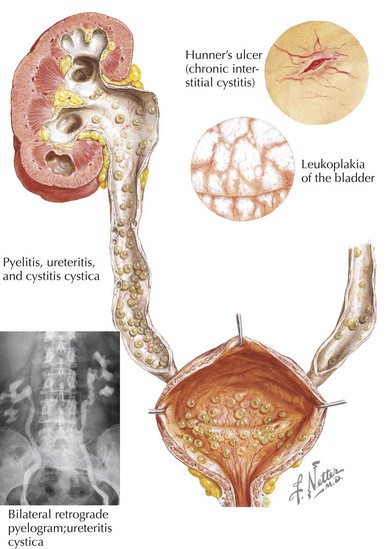
Infections of the urinary tract begin with ascension of flora that transiently colonize the lower urinary tract. The most common organism responsible for infection is the enteric gram-negative organism Escherichia coli. Other gram-negative organisms include Klebsiella, Proteus, Citrobacter, and Enterobacter spp. Gram-positive infections by enterococcus, Staphylococcus saprophyticus, and rarely Staphylococcus aureus are also known to occur. For infection to take place, the organisms must not only be present in the urinary tract but also needs to attach, adhere to, and invade the mucosa and epithelium. The bacteria are then able to ascend via the epithelial cells to the bladder and kidney (Figure 93-1). It is thought that genetically determined variation in the host’s defense against this process may explain the familial association of UTIs among first-degree relatives.

Clinical Presentation

UTIs can present in a variety of ways with varying differentials depending on the age of the patient. In general, the older the patient, the more specific the presentation will be. In infants and small children (younger than 2 years of age), UTIs may only present with fever. Certainly, fever without a clear source should prompt the clinician to consider a UTI, but even in infants with another potential source, evaluation of the urine may still be prudent. Other symptoms may include irritability, poor feeding, or failure to thrive. Of note, parental report of “foul-smelling urine” has not been shown to correlate with UTIs. The differential diagnosis, as with all infants with fever and an unknown source, is extensive, but the clinician should primarily be concerned about occult bacteremia, sepsis, and viral infections. Other aspects of the history that may be helpful are the degree and duration of the fever and any history of UTIs in the patient or family.

In older children, presentation is more similar to that of the adult. Symptoms may include fever, abdominal pain, urinary symptoms (dysuria, urgency, incontinence, hematuria, frequency), back pain, or vomiting. The differential diagnosis can vary greatly depending on the presentation. For the urinary symptoms, the differential diagnosis includes urinary calculi, urethritis (chemical, sexually transmitted infection), vaginal foreign body, and DES. The differential for fever and abdominal pain in older children includes group A streptococcus infections, Kawasaki’s disease, appendicitis, and gastroenteritis, the first three of which may also present with pyuria. In the patient’s history, it is also important to note any history of: UTIs in the patient or family, VUR or DES, constipation, or poor growth.

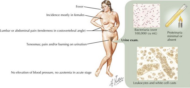
On physical examination of an infant or child with suspected UTI, it is important to note the degree of fever (if present) and any signs of cardiovascular instability that would reflect severe illness. Other findings on examination, more common in older patients, may include suprapubic or costovertebral tenderness. A complete abdominal examination should be performed to assess for any abdominal masses or abnormal distension of the bladder. A genital examination should look for any signs of urethritis or vulvovaginitis and for any anatomic abnormalities. If the patient is febrile, attention should also be paid to other potential sources of fever (Figure 93-2).

Evaluation and Management (Table 93-1)

Laboratory Evaluation

The appropriate management of a patient with UTI can vary significantly depending on the severity of illness. Those with mild to moderate illness can be effectively managed as outpatients, but those with severe illness may require inpatient or even intensive care and dialysis. Because of the often nonspecific nature of presentation, laboratory analysis of the urine is essential to confirm the diagnosis in all settings. Two important steps for the clinician as they evaluate the patient are (1) whether to obtain a urine sample and (2) how best to do so.

The decision to obtain a urine sample can be very simple, as with the febrile neonate, or in the cases of older children, it can be more complex. Important factors to consider for the clinician evaluating 3- to 24-month-old children are:

If one of these risk factors is present for girls and uncircumcised boys or two are present for circumcised boys, it is recommended to obtain a urine sample for urinalysis and culture. One should also obtain samples for girls and uncircumcised boys older than 2 years of age with any concerning symptom (abdominal pain, back pain, dysuria, frequency, high fever) and circumcised boys older than 2 years with multiple symptoms.

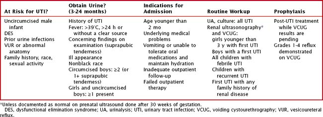
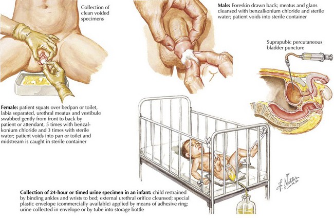
After it has been decided to obtain urine, the clinician must then determine how best to obtain it. For an infant, urine can be obtained by bag specimen, urethral catheterization, and suprapubic aspiration. The bag specimen is the most convenient but is of limited utility and generally not recommended because of the high probability of contamination from skin flora. It cannot be used for culture, and urinalysis is only useful if results are negative. Care must be taken, however, to analyze the sample promptly after voiding because the false results increase as the urine stagnates. Catheterized specimen is the primary means of obtaining urine in the infant for urine culture or enhanced urinalysis. Suprapubic aspiration can also be performed if catheterization is repeatedly unsuccessful or contraindicated, which despite its invasive nature is very well tolerated when performed correctly. For toilet-trained children, a clean catch is the primary means of collection for urinalysis and culture (Figure 93-3).

A urinalysis is a relatively rapid test that can give the provider important information and help initiate treatment while the urine culture, the gold standard test, is performed. The urinalysis exists in several forms. The simplest is the urine dipstick, which has the advantage of being affordable and can be performed in virtually any clinical setting, but it may miss some cases of infection. A urinalysis with microscopic examination can more accurately detect the presence of bacteria, white blood cells, and hematuria. An enhanced urinalysis, available in larger hospitals and academic institutions, examines uncentrifuged specimens and adds Gram staining and a hemocytometer. A positive urine dipstick result can show the presence of leukocyte esterase (higher sensitivity) or nitrites (higher specificity), the centrifuged specimen can show pyuria or bacteriuria, and the uncentrifuged specimen gives a more quantitative analysis of pyuria. Although bacteriuria and pyuria can occur without infection, the presence of both is very specific for infection in the urine.

The gold standard for diagnosis is the urine culture, which not only makes the diagnosis but also helps to guide antimicrobial management. Growth criteria for definite infection are different depending on the means of collection, with any growth significant for a suprapubic aspirate and >50,000 or >100,000 colonies for a catheterization and clean catch, respectively. Although the current American Academy of Pediatrics guidelines recognize 10,000 colonies from a catheterization specimen as significant, recent evidence suggests the 10,000 to 50,000 range be classified as indeterminate and a repeat specimen be obtained. When empiric coverage has already been initiated, however, this may not be practical, and the decision to treat will likely incorporate other factors (clinical presentation, positive or negative urinalysis). In the majority of cases, any growth of nonpathogens or mixed flora indicates contamination and the need for a new specimen (Figure 93-4).

Blood tests are usually not indicated in routine infections of the urinary tract. A blood culture is of little utility because the urine is the source of the infection, and the presence of bacteremia does not alter the course of treatment. Markers of inflammation, such as a C-reactive protein, will certainly be elevated during a UTI, but their utility in differentiating between upper tract (pyelonephritis) and lower tract infection is yet to be supported by evidence. Electrolytes are not routinely drawn if the patient does not have any clinical signs of renal failure (anuria, edema, hypertension).

Antimicrobials

It is important to begin therapy promptly in UTIs because delays in treatment can lead to complications both in the acute illness (pyelonephritis, renal abscess) and chronic sequelae (renal scarring, hypertension). When choosing empiric therapy, the clinician should consider the severity of illness, any history of prior UTIs, and the local patterns of resistance. For empiric coverage while awaiting culture results, the primary organism to consider is E. coli, although in certain circumstances (e.g., history of catheterization or manipulation), enterococcus should be covered as well. For routine outpatient care, amoxicillin is no longer adequate for coverage of E. coli, and rates of resistance to amoxicillin–clavulanate, first-generation cephalosporins, and trimethoprim−sulfamethoxazole (TMP-SMX) are increasing such that a clinician may not feel comfortable with these agents as well. Resistance patterns will dictate the empiric choice of antibiotics. Empiric therapy may be initiated with a third-generation cephalosporin such as cefixime, 8 mg/kg given once daily as a reasonable alternative. Note that this does not provide adequate coverage for enterococcus, so if this organism is of concern, amoxicillin should be added.

Inpatient management can be initiated with a third-generation cephalosporin such as cefotaxime, although the clinician also has the option of gentamicin, which is acceptable coverage for E coli. Again, neither provides adequate coverage for enterococcus, so ampicillin should be added in those cases, which many providers choose to do for all cases when hospital admission is indicated. The duration of treatment is 10 days for children younger than 2 years of age or older children with febrile UTI; for older afebrile children, a shorter course of 5 to 7 days is acceptable. If imaging studies are to be performed, it is also recommended that children begin prophylactic antibiotics until the results are available.

Imaging

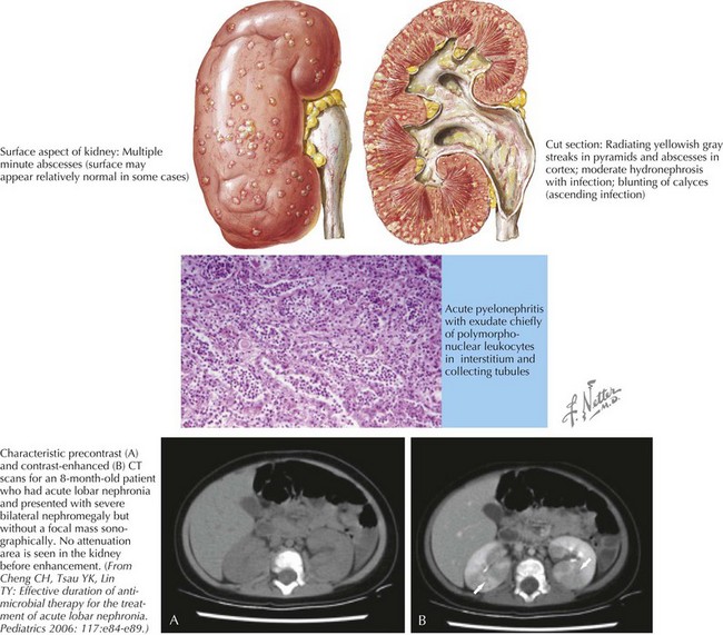
Three modalities of imaging are available to the clinician in the evaluation of UTIs: voiding cystourethrography (VCUG), renal ultrasound, and renal scintigraphy (DMSA [dimercaptosuccinic acid] scan). Intravenous pyelography (IVP) is rarely used in the pediatric population and is not discussed here. Overall, the goals of these studies are to identify any anatomic abnormalities that may predispose the patient to recurrent infections, evaluate for complications such as renal abscess or lobar nephronia (Figure 93-5), and look for evidence of renal scarring. VCUG, which involves catheterization and subsequent imaging of instilled dye during voiding, is an excellent test to evaluate for the presence and degree of VUR. Renal ultrasound can identify certain anatomic anomalies (e.g., ureteral dilatation, duplication), is noninvasive, and has the benefit of no radiation exposure. It is not able to identify renal scarring or VUR. Current recommendations are for these two studies to be performed in:

If the patient has required hospitalization, it is usually routine to complete these studies before discharge. Of note, if the patient had normal prenatal ultrasound results after 30 weeks of gestation, it does not need to be repeated, but particularly in the inpatient setting, these results may not be available, and repetition may be necessary.

DMSA scanning is used to detect acute pyelonephritis and renal scarring. It is not routinely recommended for UTIs because children with febrile UTIs are often presumed to have pyelonephritis, and treatment would not change with a positive study result. Times when it may be useful are when evaluating children with recurrent UTIs and possible renal damage or in cases where urine studies have been equivocal.