Ichthyoses
Management strategy
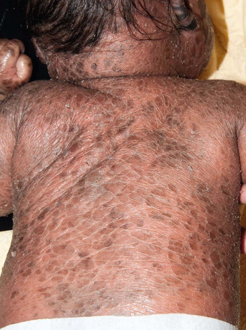
The key to management, where possible, is to establish an exact diagnosis. This provides a platform to plan therapy, discuss prognosis, and consider genetic counseling. It is important to identify the age of onset, the presence or absence of collodion membrane, blistering or erythroderma in the neonatal period, and the type, color, and distribution of scale. Family members should be examined. The Ichthyosis Support Group (ISG) provides a support network for families with resources and information sheets (www.ichthyosis.org.uk).
Specific investigations
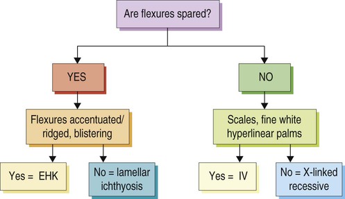
 Follow algorithm (see Figure below)
 Genetic screening: refer to www.genetests.org
 Fatty alcohol: NAD + oxidoreductase activity (Sjögren–Larsson)


 Humidification of environment
 Bathing/soaking
 Emollients
 Keratolytics (salicylic acid (avoid in children), urea, α-hydroxy acids (e.g., lactic acid), propylene glycol)
 Topical retinoids (tretinoin, tazarotene)
 Topical liarozole
 Topical calcipotriol
 Topical N-acetylcysteine
 Topical pimecrolimus 1%
 Narrowband UVB phototherapy (Netherton syndrome)
 Acitretin/etretinate
 Isotretinoin
 Alitretinoin
 Liarozole
