Hyperpituitarism, Tall Stature, and Overgrowth Syndromes

Published on 22/03/2015 by admin

Filed under Pediatrics

Last modified 22/04/2025

Print this page

rate 1 star rate 2 star rate 3 star rate 4 star rate 5 star
Your rating: none, Average: 0 (0 votes)

This article have been viewed 3269 times

Chapter 554 Hyperpituitarism, Tall Stature, and Overgrowth Syndromes

Hyperpituitarism

Primary hypersecretion of pituitary hormones rarely occurs in the pediatric population. Primary hyperpituitarism should be distinguished from secondary hyperpituitarism, which occurs in the setting of target hormone deficiencies resulting in decreased hormonal feedback, such as in hypogonadism, hypoadrenalism, or hypothyroidism. In some cases, chronic pituitary hypersecretion is accompanied by pituitary hyperplasia, which can enlarge and erode the sella, and, on rare occasions, increase intracranial pressure. Such enlargements should not be confused with primary pituitary tumors; they disappear when the underlying hormone deficiency is treated. The elevated pituitary hormone levels readily suppress to normal following replacement of end-organ hormones. Pituitary hyperplasia can also occur in response to stimulation by ectopic production of releasing hormones such as that seen occasionally in patients with Cushing syndrome secondary to corticotropin-releasing hormone excess or in children with acromegaly secondary to growth hormone-releasing hormone (GHRH) produced by a variety of systemic tumors.

Primary hypersecretion of pituitary hormones by adenoma is uncommon in childhood. The most commonly diagnosed adenoma during childhood is prolactinoma, followed by corticotropinoma, and then somatotropinoma, which secrete prolactin, corticotropin, and growth hormone, respectively. There have been a handful of case reports of thyrotropinoma in children and adolescents. There are no pediatric reports of gonadotropinoma. Hypothalamic hamartomas that secrete excess gonadotropin-releasing hormone are not an uncommon cause of precocious puberty. The monoclonal nature of most pituitary adenomas has implied that most originate from a clonal event in a single cell. It is suspected that some pituitary tumors result from stimulation with hypothalamic-releasing hormones and in other instances, as in McCune-Albright syndrome (MAS), the tumor is caused by activating mutations of the GNAS1 gene that codes for the α subunit of Gsα, a guanine nucleotide-binding protein. The clinical presentation typically depends on the pituitary hormone that is hypersecreted. Disruptions of growth regulation and/or sexual maturation are common, as a result of either hormone hypersecretion or local compression by the tumor. MAS also features polyostotic fibrous dysplasia of bone and café-au-lait spots in a distinct distribution.

Tall Stature

The normal distribution of height predicts that 2.3% of the population will be taller than 2 SD (97.7%) above the mean. The social acceptability and even desirability of tallness (heightism), makes tall stature an uncommon complaint. In North America, it is exceptionally unusual for boys and men to seek medical attention regarding excessive height, although in Europe it is somewhat more common. Even in girls and women, tall stature has become more socially acceptable, although tall girls might still approach the physician with a desire to curb their growth rate.

Differential Diagnosis

Table 554-1 lists the causes of tall stature in childhood and adolescence. Of those listed, the normal variant, familial or constitutional tall stature, is by far the most common cause. Almost invariably, a family history of tall stature can be obtained, and no organic pathology is present. The child is often taller than his or her peers throughout childhood and enjoys excellent health. The parent of the constitutionally tall adolescent might reflect unhappily upon his or her own adolescence as a tall teenager. There are no abnormalities in the physical examination, and the laboratory studies, if obtained, are negative. Additional features of overgrowth syndromes are noted in Table 554-2.
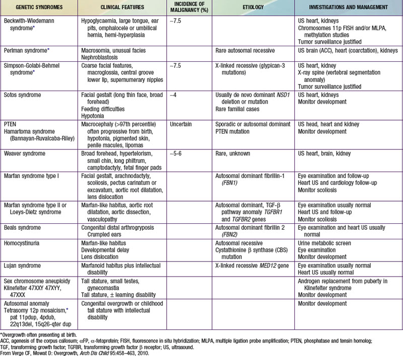
Klinefelter syndrome (XXY syndrome) is a relatively common (1 : 500-1000 live male births) abnormality associated with tall stature, learning disabilities (including requirement for speech therapy), gynecomastia, and decreased upper to lower body segment ratio. Affected boys can have hypotonia, clinodactyly, and hypertelorism. The testes are invariably small, although androgen production by Leydig cells is often in the low-normal range. Spermatogenesis and Sertoli cell function are defective, and infertility results. Other genital abnormalities including relatively small phallus, hypospadias, and cryptorchidism may be present.

XYY syndrome is associated with tall stature, problems in motor and language development, and possible antisocial behavior.

Marfan syndrome is an autosomal dominant connective tissue disorder consisting of tall stature, arachnodactyly, thin extremities, increased arm span, and decreased upper to lower body segment ratio (Chapter 693). Additional abnormalities include ocular abnormalities (e.g., lens subluxation), hypotonia, kyphoscoliosis, cardiac valvular deformities, and aortic root dilatation.

Homocystinuria is an autosomal recessive inborn error of amino acid metabolism, caused by a deficiency of the enzyme cystathionine synthetase. It is characterized by mental retardation when untreated, and many of its clinical features resemble Marfan syndrome, particularly ocular manifestations (Chapter 79). Hyperthyroidism in adolescents is associated with rapid growth but normal adult height. It is almost always caused by Graves disease and is much more common in girls (Chapter 562).

Exogenous obesity is a common condition in adolescence and may be associated with rapid linear growth and early onset of puberty. Adult height is typically normal.

The purpose of the diagnostic evaluation of tall stature is to distinguish the commonly occurring normal variant constitutional variety from the rare pathologic conditions. Often, when the history suggests familial tall stature and the physical examination is entirely normal, no laboratory tests are indicated. It is valuable to obtain a bone age radiograph to be able to predict adult height, which serves as a basis for discussions with the family and for management decisions. If the history suggests any of the aforementioned disorders or the physical examination reveals abnormalities, additional laboratory tests should be obtained. Insulin-like growth factor-1 (IGF-1) and IGF binding protein-3 (IGFBP-3) are excellent screening tests for GH excess and can be verified with a glucose suppression test. Laboratory evidence of GH excess mandates MRI evaluation of the pituitary. Chromosome analysis is useful in boys, especially when the ratio of upper to lower body segment is decreased or when developmental disability is present, to rule out Klinefelter syndrome. If Marfan syndrome or homocystinuria is suspected from the physical examination, referral to a cardiologist and an ophthalmologist should be made. Thyroid function tests are useful to diagnose or rule out hyperthyroidism when this disorder is suspected.

Precocious puberty, whether mediated centrally (increased gonadotropin secretion) or peripherally (increased secretion of androgens, estrogens, or both), results in accelerated linear growth during childhood, mimicking the pubertal growth spurt. Because skeletal maturation is also advanced, adult height is often compromised. The diagnostic evaluation and management of precocious puberty are discussed in Chapter 556.

Although delayed puberty may be associated with short stature in childhood, as with constitutional delay, failure to eventually enter puberty and complete sexual maturation can result in sustained growth during adult life, with ultimate tall stature. The report of tall stature with open epiphyses resulting from a mutation of the estrogen receptor in a man with normal male sexual maturation underscores the fundamental role of estrogen in promoting epiphyseal fusion and termination of normal skeletal growth. Aromatase deficiency leads to tall stature through similar pathways. Furthermore, androgen insensitivity is associated with tall stature in girls, demonstrating a role for androgens in this process.

Treatment

Reassurance of the family and the patients is the key to the management of normal variant tall stature. The use of the bone age to predict adult height might provide some comfort for them, as will general supportive discussions on the social acceptability of this condition. Although treatment is available for girls and boys with excessive growth, its use should be restricted to patients with predicted adult height >3-4 SD above the mean (79 inches or 200 cm in boys, 73 inches or 185 cm in girls) and evidence of significant psychosocial impairment.

For the family that feels strongly about treatment, a trial of sex steroids may be considered. Sex steroids have been used in the treatment of tall stature and is designed to accelerate puberty and to promote epiphyseal fusion; it is therefore of little benefit when given in late puberty. Therapy is initiated ideally before puberty or in early puberty. In boys, treatment should begin before the bone age reaches 14 yr. Although testosterone can be given as treatment of constitutional tall stature in boys, its use is extremely rare. In clinical practice, testosterone enanthate is used at a dose of 500 mg intramuscularly every 2 weeks for 6 months.

In girls, oral estrogens in various doses have successfully reduced the predicted height by an average of 5-10 cm. This is a direct result of the known effects of sex steroids on promoting epiphyseal fusion, thus therapy must begin before the bone age has reached 12 yr. Oral ethinyl estradiol at a dose of 0.15-0.5 mg/day until cessation of growth occurs has been used successfully in girls. If necessary, a progestational agent can be added after 1 yr of unopposed estrogen therapy. Short-term side effects have included benign breast disease, cholelithiasis, hypertension, menstrual irregularities, weight gain, nausea, limb pain, galactorrhea and thrombosis. Reduced fertility later in life may be a potential long-term complication. The lack of extensive experience with this form of therapy and the risks of estrogen treatment for tall stature should be carefully weighed and discussed with the family before embarking on therapy.

The mechanism of estrogen action involves indirect effects on the growth hormone-IGF-1 axis as well as its direct effect on the epiphysis. Estrogen mediates epiphyseal fusion in both girls and boys. In prepubertal girls, adult height is reportedly decreased by as much as 5-10 cm relative to pretreatment predictions. When therapy is initiated after the onset of puberty, the decrement in adult height will not be so large.

Therapy in boys with tall stature is even more problematic. Estrogen is likely to be most efficacious in accelerating epiphyseal fusion but is obviously undesirable in boys. Androgens also accelerate skeletal maturation, presumably via aromatization to estrogen, but are associated with virilization. For children with Klinefelter syndrome, testosterone treatment can be initiated during the puberty to facilitate development of secondary sexual characters, and this treatment can diminish adult height.

Prolactinoma

Prolactin-secreting pituitary adenomas are the most common pituitary tumors in adolescents. With the advent of MRI, more of these tumors, particularly microadenomas (<1 cm in diameter), are being detected. The most common presenting manifestations are headache, primary or secondary amenorrhea, and spontaneous or provocative galactorrhea. The disorder affects more than twice as many girls as boys; most patients have undergone normal puberty before becoming symptomatic. Only a few have delayed puberty. In some kindreds with type I multiple endocrine neoplasia (MEN), prolactinomas are the presenting feature during adolescence.

Prolactin levels may be elevated mildly (40-50 ng/mL) or markedly (10,000-15,000 ng/mL). Most prolactinomas in children are large (macroadenomas), cause the sella to enlarge, and in some cases cause visual field defects. Approximately 30% of patients with macroadenomas develop hypopituitarism, particularly GH deficiency. Alternatively, prolactin-secreting adenomas might also stain for and secrete excess GH and/or TSH.

Prolactinomas should not be confused with the hyperprolactinemia and pituitary hyperplasia that can occur in patients with primary hypothyroidism, which is readily treated with thyroid hormone (Chapter 559). Moderate elevations (<200 ng/mL) of prolactin are also associated with a variety of medications (mainly antipsychotic), with pituitary stalk dysfunction such as can occur with craniopharyngioma, and with other benign conditions. In most patients this can be effectively treated with the dopamine agonist bromocriptine or long-acting cabergoline. When dopamine agonist treatment has been unsuccessful in lowering the serum prolactin concentration or size of the adenoma, and when symptoms or signs due to hyperprolactinemia or adenoma size persist during treatment, transsphenoidal surgery should be considered.

Excess Growth Hormone Secretion and Pituitary Gigantism

In young persons with open epiphyses, overproduction of GH results in gigantism; in persons with closed epiphyses, the result is acromegaly. Often, some acromegalic features are seen with gigantism, even in children and adolescents. After closure of the epiphyses, the acromegalic features become more prominent.

Pituitary gigantism is extremely rare, and its cause is most often a pituitary adenoma, but gigantism has been observed in a 2.5 yr old boy with a hypothalamic tumor that presumably secreted GHRH. Other tumors, such as those that are part of the MEN syndromes, particularly in the pancreas, have produced acromegaly by secretion of large amounts of GHRH with resultant hyperplasia of the somatotrophs. The cardinal clinical feature of gigantism is longitudinal growth acceleration secondary to GH excess. The usual manifestations consist of coarse facial features and enlarging hands and feet. In young children, rapid growth of the head can precede linear growth. Some patients have behavioral and visual problems. In most of the recorded cases, the abnormal growth became evident at puberty, but the condition has been established as early as the newborn period in one child and at 21 mo of age in another. Giants have rarely been reported to grow to a height of over 8 ft.

Acromegalic features consist chiefly of enlargement of the distal parts of the body, but manifestations of abnormal growth involve all portions. The circumference of the skull increases, the nose becomes broad, and the tongue is often enlarged, with coarsening of the facial features. The mandible grows excessively, and the teeth become separated. Visual field defects and neurologic abnormalities are common; signs of increased intracranial pressure appear later. The fingers and toes grow chiefly in thickness. There may be dorsal kyphosis. Fatigue and lassitude are early symptoms. GH levels are elevated and occasionally exceed 100 ng/mL. There is usually no suppression of GH levels by the hyperglycemia of a glucose tolerance test. IGF-1 and IGFBP-3 levels are consistently elevated in acromegaly, whereas other growth factors are not.

Gigantism is rare, with only several hundred reported cases to date. The presentation of gigantism is usually dramatic, unlike the insidious onset of acromegaly in adults. The tumor mass itself may cause headaches, visual changes due to optic nerve compression, and hypopituitarism. About 50% of the patients also have marked hyperprolactinemia as a result of plurihormonal adenomas that secrete GH and prolactin. This is because mammosomatotrophs are the most common type of GH-secreting cells involved in childhood gigantism. GH-secreting tumors of the pituitary are typically eosinophilic or chromophobic adenomas. Adenomas can compromise other anterior pituitary function through growth or cystic degeneration. Secretion of gonadotropins, thyrotropin, or corticotropin may be impaired. Delayed sexual maturation or hypogonadism can occur. When GH hypersecretion is accompanied by gonadotropin deficiency, accelerated linear growth can persist for decades. In some cases, the tumor spreads outside the sella, invading the sphenoid bone, optic nerves, and brain. GH-secreting tumors in pediatric patients are more likely to be locally invasive or aggressive than are those in adults.

The etiology is uncertain although studies in acromegalics suggest that many cases result from mutations that generate constitutively activated G-proteins with reduced GTPase activity. The resultant increase in intracellular cyclic adenosine monophosphate in the pituitary leads to increased GH secretion. McCune-Albright syndrome (MAS), which can also be caused by mutations resulting in constitutively activated G-proteins, can also include somatotrophic tumors and excess GH secretion. Approximately 20% of patients with gigantism are those with MAS (commonly consisting of a triad of precocious puberty, café-au-lait spots, and fibrous dysplasia). GH-secreting tumors have also been reported in multiple endocrine adenomatosis and in association with neurofibromatosis, tuberous sclerosis, and Carney complex.

Activating mutations of the stimulatory Gsα proteins have been found in the pituitary lesions in MAS and are believed to be responsible for the other glandular adenomas observed in this condition as well. Somatic point mutations of the Gsα protein have also been identified in somatotrophs of up to 40% of sporadic GH-secreting pituitary adenomas.

Diagnosis

The gold standard for the diagnosis of GH excess is the failure to suppress serum GH levels to <5 ng/dL after a 1.75 g/kg oral glucose challenge (maximum, 75 g). This test measures the ability of IGF-1 to suppress GH secretion because the glucose load results in insulin secretion, leading to suppression of IGFBP-1, which results in an acute increase in free IGF-1 levels. The increased free IGF-1 suppresses GH secretion within 30-90 min. This test can be abnormal in diabetic patients. A single measurement of GH is inadequate because GH is secreted in a pulsatile manner. Therefore, the use of a random GH measurement can lead to both false-positive and false-negative results. Measurement of serum IGF-1 concentration is a sensitive screening test for GH excess. An excellent linear dose-response correlation between serum IGF-1 levels and 24-hr mean GH secretion has been demonstrated. An elevated IGF-1 level in a patient with appropriate clinical suspicion usually indicates GH excess. Potential confusion can arise in the evaluation of normal adolescents, because significantly higher IGF-1 levels occur during puberty than in adulthood; the IGF-1 level must be age- and gender-matched. Serum IGFBP-3 levels are sensitive markers of GH elevations and may be elevated despite normal IGF-1 levels. If laboratory findings suggest GH excess, the presence of a pituitary adenoma should be confirmed by MRI of brain. In rare cases, a pituitary mass is not identified. This might be due to an occult pituitary microadenoma or ectopic production of GHRH or GH. CT is acceptable when MRI is unavailable.

Treatment

The goals of therapy are to remove or shrink the pituitary mass, to restore GH and secretory patterns to normal, to restore IGF-1 and IGFBP-3 levels to normal, to retain the normal pituitary secretion of other hormones, and to prevent recurrence of disease.

For well-circumscribed pituitary adenomas, transsphenoidal surgery is the treatment of choice and may be curative. The tumor should be removed completely. The likelihood of surgical cure depends greatly on the surgeon’s expertise as well as on the size and extension of the mass. Intraoperative GH measurements can improve the results of tumor resection. Transsphenoidal surgery to resect the tumors is as safe in children as in adults. At times, a transcranial approach may be necessary. The primary goal of treatment is to normalize GH levels. GH levels (<1 ng/mL within 2 hr after a glucose load) and serum IGF-1 levels (age-adjusted normal range) are the best tests to define a biochemical cure.

If GH secretion is not normalized by surgery, the options include pituitary irradiation and medical therapy. Stereotactic radiotherapy is recommended if GH hypersecretion is not normalized by surgery. Further growth of the tumor is prevented by irradiation in >99% of patients. The main disadvantage is the delayed efficacy in decreasing GH levels. GH is reduced by approximately 50% from the initial concentration by 2 yr, by 75% by 5 yr, and approaches 90% by 15 yr. Hypopituitarism is a predictable outcome, occurring in 40-50% of patients 10 yr after irradiation.

Surgery fails to cure a significant number of patients, and therefore medical therapy has an important role in treating patients who have GH excess. Treatment is effective and well tolerated with long-acting somatostatin analogs and dopamine agonists as well as by novel GH antagonists.

The somatostatin analogs are highly effective in the treatment of patients with GH excess. Octreotide suppresses GH to <2.5 ng/mL in 65% of patients with acromegaly and normalizes IGF-1 levels in 70%. The effects of octreotide are well sustained over time. Tumor shrinkage also occurs with octreotide but is generally modest. Consistent GH suppression can be obtained with a continuous SC pump infusion of octreotide in a pubertal boy with pituitary gigantism. Long-acting formulations, including long-acting octreotide and lanreotide, produce consistent GH and IGF-1 suppression in acromegalic patients with once-monthly or biweekly IM depot injections. The sustained-release preparations have not been formally tested in children. Octreotide injection in the pediatric population has been used at doses of 1-40 µg/kg/24 hr.

For patients with both GH and prolactin oversecretion, dopamine agonists, such as bromocriptine, which bind to pituitary dopamine type 2 (D2) receptors and suppress GH secretion, should be considered, although their precise mechanism of action is unclear. Prolactin levels are often adequately suppressed; GH levels and IGF-1 levels are rarely normalized with this treatment modality. Less than 20% of patients achieve GH levels <5 ng/mL, and <10% achieve normalization of IGF-1 levels. Tumor shrinkage occurs in a minority of patients. Bromocriptine can be used as adjuvant medical treatment for GH excess. Its effectiveness may be additive to that of octreotide. Improved efficacy, defined by normal IGF-1 concentration and reduced tumor size, also occurs with other dopamine agonists such as pergolide and cabergoline. The dose of bromocriptine ranges from 10-60 mg/24 hr PO divided 4 times a day. Only a minority of patients benefit from doses >20 mg/24 hr. It has been found safe when used in children for an extended period of time, but side effects can include nausea, vomiting, abdominal pain, arrhythmias, nasal stuffiness, orthostatic hypotension, sleep disturbances, and fatigue. Octreotide long-acting release and cabergoline therapy have shown great promise in adults with acromegaly; however, pediatric experience is very limited.

Pegvisomant is a GH-receptor antagonist that competes with endogenous GH for binding to the GH receptor. It effectively suppresses GH and IGF-1 levels in patients with acromegaly due to pituitary tumors as well as ectopic GHRH hypersecretion. Normalization of IGF-1 levels occurs in up to 90% of patients treated daily with this drug for 3 months or longer. The adult dosage is 10-40 mg via subcutaneous injection once daily, although TIW protocols have also been reported as highly successful. IGF-1 levels and hepatic enzymes must be monitored. Combined therapy with somatostatin analogs and weekly pegvisomant injections also is shown to be effective. Its pediatric experience is very limited.

Sotos Syndrome (Cerebral Gigantism)

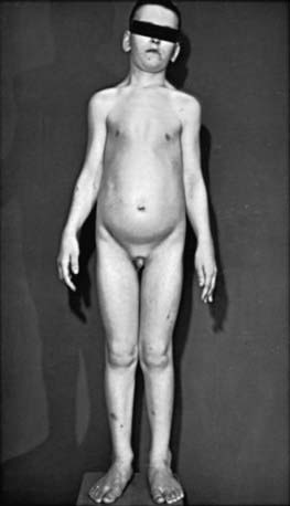
Children with cerebral gigantism (also known as Sotos syndrome) are above the 90th percentile for both length and weight at birth; they can also have macrocrania at that time. The NSD1 (nuclear receptor SET domain-containing protein 1) gene is responsible for this syndrome. Although it is characterized by rapid growth, there is no evidence that Sotos syndrome is caused by endocrine dysregulation. A hypothalamic defect has been suggested as a cause, but none has been demonstrated functionally or at necropsy. Growth is markedly rapid; by 1 yr of age, affected infants are greater than the 97th percentile in height. Accelerated growth continues for the first 4-5 yr and then returns to a normal rate (Fig. 554-1). Puberty usually occurs at the expected time but may occur slightly early. Adult height is usually in the upper normal range.

The syndrome is characterized by large (macrocephaly) dolichocephalic head, prominent forehead and jaw, hypertelorism, antimongoloid slant of the palpebral fissures, high-arched palate, and large hands and feet with thickened subcutaneous tissue. Clumsiness and awkward gait are also noted, and affected children have great difficulty in sports, in learning to ride a bicycle, and in other tasks requiring coordination. Some degree of developmental disability affects most patients; in some affected children perceptual deficiencies may predominate. Osseous maturation is usually compatible with the patient’s height although advanced bone age has been reported. GH, IGF-1, and other endocrine studies are usually normal; there is no distinctive laboratory or radiologic marker for the syndrome. Abnormal electroencephalograms are common; imaging studies often reveal a dilated ventricular system. Affected patients may be at increased risk for neoplasia, particularly hepatic carcinoma, but Wilms, ovarian, and parotid tumors have also been reported.

Overgrowth in the Fetus and Neonate

Maternal diabetes is the most common cause of infants large for gestational age (LGA). Even in the absence of clinical symptoms or a family history, the birth of an LGA infant should lead to evaluation for maternal (or gestational) diabetes.

A group of disorders associated with excessive somatic growth and growth of specific organs has been described and is collectively referred to as overgrowth syndromes. These disorders appear to be caused by excess production and availability of IGF-2 encoded by the gene Igf2. The best described of these syndromes is the Beckwith-Wiedemann syndrome (BWS), which is an overgrowth malformation syndrome that occurs with an incidence of 1 : 14,000 births. It manifests as a fetal overgrowth syndrome in which hypertrophy dominates the clinical picture. Affected infants characteristically have microsomia including macroglossia, hepatosplenomegaly, nephromegaly, and omphalocele. They also have hypoglycemia secondary to hyperinsulinemia due to pancreatic β-cell hyperplasia. Children with BWS are predisposed to a specific subset of childhood neoplasms consisting of Wilms tumor and adrenocortical carcinoma. Overexpression of IGF-2 in BWS may be caused by a number of genetic disruptions including gene duplication, loss of heterozygosity, and relaxation or loss of imprinting of the Igf2 gene. Various lines of investigation have localized “imprinted” genes involved in BWS and associated childhood tumors to chromosome 11p. These include, in addition to Igf2, the gene H19, which is involved in Igf2 suppression, as well as WT-1 (the Wilms tumor gene).

Mutations in GPC3, a glypican gene (which codes for an IGF-2 neutralizing membrane receptor), cause the related Simpson-Golabi-Behmel overgrowth syndrome.

Bibliography

Acharya SV, Gopal RA, Bandgar TR, et al. Clinical profile and long term follow up of children and adolescents with prolactinomas. Pituitary. 2009;12:186-189.

Bademci G. Pitfalls in the management of Cushing’s disease. J Clin Neurosci. 2007;14:401-408.

Bulun SE. Aromatase deficiency and estrogen resistance: from molecular genetics to clinic. Semin Reprod Med. 2000;18:31-39.

Carel JC, Blumberg J, Bougeard-Julien M, et al. Long-acting lanreotide in adolescent girls with constitutional tall stature. Horm Res. 2009;71:228-236.

Chahal HS, Stals K, Unterlander M, et al. AIP mutation in pituitary adenomas in the 18th century and today. N Engl J Med. 2011;364(l):43-50.

Drop SL, De Waal WJ, De Muinck Keizer-Schrama SM. Sex steroid treatment of constitutionally tall stature. Endocr Rev. 1998;19:540-558.

Faravelli F. NSD1 mutations in Sotos syndrome. Am J Med Genet C Semin Med Genet. 2005;137C:24-31.

Feenstra J, de Herder WW, ten Have SM, et al. Combined therapy with somatostatin analogues and weekly pegvisomant in active acromegaly. Lancet. 2005;365:1644-1646.

Horvath A, Stratakis CA. Clinical and molecular genetics of acromegaly: MEN1, Carney complex, McCune-Albright syndrome, familial acromegaly and genetic defects in sporadic tumors. Rev Endocr Metab Disord. 2008;9:1-11.

Lania A, Spada A. G-protein and signalling in pituitary tumours. Horm Res. 2009;71(Suppl 2):95-100.

Leventopoulos G, Kitsiou-Tzeli S, et al. A clinical study of Sotos syndrome patients with review of the literature. Pediatr Neurol. 2009;40:357-364.

Melmed S, Colao A, Barkan A, et al. Guidelines for acromegaly management: an update. J Clin Endocrinol Metab. 2009;94:1509-1517.

Melmed S, Cook D, Schopohl J, et al. Rapid and sustained reduction of serum growth hormone and insulin-like growth factor-1 in patients with acromegaly receiving lanreotide Autogel therapy: a randomized, placebo-controlled, multicenter study with a 52 week open extension. Pituitary. 2010;13:18-28.

Misra M, Cord J, Prabhakaran R, et al. Growth hormone suppression after an oral glucose load in children. J Clin Endocrinol Metab. 2007;92:4623-4629.

Morison IM, Becroft DM, Taniguchi T, et al. Somatic overgrowth associated with overexpression of insulin-like growth factor II. Nat Med. 1996;2:311-316.

Narayanaswamy V, Rettig KR, Bhowmick SK. Excessive growth. Clin Pediatr (Phila). 2008;47:705-708.

Prevedello DM, Pouratian N, Sherman J, et al. Management of Cushing’s disease: outcome in patients with microadenoma detected on pituitary magnetic resonance imaging. J Neurosurg. 2008;109:751-759.

Reddy R, Hope S, Wass J. Acromegaly. BMJ. 2010;341:c4189.

Reeve AE. Role of genomic imprinting in Wilms’ tumour and overgrowth disorders. Med Pediatr Oncol. 1996;27:470-475.

Riccio A, Sparago A, Verde G, et al. Inherited and sporadic epimutations at the IGF2-H19 locus in Beckwith-Wiedemann syndrome and Wilms’ tumor. Endocr Dev. 2009;14:1-9.

Rosenfield RL, Lipton RB, Drum ML. Thelarche, pubarche, and menarche attainment in children with normal and elevated body mass index. Pediatrics. 2009;123:84-88.

Sacks DA. Etiology, detection, and management of fetal macrosomia in pregnancies complicated by diabetes mellitus. Clin Obstet Gynecol. 2007;50:980-989.

Simm PJ, Bajpai A, Russo VC, et al. Estrogens and growth. Pediatr Endocrinol Rev. 2008;6:32-41.

Socin HV, Chanson P, Delemer B, et al. The changing spectrum of TSH-secreting pituitary adenomas: diagnosis and management in 43 patients. Eur J Endocrinol. 2003;148:433-442.

Sotos JF. Overgrowth: genetic syndromes and other disorders associated with overgrowth. Clin Pediatr. 1997;36:157-170.

Sotos JF, Argente J. Overgrowth disorders associated with tall stature. Adv Pediatr. 2008;55:213-254.

Tajima T, Tsubaki J, Ishizu K, et al. Case study of a 15-year-old boy with McCune-Albright syndrome combined with pituitary gigantism: effect of octreotide-long acting release (LAR) and cabergoline therapy. Endocr J. 2008;55:595-599.

Topaloglu AK, Sansaricq C, Snyderman SE. Influence of metabolic control on growth in homocystinuria due to cystathionine B-synthase deficiency. Pediatr Res. 2001;49:796-798.

Venn A, Bruinsma F, Werther G, et al. Oestrogen treatment to reduce the adult height of tall girls: long-term effects on fertility. Lancet. 2004;364:1513-1518.

Verge CF, Mowat D. Overgrowth. Arch Dis Child. 2010;95:458-463.

Zeger MP, Zinn AR, Lahlou N, et al. Effect of ascertainment and genetic features on the phenotype of Klinefelter syndrome. J Pediatr. 2008;152:716-722.