[level-membership-for-pediatrics-category]
Chapter 284 Hookworms (Necator americanus and Ancylostoma spp.)
Etiology
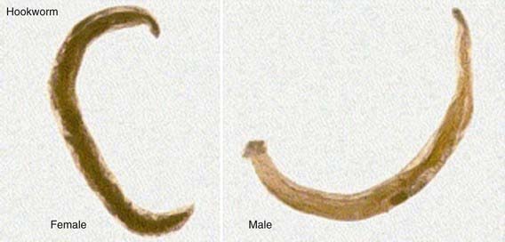
The infective larval stages of the anthropophilic hookworms live in a developmentally arrested state in warm, moist soil. Larvae infect humans either by penetrating through the skin (N. americanus and A. duodenale) or when they are ingested (A. duodenale). Larvae entering the human host by skin penetration undergo extraintestinal migration through the venous circulation and lungs before they are swallowed, whereas orally ingested larvae may undergo extraintestinal migration or remain in the gastrointestinal tract. Larvae returning to the small intestine undergo 2 molts to become adult sexually mature male and female worms ranging in length from 5 to 13 mm. The buccal capsule of the adult hookworm is armed with cutting plates (N. americanus) or teeth (A. duodenale) to facilitate attachment to the mucosa and submucosa of the small intestine. Hookworms can remain in the intestine for 1-5 yr, where they mate and produce eggs. Although approximately 2 mo is required for the larval stages of hookworms to undergo extraintestinal migration and develop into mature adults, A. duodenale larvae may remain developmentally arrested for many months before resuming development in the intestine. Mature A. duodenale female worms produce about 30,000 eggs/day; daily egg production by N. americanus is <10,000/day (Fig. 284-1). The eggs are thin shelled and ovoid, measuring approximately 40-60 µm. Eggs that are deposited on soil with adequate moisture and shade develop into 1st stage larvae and hatch. Over the ensuing several days and under appropriate conditions, the larvae molt twice to the infective stage. Infective larvae are developmentally arrested and nonfeeding. They migrate vertically in the soil until they either infect a new host or exhaust their lipid metabolic reserves and die.
Clinical Manifestations
Anthropophilic hookworm larvae elicit dermatitis sometimes referred to as ground itch when they penetrate human skin. The vesiculation and edema of ground itch are exacerbated by repeated infection. Infection with a zoonotic hookworm, especially A. braziliense, can result in lateral migration of the larvae to cause the characteristic cutaneous tracts of cutaneous larva migrans (Chapter 284.1). Cough subsequently occurs in A. duodenale and N. americanus hookworm infection when larvae migrate through the lungs to cause laryngotracheobronchitis, usually about 1 wk after exposure. Pharyngitis also can occur.
Diagnosis
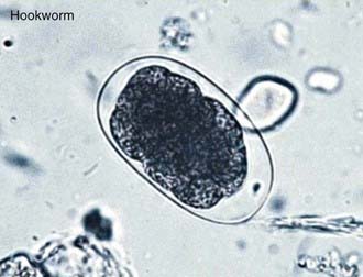
Children with hookworm release eggs that can be detected by direct fecal examination (Fig. 284-2). Quantitative methods are available to determine whether a child has a heavy worm burden that can cause hookworm disease. The eggs of N. americanus and A. duodenale are morphologically indistinguishable. Species identification typically requires egg hatching and differentiation of 3rd stage infective larvae; newer methods using polymerase chain reaction methods are under development.
284.1 Cutaneous Larva Migrans
Etiology
Cutaneous larva migrans (creeping eruption) is caused by the larvae of several nematodes, primarily hookworms, which are not usually parasitic for humans (Table 284-1). A. braziliense, a hookworm of dogs and cats, is the most common cause, but other animal hookworms may also produce the disease.
Table 284-1 ETIOLOGIES OF THE CUTANEOUS LARVA MIGRANS SYNDROME ACCORDING TO CUTANEOUS PRESENTATION
| CAUSATIVE AGENT | CUTANEOUS TRACK | OTHER CUTANEOUS SIGNS |
|---|---|---|
| Animal hookworm | 1-10 burrows, on the feet or buttocks, about 3 mm wide and up to 15-20 cm long, slow-moving (2-5 cm/day), chronic (weeks to months) | Highly pruritic, vesiculobullous lesions, impetiginization, hookworm folliculitis |
| Pelodera strongyloides | 10-100 burrows, on abdomen or buttocks, 1-2 cm long, 2-3 mm wide, may persist for months | Pruritus, follicular papules and pustules |
| Strongyloides stercoralis | Usually 1 burrow, on the abdomen or buttocks; lasts for hours only, may recur, fast-moving (larva currens) | Pruritus, urticaria |
| Gnathostoma spp. (G. hispidum, etc.) | Usually 1 burrow located anywhere, lasts for days, medium-fast-moving | Cutaneous migratory edema (eosinophilic panniculitis), cellulitis, papules, and nodules |
From Caumes E, Danis M: From creeping eruption to hookworm-related cutaneous larva migrans, Lancet Infect Dis 4:659–660, 2004.
Clinical Manifestations
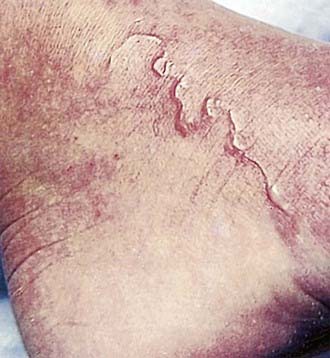
After penetrating the skin, larvae localize at the epidermal-dermal junction and migrate in this plane, moving at a rate of 1-2 cm/day. The response to the parasite is characterized by raised, erythematous, serpiginous tracks, which occasionally form bullae (Fig. 284-3). These lesions may be single or numerous and are usually localized to an extremity, although any area of the body may be affected. As the organism migrates, new areas of involvement may appear every few days. Intense localized pruritus, without any systemic symptoms, may be associated with the lesions. Bacterial superinfection can occur.
Budhatohoki S, Shah D, Bhurtyal KK, et al. Hookworm causing melaena and severe anaemia in early infancy. Ann Trop Paediatr. 2008;28:293-296.
Centers for Disease Control and Prevention. Outbreak of cutaneous larva migrans at a children’s camp—Miami, Florida, 2006. MMWR Morb Mortal Wkly Rep. 2007;56:1285-1287.
Heukelbach J, Feldmeier H. Epidemiological and clinical characteristics of hookworm-related cutaneous larva migrans. Lancet Infect Dis. 2008;8:302-309.
Hotez PJ, Brindley PJ, Bethony JM, et al. Helminth infections: the great neglected tropical diseases. J Clin Invest. 2008;118:1311-1321.
Hotez PJ, Brooker S, Bethony JM, et al. Hookworm infection. N Engl J Med. 2004;351:799-807.
Keiser J, Utzinger J. Efficacy of current drugs against soil-transmitted helminth infections: systematic review and meta-analysis. JAMA. 2008;299:1937-1948.
[/level-membership-for-pediatrics-category][not-level-membership-for-pediatrics-category]
Chapter 284 Hookworms (Necator americanus and Ancylostoma spp.)
Etiology
The infective larval stages of the anthropophilic hookworms live in a developmentally arrested state in warm, moist soil. Larvae infect humans either by penetrating through the skin (N. americanus and A. duodenale) or when they are ingested (A. duodenale). Larvae entering the human host by skin penetration undergo extraintestinal migration through the venous circulation and lungs before they are swallowed, whereas orally ingested larvae may undergo extraintestinal migration or remain in the gastrointestinal tract. Larvae returning to the small intestine undergo 2 molts to become adult sexually mature male and female worms ranging in length from 5 to 13 mm. The buccal capsule of the adult hookworm is armed with cutting plates (N. americanus) or teeth (A. duodenale) to facilitate attachment to the mucosa and submucosa of the small intestine. Hookworms can remain in the intestine for 1-5 yr, where they mate and produce eggs. Although approximately 2 mo is required for the larval stages of hookworms to undergo extraintestinal migration and develop into mature adults, A. duodenale larvae may remain developmentally arrested for many months before resuming development in the intestine. Mature A. duodenale female worms produce about 30,000 eggs/day; daily egg production by N. americanus is <10,000/day (Fig. 284-1). The eggs are thin shelled and ovoid, measuring approximately 40-60 µm. Eggs that are deposited on soil with adequate moisture and shade develop into 1st stage larvae and hatch. Over the ensuing several days and under appropriate conditions, the larvae molt twice to the infective stage. Infective larvae are developmentally arrested and nonfeeding. They migrate vertically in the soil until they either infect a new host or exhaust their lipid metabolic reserves and die.
