Hiccups, sore mouth and bad breath

Published on 13/02/2015 by admin

Filed under Gastroenterology and Hepatology

Last modified 22/04/2025

Print this page

rate 1 star rate 2 star rate 3 star rate 4 star rate 5 star
Your rating: none, Average: 0 (0 votes)

This article have been viewed 3288 times

3 Hiccups, sore mouth and bad breath

Hiccups

Pathophysiology

Electromyographic studies show the reflex involves a transient burst of intense inspiratory activity, involving excitation of the diaphragm and inspiratory intercostal muscles, with reciprocal inhibition of the expiratory intercostal muscles. The discharge burst lasts for approximately 500 ms, but the duration of the inspiratory air flow is very much shorter than this because inspiration is halted abruptly, in the presence of continued powerful inspiratory muscle contraction, by glottal closure 35 ms after the onset of inspiration. This phenomenon generates the noxious sensation and the characteristic sound of hiccups. Glottal closure persists for 1 second or more until the mechanical effect of the hiccups has subsided. Hiccups are most likely to occur during inspiration, suggesting the rate of change of lung volume may be a co-stimulus for the reflex.

The reflex is mediated by afferent fibres primarily in the phrenic and vagus nerves as well as dorsal sympathetic fibres. These fibres synapse within the dorsolateral region of the medulla. The main efferent limb of the reflex causing spasm of the diaphragm is then mediated by motor fibres of the phrenic nerve but also via fibres in the vagus, cervical and thoracic nerves. Hence, hiccups can result from direct stimulation or irritation of the afferent or efferent vagal or phrenic pathways; from lesions in the medulla or they can be secondary to metabolic disturbances.

A range of gastrointestinal stimuli can cause reflex excitation of visceral afferent vagal fibres. Vagal afferent receptors in the oesophagus can trigger the responsible medullary centres. This is the proposed mechanism for hiccups occurring during swallowed bolus impaction at the site of a benign stricture or ring, or in response to oesophageal distension by retained food and fluid in oesophageal achalasia, or in the context of pill-induced oesophageal ulceration or stricture. Similar vagal afferent stimuli might originate in cases of hiatus hernia or gastro-oesophageal reflux disease, but the evidence implicating reflux as a cause of hiccups remains inconclusive.

Although an association between hiccups and reflux has been well described, whether there is a cause-and-effect relationship remains controversial. The association may be fortuitous; reflux may cause hiccups or hiccups may cause reflux. Hiccups transiently create a pressure gradient that favours reflux of gastric content into the oesophagus. Hiccups acutely reduce intraoesophageal pressure by 20–40 mmHg and simultaneously reduce lower oesophageal sphincter pressure. Patients have been described in whom oesophageal acid infusion provoked hiccups, and in whom the treatment of reflux cured the hiccups. However, other investigators could not induce hiccups by acid infusion. Furthermore, there are numerous studies reporting failure of fundoplication to cure hiccups even in the context of demonstrable acid reflux, which argues against a causal relationship between reflux and hiccups.

Are hiccups associated with oesophageal dysmotility? An achalasia-like picture during the symptomatic phase of intractable hiccups has been reported. This pattern reverted partially to normal with resolution of the hiccups. Achalasia, presumably secondary to oesophageal distension, can present with hiccups usually during consumption of a meal—a symptom that is alleviated following pneumatic dilatation. A range of non-specific oesophageal motor abnormalities can co-exist during the symptomatic phase. Oesophageal peristaltic abnormalities have been reported by some to be present during hiccups. Several manometric studies have demonstrated oesophageal aperistalsis in response to swallows while patients were symptomatic and a return to normal motility thereafter in the absence of hiccups. However, at least one study reported normal oesophageal peristalsis during hiccups. A non-specific oesophageal motor disorder, with some features consistent with spasm, was reported in one case.

Aetiology of hiccups

The relatively common self-limiting bout of hiccups is frequently induced by gastric distension, emotion, alcohol ingestion or sudden change in temperature. However, most frequently the cause of transient hiccups is unknown. The cause of intractable hiccups can be classified into five groups (Box 3.1):

Structural or functional disturbances of the dorsolateral medulla, in the region of the vagal nuclei and the nucleus tractus solitarius, can cause hiccups. Medullary lesions causing hiccups include infarction in the territory of the posterior inferior cerebellar artery, tumour, abscess, syrinx, haemangioma, haematoma, aneurysm and demyelination. Central nervous system infections including viral encephalitis, syphilis and HIV encephalopathy are less common central causes for hiccups. Neurogenic hiccups due to brainstem disease are frequently accompanied by localising neurological signs.

Stimulation of visceral afferent vagal fibres originating in the gastrointestinal tract will cause hiccups. Gastro-oesophageal reflux, achalasia, gastric distension, oesophageal or small bowel obstruction and even pancreatic or biliary disease can provide the stimulus in these cases. Vagal afferent stimulation of the auricular branch of the vagus nerve supplying the external auditory canal has reportedly caused hiccups—in one instance the stimulus arose from an insect trapped in the auditory canal. Mediastinal disease (e.g. tumour, thoracic aortic aneurysm or diaphragmatic irritation caused by subphrenic and hepatic disease, pleural or pericardial effusion, or myocardial infarction) can similarly result in hiccups by stimulation of vagal or phrenic nerve fibres within the mediastinum or by direct irritation of the diaphragm.

Systemic and metabolic disorders including diabetes mellitus, chronic kidney disease, hypocalcaemia, hyponatraemia and Addison’s disease can cause hiccups. Drugs, most commonly alcohol and general anaesthetic agents, can cause hiccups but others include corticosteroids, benzodiazepines, barbiturates and etoposide. Psychogenic hiccups have been described, although this is speculative as the reflex is truly involuntary.

Approach to the patient with intractable hiccups

A careful history should enquire about neurological symptoms, particularly headache and brainstem symptoms such as diplopia, vertigo, nausea, vomiting, hoarseness, ataxia or clumsiness, and disordered pain sensation. Chest pain, fever and cough are clues to cardiac, respiratory or mediastinal disease. Gastrointestinal causes may be suspected if heartburn, regurgitation, chest pain, dysphagia, vomiting or abdominal pain is reported. A history of metabolic disorders and drug enquiry are also important (Box 3.2).

Physical examination should include examination of cranial nerve, long tract and cerebellar signs. Disordered cognitive function may relate to metabolic derangements or to central causes such as encephalitis. Cardiorespiratory examination should look for signs of pleural or pericardial disease and postural hypotension, which is present in Addison’s disease. The external auditory canals should be examined for foreign bodies. Abdominal examination should specifically look for signs of gastric stasis (e.g. succussion splash), bowel obstruction or tender hepatomegaly, which may indicate hepatic enlargement, or an intrahepatic lesion such as an abscess.

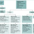
The priorities with respect to investigations will be dictated by the historical and physical findings. Serum electrolytes including sodium, calcium, blood sugar level and liver function tests should be done. A leucocytosis may indicate an underlying infective process. Chest x-ray and electrocardiogram are important to detect pericardial, plural or myocardial disease such as myocardial infarction. A thoracic computed tomography (CT) scan can be performed if mediastinal disease is suspected. Imaging of the abdomen is indicated if a subdiaphragmatic abscess is suspected. Endoscopy is indicated if oesophageal disease or gastric stasis is apparent clinically. The approach to intractable hiccups is outlined in Box 3.2.

Treatment

Treatments for intractable hiccups are many and varied and, due to the nature of the condition, virtually none have been subjected to randomised controlled therapeutic trial. There are at least 100 physical and pharmacological therapies described. Many of these cures are purely anecdotal and have included prayers to St Jude, the Patron Saint of Hopeless Causes; anger; sexual intercourse; pressing a finger firmly into each external auditory canal (to stimulate the auricular branch of vagus nerve); pharyngal stimulation by swallowing coarse-grain sugar; and laryngotracheal stimulation induced by a burst of coughing. Physical measures such as breath holding have been attributed to Hippocrates. There is some physiological basis for this as increasing the partial pressure of CO2 by rebreathing does reduce hiccup frequency. Other noxious afferent stimuli include strong traction on the tongue or phrenic nerve stimulation.

Primary therapy of any identified underlying cause is appropriate and, in most instances, will lead to rapid resolution of the hiccups. In refractory or idiopathic cases, there are no established guidelines regarding drug therapy for hiccups. A very extensive list of drugs has been described with variable success in hiccups. Notwithstanding, there are numerous reported failures in response to a number of agents and therapy is largely a matter of trial and error. Efficacy studies are largely single case reports and uncontrolled. There is only one randomised, controlled therapeutic trial in hiccups. This study found the GABAB receptor agonist, baclofen, to be effective in four patients. There is little objective evidence suggesting that one pharmacological agent is superior to another. Substantial numbers of favourable case reports support the efficacy of dopaminergic antagonists such as chlorpromazine, haloperidol and metoclopramide in intractable hiccups. Other drugs most frequently reported to be effective include anticonvulsants (phenytoin, sodium valproate and carbamazepine); benzodiazepines (clonazepam); calcium channel blockers (nifedipine and nimodipine), anaesthetic agents (ketamine and lignocaine), and amitriptyline (Box 3.3).

Finally, if pharmacotherapy is unsuccessful, disruption of phrenic nerve traffic by transcutaneous electrical stimulation of cervical phrenic nerve, or left phrenic nerve block may be necessary in severe, refractory cases.

Sore Mouth

The terminology describing oral sensations varies widely because there is no unanimity in relation to the terms applied. The terms used include glossodynia, glossopyrosis, stomatodynia and oral dysaesthesia. Soreness in the mouth can be due to objective alterations to the oral mucosa due to ulceration or inflammation. Very commonly, however, the symptoms are variable and there is no evidence of mucosal disease or any underlying pathology to account for symptoms. The latter situation is classified as the burning mouth syndrome. In contrast to conditions causing oral ulceration in which pain is aggravated by eating, the pain of burning mouth syndrome is often alleviated by eating.

Oral ulceration

Oral ulcers are so common as to be almost universally experienced at one time or another. Serious or recurrent and painful oral ulceration can be an indicator of underlying systemic or gastrointestinal disease.

Aphthous ulceration usually takes the form of multiple, small, painful, punched-out shallow ulcers and can affect the lips, buccal mucosa and tongue. These lesions can also be quite large, in which case they are usually single. Immunological responses seem to contribute to ulcer formation although trauma to the mucosa is also an important precipitant. The lesions are usually relatively minor and heal over a period of a few days up to 2 weeks. There is little controlled evidence that topical therapy is effective. However, topical steroid preparations, such as triamcinolone acetonide in dental base, or hydrocortisone hemisuccinate lozenges are often used with apparent benefit. Severe, persistent cases may respond to colchicine, dapsone or thalidomide.

Herpes simplex ulcers are located primarily on the hard palate, gingival and alveolar ridges. They begin as small clusters of vesicles, which transform into small punctate ulcers. Treatment is analgesics in immunocompetent patients. If ulcers are frequently recurrent, oral acyclovir can be used to reduce recurrence.

There are other causes of oral ulceration. Crohn’s disease and coeliac disease may present with aphthous ulceration. Erythema multiforme is preceded by vesicles. Pemphigus and pemphigoid are preceded by bullae. Behçet’s syndrome is a rare, multisystem disease in which recurrent oral aphthous ulceration occurs in conjunction with genital ulceration; the skin, eyes, joints and the gut can also be affected. Vitamin B deficiency, neutropenia, HIV infection and antimetabolites (e.g. methotrexate) can also cause frequent oral ulcers.

Burning mouth syndrome

Burning mouth syndrome (glossodynia or glossopyrosis) is characterised by the prolonged sensation of unexplained pain or burning inside the oral cavity, often accompanied by other symptoms, such as dryness, paraesthesia, altered sense of taste (cacogeusia or dysgeusia) or smell. It is at least two or three times more common in women and is most prevalent among middle-aged women. The oral mucosa looks normal, and neither local nor systemic disease is present.

Aetiology

Dentures are commonly implicated in glossodynia. Ill-fitting dentures are reported in 50% of patients with this symptom. It has been proposed that pressure is exerted by the dentures on oral tissues or that muscular tension influences sensory innervation and oral sensory perception. Monomers in the denture base material have also been linked to some cases.

Deficiency states cause macroscopic mucosal alterations, but glossodynia can precede these mucosal changes. Glossitis is a characteristic feature of advanced deficiency of vitamins such as folate and B12 (Box 3.4).

Glossodynia in diabetes mellitus can be caused by oral candidiasis; however, a form of oral sensory neuropathy is believed to be a factor in some cases in whom Candida infection is not a feature.

Several neurogenic causes of glossodynia, including trauma to the lingual nerve, are described. Glossopharyngeal neuralgia is characterised by paroxysmal pain, not only in the tongue, but also in the entire distribution of the nerve. Glossopharyngeal neuralgia can be elicited by stimulation of trigger zones in the tonsils, pharynx, tongue base and ear.

Gastro-oesophageal reflux is thought in some cases to be associated with glossodynia, but there is little objective evidence to suggest that reflux causes it. Furthermore, the observation that reflux is extremely common and glossodynia relatively uncommon argues against a strong association. Anecdotal experience suggests potent antireflux therapy is ineffective in the treatment of glossodynia.

Xerostomia can be associated with a burning sensation in the tongue. Salivary secretion tends to fall with age but xerostomia is commonly caused by drugs with anticholinergic effects and is a significant problem after head and neck radiotherapy.

Bad Breath

Halitosis (bad breath), a disagreeable odour detectable in exhaled breath, is common in healthy individuals, particularly after sleep (morning breath) or after consumption of certain foods. Halitosis is most commonly caused by the action of oral microflora on oral debris located in gingival crevices, within tongue coating and periodontal pockets. Plaque organisms, particularly Porphyromonas gingivalis, fusobacteria and other anaerobes, cause putrefaction and subsequent release of volatile chemicals, particularly sulfide compounds. Halitosis may be physiological or pathological. Physiological halitosis is usually transient and an identifying agent, usually a food or drug, is apparent. For example, high sulfur-containing foods such as garlic, onions, broccoli, radishes, leeks, or drugs such as isosorbide dinitrate, disulfiram and dimethyl sulfoxide can cause halitosis, as can dehydration or starvation. Halitosis may also be a manifestation of significant underlying local or systemic disease, a search for which may be necessary if an oral cause cannot be identified.

Patients frequently do not perceive their own oral malodour, which may be apparent to others. The senses of taste and olfaction are subject to the phenomenon of adaptation, making the subject insensitive over time to the offensive odour or taste. Also, chemical senses can be affected by many factors that themselves can be associated with halitosis, such as normal ageing, poor oral hygiene, xerostomia, craniofacial abnormalities, psychiatric disorders and neoplasm.

Pathogenesis

The majority of cases of halitosis arise from oral conditions (see Box 3.6). The odour is a result of sulfur-containing proteins and peptides being hydrolysed by gram-negative bacteria in the alkaline environment of the mouth. The resulting volatile sulfur-containing end-products include hydrogen sulfide and methyl mercaptan. Surprisingly, the usual compounds associated with putrefaction of biological tissues (ammonia, putrescine, indole, skatole and cadaverine) are not contributors to oral malodour. Under normal circumstances the odour of the oral cavity is not static and varies throughout the day and as a function of age, gender, hunger state and, perhaps, menstruation. It is affected by multiple factors, many of which are interdependent, including oral flora, salivary flow, pH, oral musculature and the presence of appropriate substrates. Conditions favouring production of putrid odours include low ambient oxygen concentration, a shift from gram-positive to gram-negative bacterial colonisation, reduced carbohydrates available as bacterial substrates, an alkaline oral pH and reduced salivary flow.

Any oral inflammatory condition, by its association with tissue degeneration and necrosis, will promote bacterial putrefaction. Gingivitis and periodontal disease favour bacterial putrefaction by increasing local growth factors and substrates available from the inflammatory process. These two conditions produce a very potent halitosis and, because of the increased incidence of these two conditions in the ageing population, they may account for the oral malodour ascribed to ageing.

The surface structure of the tongue provides a suitable environment for a biofilm serving as a repository for periodontal and other bacteria, including anaerobes. Deep fissures on the tongue dorsum, present in around 25% of the population, has been associated with higher mouth and tongue odour scores, although proof of higher bacterial counts in this population is lacking. The degree of tongue coating (containing a mix of desquamated cells, debris and bacteria) has some correlation with tongue bacterial counts and with halitosis. Nasal disease uncommonly causes halitosus (with a slight cheesy character); sinusitus or nasal polyps may or may not be present. Trimethylaminuria causes a fishy odour and is genetic (autosomal-recessive).

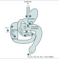
Approach to the patient

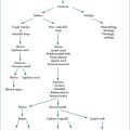
The approach to the patient is detailed in Box 3.7. The first step is to determine whether the patient does actually have halitosis, by wafting the expired air towards the examiner’s nose with the palm of the hand. It is important to be aware, however, that halitosis can be intermittent or reduced by recent antibiotic use. Therefore, if it is not apparent to the clinician, corroborative evidence should be sought from family members or close contacts. If the problem cannot be substantiated, the patient may have disordered chemoreception or the problem may be psychogenic (halitophobic). Neurosis can lead to compulsive use of mints, mouthwashes and oral deodorants in the absence of objective malodour. In such cases, halitosis is not physically based and counselling or psychiatric help may be needed.

The next step is to determine whether the odour is perceived predominantly during exhalation via the mouth or via the nose. Ask the patient to breathe out each time through the mouth or the nose and count to 20; place your nose 10 cm away. A stronger odour from nasally expired air indicates a lesion or disease of the nose, nasopharynx, sinuses or respiratory tract. Common causes include local tumours, rhinitis, sinusitis, or nasal foreign body (Box 3.6). Bronchiectasis can cause halitosis. Other respiratory disorders, such as asthma requiring systemic or inhaled corticosteroids, can result in oropharyngeal candidiasis that causes halitosis. Conditions in which oral candidiasis is common include cancer, immune deficiency states, diabetes mellitus and xerostomia. Tongue odour can be tested by using a plastic spoon to scrape the back of the tongue. Check whether the dentures smell foul.

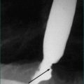
A more pronounced odour emanating from oral exhalation indicates an oral, oropharyngeal, hypopharyngeal or oesophagogastric source. Halitosis is rarely caused by gastrointestinal disorders. Gases from the upper gastrointestinal tract do not normally mix with those of the respiratory tract, but they can in the context of eructation, regurgitation or vomiting. Stagnation of food in a pharyngeal pouch or a dilated oesophagus in achalasia can cause regurgitation or eructation of partially fermented food and secretions.

If the expired air is equally offensive whether exhaled by nose or mouth, a systemic or metabolic cause is suspected.

Finally, a number of patients can present with halitosis and/or altered chemoreception for which no underlying cause can be found. Sophisticated breath analysis (gas chromatography and mass spectrometry) in some of these patients has identified higher than normal levels of volatile sulfur compounds. While some of these individuals have enzymatic or transport abnormalities, it is not known whether a subset of the remainder are heterozygous for sulfur-containing amino acidurias.

Treatment