Hereditary Motor-Sensory Neuropathies

Published on 25/03/2015 by admin

Filed under Pediatrics

Last modified 22/04/2025

Print this page

rate 1 star rate 2 star rate 3 star rate 4 star rate 5 star
Your rating: none, Average: 0 (0 votes)

This article have been viewed 1876 times

Chapter 605 Hereditary Motor-Sensory Neuropathies

The hereditary motor-sensory neuropathies (HMSNs) are a group of progressive diseases of peripheral nerves. Motor components generally dominate the clinical picture, but sensory and autonomic involvement is expressed later.

605.1 Peroneal Muscular Atrophy (Charcot-Marie-Tooth Disease; HMSN Type I)

Harvey B. Sarnat

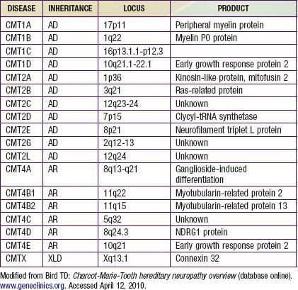
Charcot-Marie-Tooth disease is the most common genetically determined neuropathy and has an overall prevalence of 3.8/100,000. It is transmitted as an autosomal dominant trait with 83% expressivity; the 17p11.2 locus is the site of the abnormal gene. Autosomal recessive transmission also is described but is rarer. The gene product is peripheral myelin protein 22 (PMP22). A much rarer X-linked HMSN type I results from a defect at the Xq13.l locus, causing mutations in the gap junction protein connexin-32. Other forms have been reported (Table 605-1).

Clinical Manifestations

Most patients are asymptomatic until late childhood or early adolescence, but young children sometimes manifest gait disturbance as early as the 2nd year of life. The peroneal and tibial nerves are the earliest and most severely affected. Children with the disorder are often described as being clumsy, falling easily, or tripping over their own feet. The onset of symptoms may be delayed until after the 5th decade.

Muscles of the anterior compartment of the lower legs become wasted, and the legs have a characteristic stork-like contour. The muscular atrophy is accompanied by progressive weakness of dorsiflexion of the ankle and eventual footdrop. The process is bilateral but may be slightly asymmetric. Pes cavus deformities invariably develop due to denervation of intrinsic foot muscles, further destabilizing the gait. Atrophy of muscles of the forearms and hands is usually not as severe as that of the lower extremities, but in advanced cases contractures of the wrists and fingers produce a claw hand. Proximal muscle weakness is a late manifestation and is usually mild. Axial muscles are not involved.

The disease is slowly progressive throughout life, but patients occasionally show accelerated deterioration of function over a few years. Most patients remain ambulatory and have normal longevity, although orthotic appliances are required to stabilize the ankles.

Sensory involvement mainly affects large myelinated nerve fibers that convey proprioceptive information and vibratory sense, but the threshold for pain and temperature can also increase. Some children complain of tingling or burning sensations of the feet, but pain is rare. Because the muscle mass is reduced, the nerves are more vulnerable to trauma or compression. Autonomic manifestations may be expressed as poor vasomotor control with blotching or pallor of the skin of the feet and inappropriately cold feet.

Nerves often become palpably enlarged. Tendon stretch reflexes are lost distally. Cranial nerves are not affected. Sphincter control remains well preserved. Autonomic neuropathy does not affect the heart, gastrointestinal tract, or bladder. Intelligence is normal. A unique point mutation in PMP22 causes progressive auditory nerve deafness in addition, but this is usually later in onset than the peripheral neuropathy.

Davidenkow syndrome is a variant of HMSN type I with a scapuloperoneal distribution.
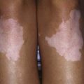
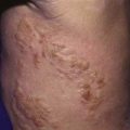
605.6 Fabry Disease

Harvey B. Sarnat

(See Chapter 80.4.)

Fabry disease, a rare X-linked recessive trait, results in storage of ceramide trihexose due to deficiency of the enzyme ceramide trihexosidase, which cleaves the terminal galactose from ceramide trihexose (ceramide-glucose-galactose-galactose), resulting in tissue accumulation of this trihexose lipid in central nervous system (CNS) neurons, Schwann cells and perineurial cells, ganglion cells of the myenteric plexus, skin, kidneys, blood vessel endothelial and smooth muscle cells, heart, sweat glands, cornea, and bone marrow. It results from a missense mutation disrupting the crystallographic structure of α-galactosidase A.

Treatment

(See Chapter 80.4 for specific therapy of Fabry disease, including enzyme replacement.)

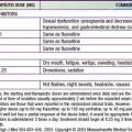
Medical therapy of painful neuropathies includes management of the initiating disease and therapy directed to the neuropathic pain independent of etiology. Pain may be burning or associated with paresthesias, hyperalgesia (abnormal response to noxious stimuli), or allodynia (induced by non-noxious stimuli; Chapter 71). Neuropathic pain is often successfully managed by tricyclic antidepressants; selective serotonin reuptake inhibitors are less effective. Anticonvulsants (carbamazepine, phenytoin, gabapentin, lamotrigine) are effective, as are narcotic and non-narcotic analgesics. Enzyme replacement therapy has improved the short and long term prognosis.

605.7 Giant Axonal Neuropathy

Harvey B. Sarnat

Giant axonal neuropathy is a rare autosomal recessive disease with onset in early childhood. It is a progressive mixed peripheral neuropathy and degeneration of central white matter, similar to the leukodystrophies. Ataxia and nystagmus are accompanied by signs of progressive peripheral neuropathy. A large majority of affected children have frizzy hair, which microscopically shows variation in diameter of the shaft and twisting, similar to that in Menkes disease; hence, microscopic examination of a few scalp hairs provides a simple screening tool in suspected cases. Focal axonal enlargements are seen in both the peripheral nervous system and the CNS, but the myelin sheath is intact. The disease is a general proliferation of intermediate filaments, including neurofilaments in axons, glial filaments (i.e., Rosenthal fibers) in brain, cytokeratin in hair, and vimentin in Schwann cells and fibroblasts.

Nonsense and missense mutations or deletions occur in the GAN gene, with allelic heterogeneity, at 16q24. These mutations are responsible for defective synthesis of the protein gygaxonin, a member of the cytoskeletal BTB/kelch superfamily, crucial to linkage between intermediate proteins and the cell membrane. MRI shows white matter lesions of the brain similar to leukodystrophies, and magnetic resonance spectroscopy (MRS) demonstrates increased ratios of choline : creatine and myoinositol : creatine, with a normally preserved ratio of N-acetyl aspartate : creatine, indicating demyelination and glial proliferation without axonal loss. Gygaxonin is expressed in a wide variety of neuronal cell types and is localized to the Golgi apparatus and endoplasmic reticulum.

The diagnosis is established by microscopy of scalp hair and by MRI and MRS of the brain; it is confirmed by sural nerve biopsy and/or by genetic studies, if available, of the GAN gene.

605.8 Congenital Hypomyelinating Neuropathy

Harvey B. Sarnat

Congenital hypomyelinating neuropathy is a lack of normal myelination of motor and sensory peripheral nerves but not of CNS white matter. It is not a degeneration or loss of previously formed myelin, thus differentiating it from a leukodystrophy. Schwann cells are preserved, and axons are normal. Cases in siblings suggest autosomal recessive inheritance. Mutations in the MTMR2, PMP22, EGR2, and MPZ genes have been demonstrated in various children with this neuropathy; hence, it is a syndrome rather than a single disease.

The condition is present from birth; hypotonia and developmental delay are the hallmark clinical findings. Many patients present clinically as having congenital insensitivity to pain. Cranial nerves are inconsistently involved, and respiratory distress and dysphagia are rare complications. Tendon reflexes are absent. Arthrogryposis is present at birth in at least half the cases. It is uncertain whether the condition is progressive; myelination of nerves proceeds at a slow rate and remains incomplete. Motor and sensory nerve conduction velocities are slow. The diagnosis is confirmed by sural nerve biopsy, which shows lack of myelination of large and small fibers and sometimes interstitial hypertrophic reactive changes. It is useful in distinguishing this condition from several hereditary demyelinating neuropathies without resorting to batteries of expensive genetic markers. Muscle biopsy can show mild neurogenic atrophy but not the characteristic alterations of spinal muscular atrophy. No inflammation is demonstrated in muscle or nerve. Treatment is supportive.

Bibliography

Bruno C, Bertini E, Federico A, et al. Clinical and molecular findings in patients with giant axonal neuropathy (GAN). Neurology. 2004;62:13-16.

Chance PF. Inherited focal, episodic neuropathies: hereditary neuropathy with liability to pressure palsies and hereditary neuralgic amyotrophy. Neuromolec Med. 2006;8:159-174.

Chance PF, Alderson MK, Leppig KA, et al. DNA deletion associated with hereditary neuropathy with liability to pressure palsies. Cell. 1993;72:143-151.

Evgrafov OV, Mersiyanova I, Irobi J, et al. Mutant small heat-shock protein 27 causes axonal Charcot-Marie-Tooth disease and distal hereditary motor neuropathy. Nat Genet. 2004;36:602-606.

Gordon N. Giant axonal neuropathy. Dev Med Child Neurol. 2004;46:717-719.

Houlden H, Blake J, Reilly MM. Hereditary sensory neuropathies. Curr Opin Neurol. 2004;17:569-577.

Kochanski A, Drac H, Kabzinska D, et al. A novel MPZ gene mutation in congenital neuropathy with hypomyelination. Neurology. 2004;62:2122-2123.

Kovach MJ, Lin JP, Boyadjiev S, et al. A unique point mutation in the PMP22 gene is associated with Charcot-Marie-Tooth disease and deafness. Am J Hum Genet. 1999;64:1580-1593.

Landouré G, Zdebik AA, Martinez TL, et al. Mutations in TRPV4 cause Charcot-Marie-tooth disease type 2C. Nat Genet. 2010;42(2):170-174.

Lupski JR, Reid JG, Gonzaga-Jauregui C, et al. Whole-genome sequencing in a patient with Charcot-Marine-tooth neuropathy. N Engl J Med. 2010;362(13):1181-1190.

Mendell JR, Sahenk Z. Painful sensory neuropathy. N Engl J Med. 2003;348:1243-1255.

Pleasure D. New treatments for denervating diseases. J Child Neurol. 2005;20:258-262.

Ries M, Gupta S, Moore DF, et al. Pediatric Fabry disease. Pediatrics. 2005;115:344-355.

Seithi PK, Khandelwal D, Sethi NK, et al. Hereditary infantile motor axonal neuropathy mimicking SMA: a case report. J Pediatr Neurol. 2009;7:175-179.

Shy ME. Charcot-Marie-Tooth disease: An update. Curr Opin Neurol. 2004;17:579-585.