Heart Failure

Published on 25/03/2015 by admin

Filed under Pediatrics

Last modified 22/04/2025

Print this page

rate 1 star rate 2 star rate 3 star rate 4 star rate 5 star
Your rating: none, Average: 0 (0 votes)

This article have been viewed 1430 times

Chapter 436 Heart Failure

Heart failure occurs when the heart cannot deliver adequate cardiac output to meet the metabolic needs of the body. In the early stages of heart failure, various compensatory mechanisms are evoked to maintain normal metabolic function. When these mechanisms become ineffective, increasingly severe clinical manifestations result (Chapter 64).

Pathophysiology

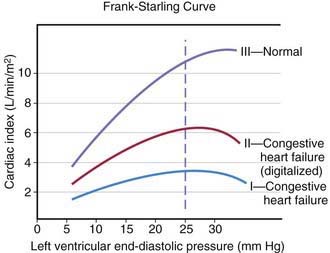
The heart can be viewed as a pump with an output proportional to its filling volume and inversely proportional to the resistance against which it pumps. As ventricular end-diastolic volume increases, a healthy heart increases cardiac output until a maximum is reached and cardiac output can no longer be augmented (the Frank-Starling principle; Fig. 436-1). The increased stroke volume obtained in this manner is due to stretching of myocardial fibers, but it also results in increased wall tension, which elevates myocardial oxygen consumption. Hearts working under various types of stress function along different Frank-Starling curves. Cardiac muscle with compromised intrinsic contractility requires a greater degree of dilatation to produce increased stroke volume and does not achieve the same maximal cardiac output as normal myocardium does. If a cardiac chamber is already dilated because of a lesion causing increased preload (e.g., a left-to-right shunt or valvular insufficiency), there is little room for further dilatation as a means of augmenting cardiac output. The presence of lesions that result in increased afterload to the ventricle (aortic or pulmonic stenosis, coarctation of the aorta) decreases cardiac performance, thereby resulting in a depressed Frank-Starling relationship. The ability of an immature heart to increase cardiac output in response to increased preload is less than that of a mature heart. Thus, premature infants are more compromised by a left-to-right shunt than full-term infants are.

Systemic oxygen transport is calculated as the product of cardiac output and systemic oxygen content. Cardiac output can be calculated as the product of heart rate and stroke volume. The primary determinants of stroke volume are the afterload (pressure work), preload (volume work), and contractility (intrinsic myocardial function). Abnormalities in heart rate can also compromise cardiac output; for example, tachyarrhythmias shorten the diastolic time interval for ventricular filling. Alterations in the oxygen-carrying capacity of blood (e.g., anemia or hypoxemia) also lead to a decrease in systemic oxygen transport and, if compensatory mechanisms are inadequate, can result in decreased delivery of substrate to tissues.

In some cases of heart failure, cardiac output is normal or increased, yet because of decreased systemic oxygen content (secondary to anemia) or increased oxygen demands (secondary to hyperventilation, hyperthyroidism, or hypermetabolism), an inadequate amount of oxygen is delivered to meet the body’s needs. This condition, high-output failure, results in the development of signs and symptoms of heart failure when there is no basic abnormality in myocardial function and cardiac output is greater than normal. It is also seen with large systemic arteriovenous fistulas. These conditions reduce peripheral vascular resistance and cardiac afterload and increase myocardial contractility. Heart “failure” results when the demand for cardiac output exceeds the ability of the heart to respond. Chronic severe high-output failure may eventually result in a decrease in myocardial performance as the metabolic requirements of the myocardium are not met.

There are multiple systemic compensatory mechanisms used by the body to adapt to chronic heart failure. Some are mediated at the molecular/cellular level, such as upregulation or downregulation of various metabolic pathway components leading to changes in efficiency of oxygen and other substrate utilization. Others are mediated by neurohormones such as the renin-angiotensin system and the sympathoadrenal axis. One of the principal mechanisms for increasing cardiac output is an increase in sympathetic tone secondary to increased secretion of circulating epinephrine by the adrenals and increased release of norepinephrine at the neuromuscular junction. The initial beneficial effects of sympathetic stimulation include an increase in heart rate and myocardial contractility, mediated by these hormones’ action on cardiac β-adrenergic receptors, increasing cardiac output. These hormones also cause vasoconstriction, mediated by their action on peripheral arterial α-adrenergic receptors. Some vascular beds may constrict more readily than others, so that blood flow is redistributed from the cutaneous, visceral, and renal beds to the heart and brain. Whereas these acute effects are beneficial, chronically increased sympathetic stimulation can have deleterious effects, including hypermetabolism, increased afterload, arrhythmogenesis, and increased myocardial oxygen requirements. Peripheral vasoconstriction can result in decreased renal, hepatic, and gastrointestinal tract function. Chronic exposure to circulating catecholamines leads to a decrease in the number of cardiac β-adrenergic receptors (downregulation) and also causes direct myocardial cell damage. Thus, therapeutic agents for heart failure are directed at restoring balance to these neuroendocrine systems. Single nucleotide differences (known as polymorphisms [SNPs]) in the genes encoding proteins involved in sympathetic signaling can alter a patient’s response to medical therapy and may predict risk of worsening heart failure, hospitalization, or death. These pharmacogenomic studies may allow us to tailor our therapies to the individual patient, based on their genetic makeup.

Clinical Manifestations

The clinical manifestations of heart failure depend on the degree of the child’s cardiac reserve. A critically ill infant or child who has exhausted the compensatory mechanisms to the point that cardiac output is no longer sufficient to meet the basal metabolic needs of the body will be symptomatic at rest. Other patients may be comfortable when quiet but are incapable of increasing cardiac output in response to even mild activity without experiencing significant symptoms. Conversely, it may take rather vigorous exercise to compromise cardiac function in children who have less severe heart disease. A thorough history is extremely important in making the diagnosis of heart failure and in evaluating the possible causes. Parents who observe their child on a daily basis may not recognize subtle changes that have occurred over the course of days or weeks. Gradually worsening perfusion or increasing respiratory effort may not be recognized as an abnormal finding. Edema may be passed off as normal weight gain, and exercise intolerance as lack of interest in an activity. The history of a young infant should also focus on feeding (Chapter 416). An infant with heart failure often takes less volume per feeding, becomes dyspneic while sucking, and may perspire profusely. Eliciting a history of fatigue in an older child requires detailed questions about activity level and its course over several months.

In children, the signs and symptoms of heart failure may be similar to those in adults and include fatigue, effort intolerance, anorexia, dyspnea, and cough. Many children, however, especially adolescents, may have primarily abdominal symptoms (abdominal pain, nausea, anorexia) and a surprising lack of respiratory complaints. Attention to the cardiovascular system may come only after an abdominal roentgenogram unexpectedly catches the lower end of an enlarged heart. The elevation in systemic venous pressure may be gauged by clinical assessment of jugular venous pressure and liver enlargement. Orthopnea and basilar rales are variably present; edema is usually discernible in dependent portions of the body, or anasarca may be present. Cardiomegaly is invariably noted. A gallop rhythm is common; when ventricular dilatation is advanced, the holosystolic murmur of mitral or tricuspid valve regurgitation may be heard.

In infants, heart failure may be difficult to distinguish from other causes of respiratory distress. Prominent manifestations include tachypnea, feeding difficulties, poor weight gain, excessive perspiration, irritability, weak cry, and noisy, labored respirations with intercostal and subcostal retractions, as well as flaring of the alae nasi. The signs of cardiac-induced pulmonary congestion may be indistinguishable from those of bronchiolitis; wheezing is often a more prominent finding in young infants with heart failure than rales. Pneumonitis with or without atelectasis is common, especially in the right middle and lower lobes, due to bronchial compression by the enlarged heart. Hepatomegaly usually occurs, and cardiomegaly is invariably present. In spite of pronounced tachycardia, a gallop rhythm can frequently be recognized. The other auscultatory signs are those produced by the underlying cardiac lesion. Clinical assessment of jugular venous pressure in infants may be difficult because of the shortness of the neck and the difficulty of observing a relaxed state; palpation of an enlarged liver is a more reliable sign. Edema may be generalized and usually involves the eyelids as well as the sacrum and less often the legs and feet. The differential diagnosis is age dependent (Table 436-1).

Diagnosis

X-rays of the chest show cardiac enlargement. Pulmonary vascularity is variable and depends on the cause of the heart failure. Infants and children with large left-to-right shunts have exaggeration of the pulmonary arterial vessels to the periphery of the lung fields, whereas patients with cardiomyopathy may have a relatively normal pulmonary vascular bed early in the course of disease. Fluffy perihilar pulmonary markings suggestive of venous congestion and acute pulmonary edema are seen only with more severe degrees of heart failure. Cardiac enlargement is often noted as an unexpected finding on a chest roentgenogram performed to evaluate for a possible pulmonary infection, bronchiolitis, or asthma.

Chamber hypertrophy noted by electrocardiography may be helpful in assessing the cause of heart failure but does not establish the diagnosis. In cardiomyopathies, left or right ventricular ischemic changes may correlate with other noninvasive parameters of ventricular function. Low-voltage QRS morphologic characteristics with ST-T wave abnormalities may also suggest myocardial inflammatory disease but can be seen with pericarditis as well. The electrocardiogram is the best tool for evaluating rhythm disorders as a potential cause of heart failure, especially tachyarrhythmias.

Echocardiography is the standard technique for assessing ventricular function. The most commonly used parameter in children is fractional shortening (a single dimensional variable), determined as the difference between end-systolic and end-diastolic diameter divided by end-diastolic diameter. Normal fractional shortening is between approximately 28% and 42%. In adults, the most commonly used parameter is ejection fraction (which uses two-dimensional data to calculate a three-dimensional volume) and the normal range is 55-65%. In children with right ventricular enlargement or other cardiac pathology resulting in flattening of the interventricular septum, ejection fraction is used since fractional shortening measured in the standard echocardiographic short-axis view will not be accurate. Doppler studies can also be used to estimate cardiac output. Doppler tissue imaging is a new technique which can assess not only cardiac function, but wall motion abnormalities that can interfere with normal synchronous cardiac contraction. Magnetic resonance angiography (MRA) is also very useful in quantifying left and right ventricular function, volume and mass. If valvar regurgitation is present, MRA can quantify the regurgitant fraction.

Arterial oxygen levels may be decreased when ventilation-perfusion inequalities occur secondary to pulmonary edema. When heart failure is severe, respiratory or metabolic acidosis, or both, may be present. Infants with heart failure often display hyponatremia as a result of renal water retention. Chronic diuretic treatment can decrease serum sodium levels further. Serum B-type natriuretic peptide (BNP), a cardiac neurohormone released in response to increased ventricular wall tension, is elevated in adult patients with congestive heart failure. In children, BNP may be elevated in patients with heart failure due to systolic dysfunction (cardiomyopathy) as well as in children with volume overload (left-to-right shunts such as ventricular septal defect).

Treatment

The underlying cause of cardiac failure must be removed or alleviated if possible. If the cause is a congenital cardiac anomaly amenable to surgery, medical treatment of the heart failure is indicated to prepare the patient for surgery. With today’s excellent outcomes of primary surgical repair of congenital heart defects, even in the neonatal period, few children require aggressive heart failure management to “grow big enough for surgery.” In contrast, if the cause of heart failure is cardiomyopathy, medical management provides temporary relief from symptoms and may allow the patient to recover if the insult is reversible (e.g., myocarditis). If the lesion is not reversible, heart failure management usually allows the child to return to normal activities for some period and delay, sometimes for months or years, the need for heart transplantation.

General Measures

Strict bed rest is rarely necessary except in extreme cases, but it is important that the child be allowed to rest during the day as needed and sleep adequately at night. Some older patients feel better sleeping in a semi-upright position, using several pillows (orthopnea). For infants with heart failure, an infant chair may be advisable. After patients begin to respond to treatment, restrictions on activities can often be modified within the context of the specific diagnosis and the patient’s ability. Competitive and strenuous sports activities are usually contraindicated. Formal cardiopulmonary exercise testing can be used to assess the patient’s ability to perform exercise in a controlled environment and is useful for recommending rational exercise restrictions. For patients with pulmonary edema, positive pressure ventilation may be required along with other drug therapy. For those in low-output heart failure, positive pressure ventilation can significantly reduce total body oxygen consumption by eliminating the work of breathing, and help to reverse metabolic acidosis. β-Adrenergic agonists such as dopamine, dobutamine, and epinephrine are usually used in combination with phosphodiesterase inhibitors such as milrinone. If the blood pressure will allow, afterload-reducing agents (nitroprusside, angiotensin-converting enzyme [ACE] inhibitors or angiotensin receptor blockers [ARBs]) may be beneficial. These agents are usually initiated in an intensive care setting, with proper invasive monitoring of central venous and arterial blood pressure.

Diet

Infants with heart failure usually fail to thrive because of a combination of increased metabolic demands and decreased caloric intake. Increasing daily calories is an important aspect of their management. Increasing the number of calories per ounce of infant formula (or supplementing breast-feeding) may be beneficial. Many infants do not tolerate an increase beyond 24 calories/oz because of diarrhea or because these formulas provide too large a solute load for compromised kidneys.

Severely ill infants and children may lack sufficient strength for effective sucking because of extreme fatigue, rapid respirations, and generalized weakness. In these circumstances, nasogastric feedings may be helpful. In many patients with cardiac enlargement, gastroesophageal reflux is a major problem. The use of continuous drip nasogastric feedings at night, administered by pump, may improve caloric intake while decreasing problems with reflux. Occasionally, especially in infants with heart failure due to complex congenital heart disease, medical or surgical intervention to correct reflux is necessary (Nissen fundoplication). Continued malnutrition may be an important factor in the decision to undertake earlier surgical intervention in patients who have an operable congenital heart lesion or for listing for transplantation in patients with cardiomyopathy.

The use of low sodium formulas in the routine management of infants with heart failure is not recommended because these preparations are often poorly tolerated and may exacerbate diuretic-induced hyponatremia. Human breast milk is the ideal low sodium nutritional source. The use of more potent diuretic agents allows more palatable standard formulas to be used for nutrition while controlling salt and water balance by chronic diuretic administration. Most older children can be managed with “no added salt” diets and abstinence from foods containing large amounts of sodium. A strict, extremely low sodium diet is rarely required, and rarely adhered to.

Diuretics

These agents interfere with reabsorption of water and sodium by the kidneys, which results in a reduction in circulating blood volume and thereby reduces pulmonary fluid overload and ventricular filling pressure. Diuretics are usually the first mode of therapy initiated in patients with congestive heart failure.

Furosemide is the most commonly used diuretic in pediatric patients with heart failure. It inhibits the reabsorption of sodium and chloride in the distal tubules and the loop of Henle. Patients requiring acute diuresis should be given intravenous or intramuscular furosemide at an initial dose of 1-2 mg/kg, which usually results in rapid diuresis and prompt improvement in clinical status, particularly if symptoms of pulmonary congestion are present. Chronic furosemide therapy is then prescribed at a dose of 1-4 mg/kg/24 hr given between 1 and 4 times a day. Careful monitoring of electrolytes is necessary with long-term furosemide therapy because of the potential for significant loss of potassium. Potassium chloride supplementation is usually required unless the potassium-sparing diuretic spironolactone is given concomitantly. Chronic administration of furosemide may cause contraction of the extracellular fluid compartment and result in “contraction alkalosis” (Chapter 52.7). Diuretic-induced hyponatremia may become difficult to manage in patients with severe heart failure.

Spironolactone is an inhibitor of aldosterone and enhances potassium retention, often eliminating the need for oral potassium supplementation, which is frequently poorly tolerated. This drug is usually given orally in 2 divided doses of 2 mg/kg/24 hr. Combinations of spironolactone and chlorothiazide are sometimes used for convenience. Adults with heart failure have improved survival when an aldosterone inhibitor is included in the diuretic regimen.

Chlorothiazide is also used for diuresis in children with heart failure. It is less immediate in action and less potent than furosemide, and it affects the reabsorption of electrolytes in the renal tubules only. The usual dose is 10-40 mg/kg/24 hr in 2 divided doses. Potassium supplementation is often required if this agent is used alone.

Afterload Reducers, Including Angiotensin-Converting Enzyme Inhibitors (ACEIs) and Angiotensin II Receptor Blockers (ARBs)

These 2 groups of drugs reduce ventricular afterload by decreasing peripheral vascular resistance and thereby improving myocardial performance. Some of these agents also decrease systemic venous tone, which significantly reduces preload. Afterload reducers are especially useful in children with heart failure secondary to cardiomyopathy and in patients with severe mitral or aortic insufficiency. They may also be effective in patients with heart failure caused by left-to-right shunts. They are not generally used in the presence of stenotic lesions of the left ventricular outflow tract because of concern over coronary perfusion. ACEIs and ARBs may have additional beneficial effects on cardiac remodeling independent of their influence on afterload by directly influencing cardiac intracellular signaling pathways. In adult patients with dilated cardiomyopathy, the addition of an ACEI to standard medical therapy reduces both morbidity and mortality. Afterload-reducing agents are most often used in conjunction with other anticongestive drugs such as diuretics and digoxin.

Intravenously administered agents such as nitroprusside should be administered only in an intensive care setting and for as short a time as possible. Nitroprusside’s short intravenous half-life makes it ideal for titrating the dose in critically ill patients. Peripheral arterial vasodilation and afterload reduction are the major effects, but venodilation causing a decrease in venous return to the heart may also be beneficial. Blood pressure must be continuously monitored because sudden hypotension can occur. Nitroprusside is therefore contraindicated in patients with pre-existing hypotension. As the drug is metabolized, small amounts of circulating cyanide are produced and detoxified in the liver to thiocyanate, which is excreted in urine. When high doses of nitroprusside are administered for several days, toxic symptoms related to thiocyanate poisoning may occur (fatigue, nausea, disorientation, acidosis, and muscular spasm). If nitroprusside use is prolonged, blood thiocyanate levels should be monitored. Phosphodiesterase inhibitors (see later) are also excellent, although somewhat less potent afterload-reducing agents, without the toxicity of nitroprusside.

The orally active ACEIs captopril and enalapril produce arterial dilatation by blocking the production of angiotensin II, thereby resulting in significant afterload reduction. Venodilation and consequent preload reduction have also been reported. In addition, these agents interferes with aldosterone production and therefore also help control salt and water retention. ACEIs have additional beneficial effects on cardiac structure and function that may be independent of their effect on afterload. The oral dose for captopril is 0.3-6 mg/kg/24 hr given in 3 divided doses; for enalapril 0.05-0.5 mg/kg/24 hr given in 1 or 2 daily doses. Adverse reactions to ACEIs include hypotension and its sequelae (weakness, dizziness, syncope) and hyperkalemia. A maculopapular pruritic rash is encountered in a small number of patients, but the drug may be continued because the rash often disappears spontaneously with time. Neutropenia, renal toxicity, and chronic cough also occur.

ARBs have recently been introduced into management protocols for adults with heart failure and have seen extensive use in pediatric patients with renal disease. However, data on these agents in children with heart failure are limited.

Digitalis Glycosides

Digoxin, once the mainstay of heart failure management in both children and adults, is currently used less frequently, as a result of the introduction of newer therapies and the recognition of its potential toxicities. Many cardiologists will use digitalis as an adjunct to ACEIs and diuretics in patients with symptomatic heart failure, whereas others have moved away from its use altogether. Despite multiple clinical studies, predominantly in adults, the controversy over digitalis remains.

Digoxin is the digitalis glycoside used most often in pediatric patients. It has a half-life of 36 hr and it is absorbed well by the gastrointestinal tract (60-85%), even in infants. An initial effect can be seen as early as 30 min after administration, and the peak effect for oral digoxin occurs at ≈2-6 hr. When the drug is administered intravenously, the initial effect is seen in 15-30 min, and the peak effect occurs at 1-4 hr. The kidney eliminates digoxin, so dosing must be adjusted according to the patient’s renal function. The half-life of digoxin may be up to 6 days in patients with anuria because slower hepatic excretion pathways are used in these patients.

Rapid digitalization of infants and children in heart failure may be carried out intravenously. The dose depends on the patient’s age (Table 436-2). The recommended digitalization schedule is to give half the total digitalizing dose immediately and the succeeding 2 one-quarter doses at 12-hr intervals later. The electrocardiogram must be closely monitored and rhythm strips obtained before each of the 3 digitalizing doses. Digoxin should be discontinued if a new rhythm disturbance is noted. Prolongation of the P-R interval is not necessarily an indication to withhold digitalis, but a delay in administering the next dose or a reduction in the dosage should be considered, depending on the patient’s clinical status. Minor ST segment or T-wave changes are commonly noted with digitalis administration and should not affect the digitalization regimen. Baseline serum electrolyte levels should be measured before and after digitalization. Hypokalemia and hypercalcemia exacerbate digitalis toxicity. Because hypokalemia is relatively common in patients receiving diuretics, potassium levels should be monitored closely in those receiving a potassium-wasting diuretic in combination with digitalis. In patients with active myocarditis, some cardiologists recommend avoiding digitalis altogether and if used, maintenance digitalis should be started at half the normal dose without digitalization due to the increased risk of arrhythmia in these patients.

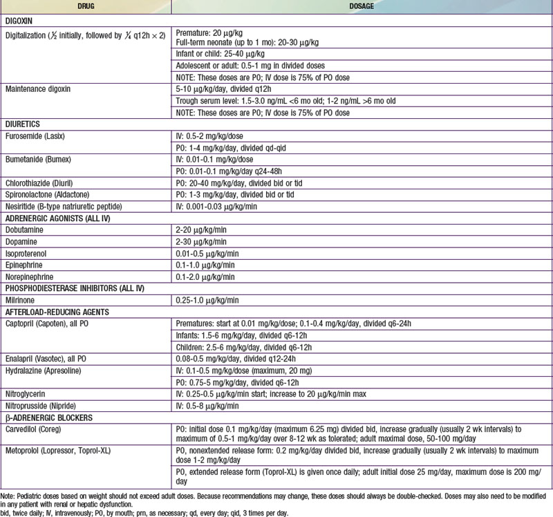
Table 436-2 DOSAGE OF DRUGS COMMONLY USED FOR THE TREATMENT OF CONGESTIVE HEART FAILURE

image

Maintenance digitalis therapy is started ≈12 hr after full digitalization. The daily dosage, one quarter of the total digitalizing dose, is divided in 2 and given at 12-hr intervals. The oral maintenance dose is usually 20-25% higher than when digoxin is used parenterally (see Table 436-2). The normal daily dose of digoxin for older children (>5 yr of age) calculated by body weight should not exceed the usual adult dose of 0.125-0.5 mg/24 hr.

Patients who are not critically ill may be given digitalis initially by the oral route, and in most instances digitalization is completed within 24 hr. When slow digitalization is desirable, for example, in the immediate postoperative period, initiation of a maintenance digoxin schedule without a previous loading dose achieves full digitalization in 7-10 days.

Measurement of serum digoxin levels is useful under several circumstances: (1) when an unknown amount of digoxin has been administered or ingested accidentally, (2) when renal function is impaired or if drug interactions are possible, (3) when questions regarding compliance are raised, and (4) when a toxic response is suspected. Therapeutic trough blood levels are usually 2-4 ng/mL in infants and 1-2 ng/mL in older children. Exceeding these levels does not generally add significantly to the management of heart failure and only increases the risk of toxicity. In suspected toxicity, elevated serum digoxin levels are not in themselves diagnostic of toxicity but must be interpreted as an adjunct to other clinical and electrocardiographic findings (rhythm and conduction disturbances). Hypokalemia, hypomagnesemia, hypercalcemia, cardiac inflammation secondary to myocarditis, and prematurity may all potentiate digitalis toxicity. A cardiac arrhythmia that develops in a child who is taking digitalis may also be related to the primary cardiac disease rather than the drug, however, any arrhythmia occurring after the institution of digitalis therapy must be considered to be drug related until proven otherwise. There are many drugs that interact with digoxin and may increase levels or risk of toxicity, so care should be taken when a patient on digoxin is being considered for additional pharmacologic therapy of any kind.

α- and β-Adrenergic Agonists

These drugs are usually administered in an intensive care setting, where the dose can be carefully titrated to hemodynamic response. Continuous determinations of arterial blood pressure and heart rate are performed; measuring serial mixed venous oxygen saturations or cardiac output directly with a pulmonary thermodilution (Swan-Ganz) catheter may be helpful in assessing drug efficacy. Though extremely efficacious in the acute intensive care setting, long-term administration of adrenergic agonists has been shown to increase morbidity and mortality in adults with heart failure and is usually avoided unless the patient is totally dependent on these agents.

Dopamine is a predominantly β-adrenergic receptor agonist, but it has α-adrenergic effects at higher doses. Dopamine has less chronotropic and arrhythmogenic effect than the pure β-agonist isoproterenol does. In addition, it results in selective renal vasodilation because of its interaction with renal dopamine receptors, which is particularly useful in patients with the compromised kidney function that is often associated with low cardiac output, although some recent studies in adults question the efficacy of dopamine for this indication. At a dose of 2-10 µg/kg/min, dopamine results in increased contractility with little peripheral vasoconstrictive effect. If the dose is increased beyond 15 µg/kg/min, however, its peripheral α-adrenergic effects may result in vasoconstriction. Fenoldopam is a dopamine DA1 receptor agonist and is used at a low dose (0.03 µg/kg/min) to increase renal blood flow and urine output. It can cause hypotension, so blood pressure should be carefully monitored.

Dobutamine, a derivative of dopamine, is also useful in treating low cardiac output. It has direct inotropic effects and causes a moderate reduction in peripheral vascular resistance. Dobutamine can be used alone or as an adjunct to dopamine therapy to avoid the vasoconstrictive effects of higher-dose dopamine. Dobutamine is also less likely to cause cardiac rhythm disturbances than isoproterenol is. The usual dose is 2-20 µg/kg/min.

Isoproterenol is a pure β-adrenergic agonist that has a marked chronotropic effect; it is most effective in patients with slow heart rates and is less commonly used in patients with heart failure and normal or increased heart rates, due to the increased risk of arrhythmias.

Epinephrine is a mixed α- and β-adrenergic receptor agonist that is usually reserved for patients with cardiogenic shock and low arterial blood pressure. Although epinephrine can raise blood pressure effectively, it also increases systemic vascular resistance and therefore increases the afterload against which the heart has to work, and is associated with an increased risk of arrhythmia.

436.1 Cardiogenic Shock

Daniel Bernstein

Cardiogenic shock (Chapter 64) may occur as a complication of (1) severe cardiac dysfunction before or after cardiac surgery, (2) septicemia, (3) severe burns, (4) anaphylaxis, (5) cardiomyopathy, (6) myocarditis, (7) myocardial infarction or stunning, and (8) acute central nervous system disorders. It is characterized by low cardiac output and hypotension and therefore results in inadequate tissue perfusion.

Treatment is aimed at reinstitution of adequate cardiac output to prevent the untoward effects of prolonged ischemia on vital organs, as well as management of the underlying cause. Under normal physiologic conditions, cardiac output is increased as a result of sympathetic stimulation, which increases both contractility and heart rate. If contractility is depressed, cardiac output can be improved by increasing heart rate, increasing ventricular filling pressure (preload) through the Frank-Starling mechanism, or by decreasing systemic vascular resistance (afterload). Optimal filling pressure is variable and depends on a number of extracardiac factors, including ventilatory support and intra-abdominal pressure. The increased pressure necessary to fill a relatively noncompliant ventricle should also be considered, particularly after open heart surgery, or in patients with restrictive or hypertrophic cardiomyopathies. If carefully administered incremental fluid does not result in improved cardiac output, abnormal myocardial contractility or an abnormally high afterload, or both, must be implicated as the cause of the low cardiac output. Although tachycardia is one mechanism to increase cardiac output, an excessive increase in heart rate may reduce cardiac output because of decreased time for diastolic filling.

Myocardial contractility usually improves when treatment of the basic cause of shock is instituted, hypoxia is eliminated, and acidosis is corrected. β-Adrenergic agonists such as dopamine, epinephrine, and dobutamine improve cardiac contractility, increase heart rate, and ultimately increase cardiac output. However, some of these agents also have α-adrenergic effects, which cause peripheral vasoconstriction and increase afterload, so careful consideration of the balance of these effects in an individual patient is important. The use of cardiac glycosides to treat acute low cardiac output states should be avoided.

Patients in cardiogenic shock may have a marked increase in systemic vascular resistance resulting in high afterload and poor peripheral perfusion. If the increased systemic vascular resistance is persistent and the administration of positive inotropic agents alone does not improve tissue perfusion, the use of afterload-reducing agents may be appropriate, for example, nitroprusside or milrinone in combination with a β-adrenergic agonist. Milrinone, which acts through inhibition of phosphodiesterase is also a positive inotropic agent, and combined with a β-adrenergic agonist, works synergistically to increase levels of myocardial cAMP.

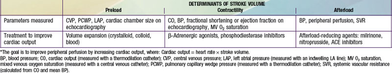
Sequential evaluation and management of cardiovascular shock are mandatory (Chapter 64). Table 436-3 outlines the general treatment principles for acute cardiac circulatory failure under most circumstances. Treatment of infants and children with low cardiac output after cardiac surgery also depends on the nature of the operative procedure, any intraoperative complications, and the physiology of the circulation after repair or palliation (Chapter 428).

Patients with deteriorating cardiogenic shock may benefit from a left ventricular assist device (LVAD). These devices have been used successfully in children and adolescents as a bridge to cardiac transplantation. Advances have produced devices small enough to support young infants. In some cases, both right and left ventricular assist is necessary (BiVAD). Once implanted, assist devices allow patients to recover sufficiently to be extubated, become ambulatory, and often leave the intensive care setting while awaiting transplantation. However, the morbidity from these devices is not insubstantial, especially in infants, and therefore the benefits and risks of their use should be carefully weighed for each individual patient. Patients with reversible ventricular failure, for example, those in the immediate postoperative state or those with acute myocarditis, may also benefit from extracorporeal membrane oxygenation (ECMO). When ventricular function is not expected to recover, heart transplantation should be considered (Chapter 437.1).

Bibliography

Anker SD, Colet JC, Filippatos G, et al. Ferric carboxymaltose in patients with heart failure and iron deficiency. N Engl J Med. 2009;361:2436-2448.

Aurigemma GP, Gaasch WH. Diastolic heart failure. N Engl J Med. 2004;351:1097-1105.

Bristow M. Antiadrenergic therapy of chronic heart failure: surprises and new opportunities. Circulation. 2003;107:1100-1102.

Bristow MR, Saxon LA, Boehmer J, et al. Cardiac-resynchronization therapy with or without an implantable defibrillator in advanced chronic heart failure. N Engl J Med. 2004;350:2140-2150.

Cheng JM, den Uil CA, Hoeks SE, et al. Percutaneous left ventricular assist devices vs. intra-aortic balloon pump counterpulsation for treatment of cardiogenic shock: a meta-analysis of controlled trials. Eur Heart J. 2009;30:2102-2108.

Dubin AM, Berul CI. Electrophysiological interventions for treatment of congestive heart failure in pediatrics and congenital heart disease. Expert Rev Cardiovasc Ther. 2007;5:111-118.

Dubin AM, Berul CI, Bevilacqua LM, et al. The use of implantable cardioverter-defibrillators in pediatric patients awaiting heart transplantation. J Card Fail. 2003;9:375-379.

Fried I, Bar-Oz B, Perles Z, et al. N-Terminal pro-B-type natriuretic peptide levels in acute versus chronic left ventricular dysfunction. J Pediatr. 2006;149:28-31.

Gheorghiade M, Braunwald E. Reconsidering the role of digoxin in the management of acute heart failure syndromes. JAMA. 2009;302:2146-2147.

Hoffman TM, Wernovsky G, Atz AM, et al. Efficacy and safety of milrinone in preventing low cardiac output syndrome in infants and children after corrective surgery for congenital heart disease. Circulation. 2003;107:996-1002.

Kaufman BD, Shaddy RE, Shirali GS, et al. Assessment and management of the failing heart in children. Cardiol Young. 2008;18(Suppl 3):63-71.

Krum H, Abraham WT. Heart failure. Lancet. 2009;373:941-952.

Law YM, Hoyer AW, Reller MD, et al. Accuracy of plasma B-type natriuretic peptide to diagnose significant cardiovascular disease in children. J Am Coll Cardiol. 2009;54:1467-1475.

Liggett SB. Polymorphisms of beta-adrenergic receptors in heart failure. Am J Med. 2004;117:525-527.

Maisel AS, Krishnaswamy P, Nowak RM, et al. Rapid measurement of B-type natriuretic peptide in the emergency diagnosis of heart failure. N Engl J Med. 2002;347:161-166.

McMurphy JJV. Heart failure. Lancet. 2005;365:1877-1889.

Neubauer S. The failing heart: an engine out of fuel. N Engl J Med. 2007;356:1140-1150.

Port JD, Bristow MR. Altered beta-adrenergic receptor gene regulation and signaling in chronic heart failure. J Mol Cell Cardiol. 2001;33:887-905.

Rosenthal D, Chrisant MR, Edens E, et al. International Society for Heart and Lung Transplantation: practice guidelines for management of heart failure in children. J Heart Lung Transplant. 2004;23:1313-1333.

Shaddy RE. Optimizing treatment for chronic congestive heart failure in children. Crit Care Med. 2001;29(Suppl):237-240.

Shaddy RE, Boucek MM, Hsu DT, et al. Carvedilol for children and adolescents with heart failure. JAMA. 2007;298:1171-1179.

Trani JF. Optimizing management of chronic heart failure. Lancet. 2009;374:1808-1809.

Zannad F, McMurray JJV, Krum H, et al. Eplerenone in patients with systolic heart failure and mild symptoms. N Engl J Med. 2011;364:11-21.