Chapter 16 Hamstring Anterior Cruciate Ligament Reconstruction with a Quadrupled or Tripled Semitendinosus Tendon Graft
Introduction
Currently, most surgeons use either the hamstring graft or the bone–patellar tendon–bone (BPTB) graft for ACL reconstruction. Previous studies have demonstrated the advantages and disadvantages of using one type of graft over the other. However, recent investigations have confirmed that comparable outcomes can be achieved with either of these two graft types.1–3
Inherent advantages cited with the use of hamstring grafts include its strength, decreased incidence of donor site morbidity, easier rehabilitation, smaller incisions, and better cosmesis.1,2,4 With BPTB graft, the strong bone-to-bone fixation and the faster healing achieved with the bone plugs at the graft’s end1,5 remain important advantages.
Scientific Rationale for a Quadrupled Construct
Hamstring grafts have gained popularity among surgeons due to the well-documented higher donor site morbidity when patellar tendon graft is used.6–8 Although prospective randomized studies comparing patellar tendon and hamstring grafts demonstrated no significant difference in final outcome, the apparent advantages offered by hamstring grafts remain appealing to surgeons. Previous concerns related to the hamstring tendon’s viability have long been dismissed, and studies comparing different graft types and configurations have demonstrated that failure load and stiffness values for four-stranded hamstring tendon grafts are higher than values reported for the natural ACL (2160N, 242 N/mm), 10-mm-wide patellar tendon grafts (2977N, 455 N/mm), and 10-mm-wide quadriceps tendon grafts (2353N, 326 N/mm).9,10
On the other hand, concerns related to hamstring graft incorporation within the tunnel was addressed with Morgan’s11 introduction of an “all inside” technique using bone–hamstring–bone composite graft. Therefore to address the concerns related to morbidity and delayed graft incorporation, we developed a technique that combines the advantages of a decreased donor site morbidity by using only one hamstring tendon (semitendinosus) with the possibility of achieving faster graft–tunnel incorporation by including a bone block with the distal limb of the semitendinosus tendon during harvest.1,12,13
Surgical Technique
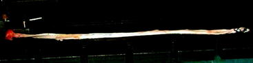
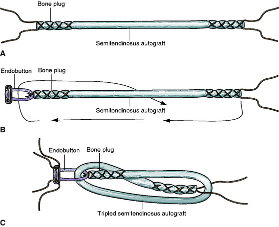
A 3-cm vertical incision centered approximately 5 cm below the medial joint line, midway between the tibial tubercle and the posteromedial aspect of the tibia, is performed. The sartorial fascia is incised, and the semitendinosus tendon is dissected and detached proximally with a tendon stripper. The distal limb of the tendon is detached along with a tibial bone plug and periosteum with the use of an osteotome. To achieve the desired 7-cm quadrupled graft construct (2 cm inserted in the femoral tunnel, 3 cm intraarticular, and 2 cm inserted in the tibial tunnel), the required minimum tendon length would be 28 cm (range 28–30 cm) (Fig. 16-1). Alternatively, semitendinosus tendons that are shorter than 28 cm can be prepared in a tripled configuration.
Graft Preparation
Quadrupled Semitendinosus Graft
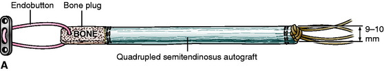
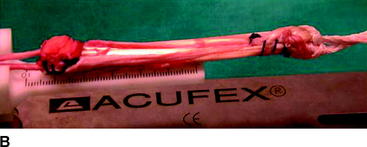
At the back table, all the muscle tissues attached to the tendon are removed with the use of a curette. Once devoid of excess tissues, the tendon is folded in a quadrupled fashion with the bone plug tied outside. Prior to suture placement on the tendon construct, the depth of the femoral tunnel is measured to determine the appropriate size of the Endobutton-CL (Smith & Nephew, Endoscopy, Andover, MA) to be used. Once the proper size is chosen, the Endobutton is then positioned in the quadrupled construct’s end where the bone block is located. Both ends of the graft are then whipstitched using #5 nonabsorbable sutures. A polyester tape is then knotted at the other end of the graft (Fig. 16-2, A, B). Measurement of the graft diameter follows, using 0.5-mm increment sizers to match this with the diameter of the femoral and tibial tunnels. Once in place, the grafts are pretensioned and preconditioned prior to fixation with cyclical flexion and extension of the knee under maximum manual tension.1,6
Tripled Semitendinosus Graft (Alternative Option for Short Semitendinosus Grafts)
Harvested semitendinosus tendons with a total length of less than 28 cm can be prepared in a tripled configuration. Once the excess tissues are removed, both ends of the semitendinosus tendon are whipstitched using #5 nonabsorbable sutures (Fig. 16-3, A). The tendon is then folded in three parts (three limbs) to determine the graft’s length and to approximate the size of the Endobutton-CL to be used. In general, we usually use either a 20- or 25-mm Endobutton-CL, considering that we have a tunnel length of about 40 to 45 mm. On the end of the graft where the bone plug is located, the free ends of the suture are used to tie a knot around the Endobutton-CL so that it becomes attached to the graft (Fig. 16-3, B). The other end of the graft is then passed through the loop of the Endobutton-CL as the tendon is folded in three parts. After passing through the Endobutton-CL, the suture at the free end of the graft is separated and positioned in such a way that it would catch the looped tendon at the opposite end (Fig. 16-3, C). With this configuration the diameter of this tripled semitendinosus is measured to make sure that it corresponds with the femoral and tibial tunnels. Prior to the final fixation, routine pretensioning and preconditioning of the graft are performed.
Arthroscopic Anterior Cruciate Ligament Reconstruction
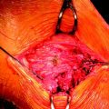
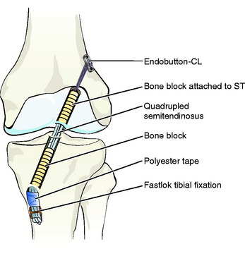
A standard anterolateral portal is created through which the arthroscope is inserted followed by an anteromedial portal where instruments can be introduced. While the graft is being prepared at the back table, tunnel preparations are completed. The tibial tunnel is prepared with the Acufex aimer set at 45 degrees with 70 degrees of inclination from the sagittal plane. During tibial tunnel reaming, a bone plug is obtained through the coring system used. On the other hand, the femoral tunnel is drilled in the 10:30 position for the right knee. Femoral fixation is achieved with the Endobutton connected to the graft while tibial fixation is obtained with an 8-mm titanium Fastlok device (Neoligaments, Leeds, United Kingdom), which is also connected to the graft with a quadrupled polyester tape. Finally, the bone block previously obtained from reaming the tibia is press-fitted in the tibial tunnel (Fig. 16-4). Postoperatively, rehabilitation is commenced according to the protocol described by Rosenberg and Pazik.14
Clinical Results
In a previous study10 of 100 patients who underwent ACL reconstruction using this technique, it was demonstrated that the average postoperative VAS pain score was 5 (range 2–7), with 90% of the patients discharged within 24 hours following the procedure. This finding was consistent with the subjective IKDC scores in which an average rating of 80% was obtained. Six months following the procedure, 10% of patients had noted pain over the tibial hardware with associated hypoesthesia over the surgical incision. Clinical examination at final evaluation demonstrated 90 patients with less than 1 cm difference in thigh circumference, two patients with extension lag of 6 degrees, and another two patients with flexion loss of 10 degrees. Kneeling test was positive only in 7% of these patients, while the postoperative Lachman test was negative in 90% (+1 in nine cases and +2 in one case). Sensory changes were evident in 30% of patients at 3 months with only 10% having localized hypoesthesia at the proximal third of the tibia at final evaluation.
1 Gobbi A, Zanazzo M, Tuy B, et al. Patellar tendon versus quadrupled bone semitendinosus ACL reconstruction: a prospective investigation in athletes. Arthroscopy. 2003;19:592-601.
2 Aune AK, Holm I, Risberg MA, et al. Four-strand hamstring tendon autograft compared with patellar tendon autograft for anterior cruciate ligament reconstruction: a randomized study with two year follow-up. Am J Sports Med. 2001;29:722-728.
3 Prodromos CC, Han YS, Keller BL, et al. Stability results of hamstring anterior cruciate ligament reconstruction at 2- to 8-year follow-up. Arthroscopy. 2005;21:138-146.
4 Shelbourne KD. Donor site problems after anterior cruciate ligament reconstruction using the patellar tendon graft. J Sports Traumatol Rel Res. 1995;17:120-128.
5 Pinczewski LA, Clingeleffer AJ, Otto BD, et al. Case report: integration of hamstring tendon graft with bone in reconstruction of the anterior cruciate ligament. Arthroscopy. 1997;13:641-643.
6 Cooley VJ, Deffner KT, Rosenberg TD. Quadrupled semitendinosus anterior cruciate ligament reconstruction: 5 year results in patients without meniscus loss. Arthroscopy. 2001;17:795-800.
7 Corry IS, Webb JM, Clingeleffer AJ, et al. Arthroscopic reconstruction of the anterior cruciate ligament. A comparison of patellar tendon autograft and four-strand hamstring tendon autograft. Am J Sports Med. 1999;27:444-454.
8 Maeda A, Shino K, Horibe S. Anterior cruciate ligament reconstruction with multi stranded autogenous semitendonosus tendon. Am J Sports Med. 1996;24:504-509.
9 Brown CHJr, Sklar JH. Endoscopic anterior cruciate ligament reconstruction using quadrupled hamstring tendons and Endobutton femoral fixation. Tech Orthop. 1998;13:281-298.
10 Weiler A, Scheffler S, Gockenjau A, et al. Different hamstring tendon graft fixation techniques under incremental loading conditions (abstract). Arthroscopy. 1998;14:425-426.
11 Morgan C. The bone-hamstring-bone composite autograft for ACL reconstruction. New Orleans, Month: Presented at the AAOS, 1994.
12 Gobbi A, Panuncialman I. Quadrupled bone-semitendinosus ACL reconstruction: a prospective clinical investigation in 100 patients. J Orthopaed Traumatol. 2003;3:120-125.
13 Gobbi A, Domzalski M, Pascual J, et al. Hamstring anterior cruciate ligament reconstruction: is it necessary to sacrifice the gracilis? Arthroscopy. 2005;21:275-280.
14 Rosenberg TD, Pazik JT. Anterior cruciate ligament reconstruction with quadrupled semitendinosus autograft. In: Parisen JS, editor. Current techniques in arthroscopy. Current medicine. Philadelphia: Churchill Livingstone; 1996:77-78.