Chapter 19 GYNECOMASTIA
Causes of Gynecomastia


Key Physical Findings
 Differentiate pseudogynecomastia (fatty tissue) from true gynecomastia

 Evidence of systemic disease, such as thyrotoxicosis, liver disease, or adrenal disease
Suggested Work-up
| Thyroid-stimulating hormone (TSH) | To evaluate for hyperthyroidism |
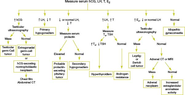
| Luteinizing hormone | See Figure 19-1 |
| Testosterone level | See Figure 19-1 |
| Estradiol level | To evaluate for testicular Leydig-cell tumor or feminizing adrenocortical neoplasm |
| Human chorionic gonadotropin (hCG) level | To evaluate for testicular germ cell tumor, extragonadal germ cell tumor, or hCG-secreting nontrophoblastic neoplasm |
| See Figure 19-1 | |
| Serum electrolytes | To evaluate for adrenal disease |
| Blood urea nitrogen (BUN) and creatinine | To evaluate for renal disease |
| Liver function tests | To evaluate for liver disease |
Additional Work-up
| Prolactin level | If gynecomastia is associated with galactorrhea to evaluate for prolactinoma |
| HIV test | If HIV infection is suspected or the patient has risk factors for infection |
| Testicular ultrasonongraphy | If testicular examination is abnormal or if estradiol is increased with a decreased or normal luteinizing hormone |
| Adrenal computed tomography (CT) or magnetic resonance imaging (MRI) | If adrenal neoplasm is suspected |
1. Braunstein G.D. Gynecomastia. N Engl J Med. 1993;328:490–495.
2. Einav-Bachar R., Phillip M., Aurbach-Klipper Y., Lazar L. Prepubertal gynaecomastia: aetiology, course, and outcome. Clin Endocrinol (Oxf). 2004;61:55–60.
3. Henley D.V., Lipson N., Korach K.S., Bloch C.A. Prepubertal gynecomastia linked to lavender and tea tree oils. N Engl J Med. 2007;356:479–485.
4. Rapaport R. Gynecomastia. In Behrman R.E., Kliegman R.M., Jenson H.B., editors: Nelson Textbook of Pediatrics, 17th ed., Philadelphia: WB Saunders, 2004.
5. Wilson J.D. Endocrine disorders of the breast. In: Isselbacher K.J., Braunwald E., Wilson J.D., editors. Harrisons’s Textbook of Internal Medicine. 13th ed. New York: McGraw-Hill; 1994:2037–2039.
6. Wise G.J., Roorda A.K., Kalter R. Male breast disease. J Am Coll Surg. 2005;200:255–269.