Scrotal masses

Published on 21/03/2015 by admin

Filed under Pediatrics

Last modified 21/03/2015

Print this page

rate 1 star rate 2 star rate 3 star rate 4 star rate 5 star
Your rating: none, Average: 0 (0 votes)

This article have been viewed 3280 times

Chapter 38 SCROTAL MASSES

Theodore X. O’Connell

General Discussion

Evaluation of a scrotal mass should include classifying it as extratesticular or intratesticular, solid or cystic, and painless or painful. A painful scrotal mass requires immediate action because torsion of the spermatic cord is a urologic emergency. The dictum that a painful scrotal mass is torsion until proven otherwise should be heeded. Other painful scrotal lesions include orchitis, epididymitis, trauma, incarcerated hernia, torsion of the appendix testis or appendix epididymis, and acute bleeding into a testicular tumor.

Masses that arise from the testicle are more likely to represent malignancies, whereas extratesticular masses are more likely to be benign. An intratesticular lesion should be considered a malignancy unless proven otherwise. Solid masses are much more likely to represent neoplastic conditions, especially when painless. Transillumination using a handheld light source may help to differentiate between solid and cystic structures. Extratesticular tumors are uncommon but do occur in the form of paratesticular rhabdomyosarcoma and ademomatoid tumors of the epididymis.

Cystic lesions of the scrotum are much more common than solid lesions. A cystic mass within the epididymis is usually a spermatocele. A cyst within the spermatic cord usually represents a hydrocele. A cystic mass that surrounds the entire testicle usually represents a hydrocele. When careful physical examination, including transillumination, fails to distinguish the exact location and nature of the lesion, then scrotal ultrasonography is of great value, especially when the testis is not palpable and a hydrocele is present. Ultrasonography can confirm the location of the mass and differentiate between solid and cystic lesions.

Most extratesticular masses are benign, and most intratesticular lesions are malignant. Sonography has been shown to have nearly 100% sensitivity for detecting testicular neoplasia. Testicular microlithiasis is a rare finding that can be associated with subsequent development of germ cell tumors of the testis. Careful follow-up is warranted if testicular microlithiasis is found on ultrasonography.

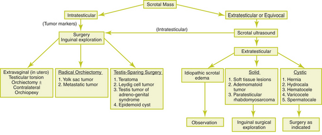
The most common painless scrotal masses in infants, children, and adolescents include indirect inguinal hernias, hydroceles, varicoceles, and spermatoceles. Testicular tumors, perinatal testicular torsion, acute idiopathic scrotal edema, and soft tissue tumors of the spermatic cord are less common causes. Figure 38-1 provides a clinical approach to the painless scrotal mass.

image

Figure 38-1 Clinical approach to the painless scrotal mass.

(From Skoog SJ. Benign and malignant pediatric scrotal masses. Pediatr Clin North Am 1997;44:1229-1250, with permission.)

A testicular tumor usually presents as a painless mass, although the patient may complain of scrotal heaviness or a dull ache. Physical examination should be performed with the patient in the upright and supine position because communicating hydroceles, hernias, and varicoceles are accentuated in the upright position and with the Valsalva maneuver. If testicular cancer is suspected, the patient should also be examined for lymphadenopathy, gynecomastia, and abdominal masses.

Cystic and Painless Extratesticular Masses

Hernias and hydroceles represent the greatest percentage of scrotal masses in pediatric patients. The incidence of inguinal hernias in the pediatric population is 10 to 20 per 1000 live births. Prematurity and low birth weight significantly increase the risk for hernias. Pediatric hernias and hydroceles are seen as bulges or swelling in the groin or scrotum and may be more visible when the child is crying.

A hydrocele is a collection of peritoneal fluid between the layers of the tunica vaginalis surrounding the testicle, with or without communication with the abdomen. A hydrocele usually presents as a painless scrotal swelling that can be transilluminated. The presence of an inguinal hernia or communicating hydrocele in the pediatric age group is an indication for surgical repair because of the risk of development of incarceration or strangulated hernia.

A varicocele is present in up to 20% of all males and is a tortuous and dilated pampiniform venous plexus and internal spermatic vein. Varicoceles often are described as a “bag of worms” superior to and distinct from the testicle. Varicoceles usually first appear near midpuberty and are rarely detected before 10 years of age. Most varicoceles occur on the left side and usually are asymptomatic. Varicoceles have been associated with male-factor infertility and may result in growth arrest of the left testicle. The dilatation and tortuosity are most noticeable when the patient is upright and may be accentuated if the patient performs a Valsalva maneuver. Once a scrotal mass has been identified as a varicocele, it is important to assess its effects on testicular size. A volume difference of greater than 2 cm3 is considered significant and serves as the minimal requirement for surgical repair of the adolescent varicocele. If a right-sided varicocele is identified, vena cava obstruction must be ruled out.

Buy Membership for Pediatrics Category to continue reading. Learn more here