[level-membership-for-endocrinology-diabetes-and-metabolism-category]CHAPTER 46
Gynecomastia
Gynecomastia is defined as the presence of palpable breast tissue in a male. True gynecomastia resulting from enlargement of glandular tissue should be distinguished from excess adipose accumulation (i.e., pseudogynecomastia).
2. How does gynecomastia manifest clinically?
Gynecomastia usually manifests as a palpable, discrete button of tissue radiating from beneath the nipple and areola. Gynecomastia feels “gritty” when the breast is pinched between the thumb and forefinger. Fatty tissue, unlike gynecomastia, will not cause resistance until the nipple is reached. If doubt remains, soap and water on the breast can facilitate the examination by decreasing skin friction.
3. What is the significance of painful gynecomastia?
Gynecomastia is frequently asymptomatic and incidentally discovered. Pain or tenderness implies recent, rapid growth of breast tissue. This may indicate a pathologic cause of the gynecomastia and should prompt further evaluation.
4. Is gynecomastia always bilateral?
The involvement tends to be bilateral, but asymmetry is common. Unilateral enlargement is present in 5% to 25% of patients and may be a preliminary stage in the development of bilateral disease. In autopsy studies, unilateral enlargement is often found to be bilateral gynecomastia histologically.
5. Summarize the pathophysiology of gynecomastia.
Gynecomastia results from an imbalance between the stimulatory effect of estrogen on ductal proliferation and the inhibitory effect of androgen on breast development. The imbalance is most commonly caused by increased production of estrogens, decreased production of testosterone, or increased conversion of androgens to estrogens in peripheral tissue. Disorders of sex hormone–binding globulin or with androgen receptor binding and function can also result in gynecomastia.
6. Where are estrogens produced in the male?
Direct testicular production of estrogens accounts for less than 15% of male estrogen production. Most estrogens come from the conversion of adrenal and testicular androgens to estrogens in peripheral tissues, particularly adipose tissue and the liver.
7. What is the most common cause of gynecomastia?
Asymptomatic palpable breast tissue is common in normal males, particularly in the neonate (60%–90%), at puberty (60%–70%, ages 12 and 15 years), and with increasing age (20%–65%, >50 years). Prevalence of histopathologically confirmed gynecomastia is up to 40% in autopsy series. Because of this high prevalence, gynecomastia is considered a relatively normal finding during these periods of life. Gynecomastia is often called physiologic or idiopathic at these ages.
8. Why does gynecomastia occur so commonly during these stages of life?
Neonatal gynecomastia results from placental transfer of estrogens. During early puberty, production of estrogens begins sooner than testosterone production, thus causing an imbalance in the ratio of estrogens to androgens. With aging, testosterone production decreases, and peripheral androgen to estrogen conversion often increases because of an age-related increase in adipose tissue. There may also be a higher prevalence of offending medications and medical conditions in the elderly population.
9. What are the other causes of gynecomastia?
Idiopathic gynecomastia and pubertal gynecomastia make up the majority of cases. Drugs account for 10% to 20% of cases and hypogonadism for another 10%. Adrenal or testicular tumors account for less than 3% of cases; gynecomastia may precede the development of the testicular tumor. Other causes combined account for less than 10% of cases and include androgen-resistant disorders, malnutrition, cirrhosis, alcohol abuse, renal disease, congenital adrenal hyperplasia, extragonadal tumors, refeeding gynecomastia, and hyperthyroidism. Case reports of atypical infectious causes have been described (e.g., tuberculosis and filariasis).
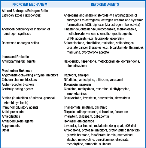
10. What drugs cause gynecomastia?
Many drugs have been implicated, some with well-characterized steroid effects and others noted in case reports and without a clearly elucidated mechanism:
11. How do testicular tumors cause gynecomastia?
Germ cell tumors can produce human chorionic gonadotropin (hCG). Like luteinizing hormone (LH), hCG increases testicular estradiol production. Leydig cell tumors may directly secrete estradiol.
12. What extragonadal tumors cause gynecomastia?
Pancreatic, gastric, and pulmonary tumors, transitional cell bladder carcinoma, and renal cell carcinoma have been associated with hCG production. Hepatomas may have increased aromatase activity that results in excess conversion of androgens to estrogens.
13. Who should undergo evaluation for gynecomastia?
History and physical examination are indicated in all cases and determine the cause in 30% to 40% of patients. Gynecomastia is so common, however, that many experts are cautious about attaching importance to the detection of a small amount of breast tissue in an otherwise asymptomatic man. In adolescents, there is no reason to consider endocrine testing unless the enlargement is massive or the gynecomastia persists longer than 2 years. Acute development of enlargement and tenderness in men who than 20 years old warrants additional evaluation, as do eccentric, hard masses and lesions larger than 4 cm.
14. What information is significant in the history?
15. What should be noted on the physical examination?
Important features include characteristics of the breast tissue (size, irregularity, firmness, eccentric location, nipple discharge), overlying skin changes (ulceration, nipple retraction), testes (size, asymmetry), abdomen (liver enlargement, ascites, spider angiomas), secondary sexual characteristics, thyroid status (goiter, tremor, reflexes), and signs of excessive cortisol (buffalo hump, central obesity, hypertension, purple striae, moon facies), and body mass index or body habitus (bodybuilder physique, obesity).
 KEY POINTS 1: GENERAL APPROACH TO GYNECOMASTIA
1. The most important differentiation is between gynecomastia and breast cancer. If doubt remains after physical examination, obtain a mammogram.
2. Most cases are bilateral, asymptomatic, and incidentally discovered. History, physical examination, and reevaluation in 3 to 6 months are appropriate for such men.
3. Rapid enlargement, size larger than 4 cm, pain, and age less than 10 years or between 20 and 50 years correlate with a systemic illness or pathologic cause of the gynecomastia. Such men should be evaluated thoroughly if the cause is not apparent after history and physical examination.
4. Malignant tumors can cause gynecomastia, although rarely. Consider testicular, pulmonary, and abdominal tumors (pancreatic, adrenal, gastric, and renal or bladder).
16. Should laboratory tests be ordered?
Some clinicians believe that hormonal testing is not cost-effective and favor checking testicular ultrasound alone to rule out the 3% incidence of feminizing tumors. Most practitioners, however, favor measuring liver enzymes, blood urea nitrogen, creatinine, thyrotropin (thyroid-stimulating hormone [TSH]), and testosterone (total and free). Estradiol, hCG, prolactin, LH, and follicle-stimulating hormone (FSH) may follow the initial screen. If the hCG or estradiol level is elevated, a testicular ultrasound scan is indicated. If this is negative, a chest radiograph and abdominal computed tomography (CT) scan should follow. For prepubertal patients, an adrenal CT scan precedes the testicular ultrasound examination.
17. What findings raise the suspicion of breast cancer?
Breast cancer is rare in men (0.2%). The risk is increased in Klinefelter’s syndrome (3%–6%) and in male relatives of young women with breast cancer. Carcinoma is usually unilateral, painless, and nontender. Bloody discharge, ulceration, firmness, fixation to the underlying tissue, eccentric location, and adenopathy are suspicious findings. If doubt remains, a mammogram or biopsy should be considered. The sensitivity and specificity of mammogram for the diagnosis of male breast cancer approach 90%. The diagnostic accuracy of fine-needle aspiration cytology is greater than 90%. Excisional biopsy or mastectomy is recommended for malignant or suspicious cytology or mammogram appearance.
18. Will gynecomastia spontaneously regress?
Gynecomastia of recent onset and less than 3 cm in size regresses in 85% of patients. It may take 18 to 36 months for gynecomastia to resolve during puberty, but resolution occurs in more than 90% of pubertal boys. Persistence is uncommon after age 17 years. Gynecomastia resulting from a medication or underlying disease should also resolve after discontinuing the inciting agent or treating the underlying disease. Persistent tissue becomes more fibrous with time, however, and is less likely to remit spontaneously if it has been present for more than 12 months. More highly developed breast tissue (Tanner stages III, IV, and V) is also less likely to regress.
19. What is the treatment when gynecomastia does not regress?
Hormonal therapy can be attempted. Tamoxifen, clomiphene, danazol, dihydrotestosterone, testolactone, and anastrozole have all been used. Although studies are small and this is an off-label use, tamoxifen has the fewest side effects and the highest response rate for both improvement in tenderness and decrease in size. Partial regression can be seen in approximately 80% of patients and complete regression in about 60%. Medication is more likely to work if gynecomastia has been present for less than 4 months and the size of the tissue is less than 3 cm. Tamoxifen is given at a dosage of 10-20 mg daily with follow-up in 3 months to assess response. For recurrent or persistent gynecomastia greater than 3 cm, surgery is the recommended therapy. Liposuction or ultrasound-guided liposuction, excision, or both may be used. Low-dose bilateral breast irradiation and tamoxifen have also been studied in trials as prophylaxis to prevent the development of gynecomastia caused by estrogens and antiandrogens used in the treatment of prostate cancer.
 KEY POINTS 2: TREATMENT OF GYNECOMASTIA
1. Most cases resolve spontaneously or after removal of the offending medication or treatment of the underlying disease.
2. Medical management with tamoxifen can be attempted for 3 to 6 months if desired.
3. The longer the tissue has been present and the larger the amount of tissue, the less likely a response to tamoxifen will be. Surgery is indicated for these cases.
Bowers, S, Pearlman, N, McIntyre, R, et al. Cost-effective management of gynecomastia. Am J Surg. 1998;176:638–641.
Braunstein, G. Gynecomastia. N Engl J Med. 2007;357:1229–1237.
Braunstein, G. Pathogenesis and diagnosis of gynecomastia. UpToDate. 2003;11:1–11.
Braunstein, G. Prevention and treatment of gynecomastia. UpToDate. 2003;11:1–9.
Carlson, HE. Approach to the patient with gynecomastia. J Clin Endocrinol Metab. 2011;96:15–21.
Ersoz, H, Onde, M, Terekeci, H, et al. Causes of gynecomastia in young adult males and factors associated with idiopathic gynecomastia. Int J Androl. 2002;25:312–316.
Evans, G, Anthony, T, Appelbaum, A, et al. The diagnostic accuracy of mammography in the evaluation of male breast disease. Am J Surg. 2001;181:96–100.
Fruhstorfer, B, Malata, C. A systematic approach to the surgical treatment of gynecomastia. Br J Plast Surg. 2003;56:237–246.
Gruntmanis, U, Braunstein, G. Treatment of gynecomastia. Curr Opin Investig Drugs. 2001;2:643–649.
Henley, DV, Lipson, N, Korach, KS, et al. Prepubertal gynecomastia linked to lavender and tea tree oils. N Engl J Med. 2007;356:479–485.
Ismail, A, Barth, J. Endocrinology of gynaecomastia. Ann Clin Biochem. 2001;38:596–607.
Khan, H, Blarney, R. Endocrine treatment of physiological gynecomastia. BMJ. 2003;327:301–302.
Koh, J, Tee, A, Images in clinical medicine. tuberculous abscess manifesting as unilateral gynecomastia. N Engl J Med 2009;361:2270.
Kolhi, K, Jain, S. Filariasis presenting as gynecomastia. Breast J,. 2012;18:83–84.
Narula, HS, Carlson, HE. Gynecomastia. Endocrinol Metab Clin North Am. 2007;36:497–519.
Widmark, A, Fossa, S, Lundmo, P, et al. Does prophylactic breast irradiation prevent antiandrogen induced gynecomastia. Urology. 2003;61:145–151.
William, MJ, Gynecomastia. its incidence, recognition and host characterization in 447 autopsy studies. Am J Med 1963;34:103–112.
Yaturu, S, Harrara, E, Nopajaroonsri, C, et al. Gynecomastia attributable to HCG secreting giant cell carcinoma of the lung. Endocr Pract. 2003;9:233–235.
[/level-membership-for-endocrinology-diabetes-and-metabolism-category][not-level-membership-for-endocrinology-diabetes-and-metabolism-category]<
[/not-level-membership-for-endocrinology-diabetes-and-metabolism-category]
