Group B Streptococcus

Published on 22/03/2015 by admin

Filed under Pediatrics

Last modified 22/04/2025

Print this page

rate 1 star rate 2 star rate 3 star rate 4 star rate 5 star
Your rating: none, Average: 0 (0 votes)

This article have been viewed 1344 times

Chapter 177 Group B Streptococcus

Group B streptococcus (GBS), or Streptococcus agalactiae, is a major cause of neonatal bacterial sepsis in the USA. While advances in prevention strategies have led to a decline in the incidence of neonatal disease, GBS remains a major pathogen for neonates, pregnant women, and nonpregnant adults.

Epidemiology

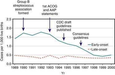
GBS emerged as a prominent neonatal pathogen in the late 1960s. For the next 2 decades, the incidence of neonatal GBS disease remained fairly constant, affecting 1.0-5.4/1,000 liveborn infants in the USA. Two patterns of disease were seen: early-onset disease, which presents at <7 days of age, and late-onset disease, which presents at 7 days of age or later. In the 1990s, widespread implementation of maternal chemoprophylaxis led to a striking 65% decrease in the incidence of early-onset neonatal GBS disease in the USA, from 1.7/1,000 live births to 0.6/1,000 live births, whereas the incidence of late-onset disease remained essentially stable at approximately 0.4/1,000 (Fig. 177-1). Release of revised guidelines in 2002 coincided with a further reduction in the incidence of early-onset neonatal disease. In other developed countries, rates of neonatal GBS disease are similar to those in the USA prior to use of GBS chemoprophylaxis. In the developing world, GBS is not a major cause of neonatal sepsis, even though the prevalence of maternal vaginal colonization with GBS (a major risk factor for neonatal disease) among women from developing countries is similar to that reported among women living in the USA. The incidence of neonatal GBS disease is higher in premature and low birthweight infants, although most cases occur in full-term infants.

Colonization by GBS in healthy adults is common. Vaginal or rectal colonization occurs in up to approximately 30% of pregnant women and is the usual source for GBS transmission to newborn infants. In the absence of maternal chemoprophylaxis, approximately 50% of infants born to colonized women acquire GBS colonization, and 1-2% of these infants develop invasive disease. Heavy maternal colonization increases the risk for infant colonization and development of early-onset disease. Additional risk factors for early-onset disease include prolonged rupture of membranes, intrapartum fever, prematurity, maternal bacteriuria during pregnancy, or previous delivery of an infant who developed GBS disease. Risk factors for late-onset disease are less well defined. Whereas late-onset disease may follow vertical transmission, horizontal acquisition from nursery or other community sources has also been described.

GBS is also an important cause of invasive disease in adults. GBS may cause urinary tract infections, bacteremia, endometritis, chorioamnionitis, and wound infection in pregnant and parturient women. In nonpregnant adults, especially those with underlying medical conditions such as diabetes mellitus, cirrhosis, or malignancy, GBS may cause serious infections such as bacteremia, skin and soft tissue infections, endocarditis, pneumonia, and meningitis. In the era of maternal chemoprophylaxis, most invasive GBS infections occur in nonpregnant adults. Unlike neonatal disease, the incidence of invasive GBS disease in adults has increased substantially, doubling between 1990 and 2007.

The serotypes most commonly associated with neonatal GBS disease are types Ia, III, V, Ib, and II. Strains of serotype III are isolated in more than 50% of cases of late-onset disease and of meningitis associated with early- or late-onset disease. The serotype distribution of colonizing and invasive strains from pregnant women is similar to that from infected newborns. In Japan, serotypes VI and VIII have been reported as common maternal colonizing serotypes, and case reports indicate that type VIII strains may cause neonatal disease indistinguishable from that caused by other serotypes.

Pathogenesis

A major risk factor for the development of early-onset neonatal GBS infection is maternal vaginal or rectal colonization by GBS. Infants acquire GBS during passage through the birth canal or, in some cases, via ascending infection. Fetal aspiration of infected amniotic fluid may occur. The incidence of early-onset GBS infection increases with the length of rupture of membranes. Infection may also occur through seemingly intact membranes. In cases of late-onset infection, GBS may be vertically transmitted or acquired later from maternal or nonmaternal sources.

Several bacterial factors are implicated in the pathophysiology of invasive GBS disease. Foremost among these is the type-specific capsular polysaccharide. Strains that are associated with invasive disease in humans elaborate more capsular polysaccharide than do colonizing isolates. All GBS capsular polysaccharides are high molecular weight polymers and contain a short side chain terminating in N-acetylneuraminic acid (sialic acid). Studies in type III GBS show that the sialic acid component of the capsular polysaccharide prevents activation of the alternative complement pathway in the absence of type-specific antibody. Thus, the capsular polysaccharide appears to exert a virulence effect by protecting the organism from opsonophagocytosis in the nonimmune host. In addition, type-specific virulence attributes are suggested by the fact that type III strains are implicated in most cases of late-onset neonatal GBS disease and meningitis. Type III strains are taken up by brain endothelial cells more efficiently in vitro than are strains of other serotypes, although studies using acapsular mutant strains demonstrate that it is not the capsule itself that facilitates cellular invasion. Other putative GBS virulence factors include GBS surface proteins, which may play a role in adhesion to host cells; C5a peptidase, which is postulated to inhibit the recruitment of polymorphonuclear cells into sites of infection; β-hemolysin, which has been associated with cell injury in vitro studies; and hyaluronidase, which has been postulated to act as a spreading factor in host tissues.

In a classic study, among pregnant women colonized with type III GBS, those who gave birth to healthy infants had higher levels of capsular polysaccharide-specific antibody than those who gave birth to infants who developed invasive disease. In addition, there is a high correlation of antibody to GBS type III in mother-infant paired sera. These observations indicate that transplacental transfer of maternal antibody is critically involved in neonatal immunity to GBS. Optimal immunity to GBS also requires an intact complement system. The classic complement pathway is an important component of GBS immunity in the absence of specific antibody; antibody-mediated opsonophagocytosis may also proceed via the alternative complement pathway. These and other results indicate that anticapsular antibody can overcome the prevention of C3 deposition on the bacterial surface by the sialic acid component of the type III capsule.

The precise steps between GBS colonization and invasive disease remain unclear. In vitro studies showing GBS entry of alveolar epithelium and pulmonary vasculature endothelial cells suggest that GBS may gain access to the bloodstream via invasion from the alveolar space, perhaps following intrapartum aspiration of infected fluid. β-Hemolysin/cytolysin may facilitate GBS entry into the bloodstream following inoculation into the lungs. However, highly encapsulated GBS strains enter eukaryotic cells poorly in vitro compared with capsule-deficient organisms, yet are associated with virulence clinically and in experimental infection models.

GBS induces the release of proinflammatory cytokines. The group B antigen and the peptidoglycan component of the GBS cell wall are potent inducers of tumor necrosis factor-α release in vitro, whereas purified type III capsular polysaccharide is not. Even though the capsule plays a central role in virulence through avoidance of immune clearance, the capsule does not directly contribute to cytokine release and the resultant inflammatory response.

The complete genome sequences of type III, V, and Ia GBS strains have been reported, emphasizing a genomic approach to better understanding GBS. Analysis of these sequences shows that GBS is closely related to Streptococcus pyogenes and Streptococcus pneumoniae. Many known and putative GBS virulence genes are clustered in pathogenicity islands that also contain mobile genetic elements, suggesting that interspecies acquisition of genetic material plays an important role in genetic diversity.

Clinical Manifestations

Two syndromes of neonatal GBS disease are distinguishable on the basis of age at presentation, epidemiologic characteristics, and clinical features (Table 177-1). Early-onset neonatal GBS disease presents within the 1st 6 days of life and is often associated with maternal obstetric complications, including chorioamnionitis, prolonged rupture of membranes, and premature labor. Infants may appear ill at the time of delivery, and most infants become ill within the 1st 24 hr of birth. In utero infection may result in septic abortion. The most common manifestations of early-onset GBS disease are sepsis (50%), pneumonia (30%), and meningitis (15%). Asymptomatic bacteremia is uncommon but can occur. In symptomatic patients, nonspecific signs such as hypothermia or fever, irritability, lethargy, apnea, and bradycardia may be present. Respiratory signs are prominent regardless of the presence of pneumonia and include cyanosis, apnea, tachypnea, grunting, flaring, and retractions. A fulminant course with hemodynamic abnormalities, including tachycardia, acidosis, and shock, may ensue. Persistent fetal circulation may develop. Clinically and radiographically, pneumonia associated with early-onset GBS disease is difficult to distinguish from respiratory distress syndrome. Patients with meningitis often present with nonspecific findings, as described for sepsis or pneumonia, with more specific signs of central nervous system (CNS) involvement initially being absent.

Table 177-1 CHARACTERISTICS OF EARLY- AND LATE-ONSET GBS DISEASE

  EARLY-ONSET DISEASE LATE-ONSET DISEASE
Age at onset 0-6 days 7-90 days
Increased risk after obstetric complications Yes No
Common clinical manifestations Sepsis, pneumonia, meningitis Bacteremia, meningitis, other focal infections
Common serotypes Ia, III, V, II, Ib III predominates
Case fatality rate 4.7% 2.8%

Adapted from Schrag SJ, Zywicki S, Farley MM, et al: Group B streptococcal disease in the era of intrapartum antibiotic prophylaxis, N Engl J Med 342:15–20, 2000.

Late-onset neonatal GBS disease occurs on or after 7 days of life and most commonly manifests as bacteremia (45-60%) and meningitis (25-35%). Focal infections involving bone and joints, skin and soft tissue, the urinary tract, or lungs have been reported in approximately 20% of patients with late-onset disease. Cellulitis and adenitis are often localized to the submandibular or parotid regions. In contrast to early-onset disease, maternal obstetric complications are not risk factors for the development of late-onset GBS disease. Infants with late-onset disease are often less severely ill on presentation than infants with early-onset disease and the disease is often less fulminant.

Invasive GBS disease in children beyond early infancy is uncommon. In a multistate surveillance study in the 1990s, 2% of all cases of invasive GBS disease were identified in children age 90 days to 14 yr. Two of the more common syndromes associated with childhood GBS disease beyond early infancy are bacteremia and endocarditis. HIV infection should be considered in children with invasive GBS disease beyond the neonatal period.

Treatment

Penicillin G is the treatment of choice of confirmed GBS infection. Initial empirical therapy of neonatal sepsis should include ampicillin and an aminoglycoside (or cefotaxime), both for the need for broad coverage pending organism identification and for synergistic bactericidal activity. Once GBS has been definitively identified and a good clinical response has occurred, therapy may be completed with penicillin alone. Especially in cases of meningitis, high doses of penicillin (450,000-500,000 U/kg/day) or ampicillin (300 mg/kg/day) are recommended because of the relatively high mean inhibitory concentration of penicillin for GBS as well as the potential for a high initial CSF inoculum. The duration of therapy varies according to the site of infection (Table 177-2) and should be guided by clinical circumstances. Extremely ill near-term patients with respiratory failure have been successfully treated with extracorporeal membrane oxygenation.

Table 177-2 RECOMMENDED DURATION OF THERAPY FOR MANIFESTATIONS OF GBS DISEASE

TREATMENT DURATION
Bacteremia without a focus 10 days
Meningitis 2-3 wk
Ventriculitis 4 wk
Osteomyelitis 4 wk

Adapted from the American Academy of Pediatrics: Group B streptococcal infections. In Pickering LK, editor: Red book: 2000 report of the Committee on Infectious Diseases, ed 25, Elk Grove Village, IL, 2000, American Academy of Pediatrics, pp 537–544.

In cases of GBS meningitis, some experts recommend that additional CSF be sampled at 24-48 hr to determine whether sterility has been achieved. Persistent GBS growth may indicate an unsuspected intracranial focus or an insufficient antibiotic dose.

For recurrent neonatal GBS disease, standard intravenous antibiotic therapy followed by attempted eradication of GBS mucosal colonization has been suggested. This suggestion is based on the findings in several studies that invasive isolates from recurrent episodes are often identical to each other and to colonizing strains from the affected infant. Rifampin has most frequently been used for this purpose, but one report demonstrates that eradication of GBS colonization in infants is not reliably achieved by rifampin therapy. Optimal management of this uncommon situation remains unclear.

Prevention

Persistent morbidity and mortality from perinatal GBS disease despite advances in neonatal care has spurred intense investigation into modes of prevention. Two basic approaches to GBS prevention have been investigated: (1) elimination of colonization from the mother or infant (chemoprophylaxis), and (2) induction of protective immunity (immunoprophylaxis).

Chemoprophylaxis

Administration of antibiotics to pregnant women before the onset of labor does not reliably eradicate maternal GBS colonization and is not an effective means of preventing neonatal GBS disease. Interruption of neonatal colonization is achievable through administration of antibiotics to the mother during labor (Chapter 103). Infants born to GBS-colonized women with premature labor or prolonged rupture of membranes who were given intrapartum chemoprophylaxis had a substantially lower rate of GBS colonization (9% vs 51%) and early-onset disease (0% vs 6%) than did the infants born to women who were not treated. Maternal postpartum febrile illness was also decreased in the treatment group.

In the mid-1990s, guidelines for chemoprophylaxis were issued that specified administration of intrapartum antibiotics to women identified as high-risk by either culture-based or risk factor–based criteria. These guidelines were revised in 2002 after epidemiologic data indicated the superior protective effect of the culture-based approach in the prevention of neonatal GBS disease (Chapter 103). According to current recommendations, vaginorectal GBS screening cultures should be performed for all pregnant women at 35-37 wk gestation. Any woman with a positive prenatal screening culture, GBS bacteriuria during pregnancy, or a previous infant with invasive GBS disease should receive intrapartum antibiotics. Women whose culture status is unknown (culture not done, incomplete, or results unknown) and who deliver prematurely (<37 wk gestation) or experience prolonged rupture of membranes (≥18 hr) or intrapartum fever (≥38.0°C) should also receive intrapartum chemoprophylaxis. If amnionitis is suspected, broad-spectrum antibiotic therapy that includes an agent active against GBS should replace GBS prophylaxis. Routine intrapartum prophylaxis is not recommended for women with GBS colonization undergoing planned cesarean delivery who have not begun labor or had rupture of membranes.

These guidelines also suggest an approach for the management of infants born to mothers who received intrapartum chemoprophylaxis (Chapter 103). Data from a large epidemiologic study indicate that the administration of maternal intrapartum antibiotics does not change the clinical spectrum or delay the onset of clinical signs in infants who developed GBS disease despite maternal prophylaxis. Thus, the Centers for Disease Control and Prevention guidelines reserve a full diagnostic evaluation for those infants who appear clinically ill or whose mothers are suspected of having chorioamnionitis.

A significant concern with maternal intrapartum prophylaxis has been that large-scale antibiotic use among parturient women might lead to increased rates of antimicrobial resistance or infection in infants with organisms other than GBS. To date, an increase in the incidence of non-GBS early-onset neonatal infections has been seen only in premature, low birthweight, and very low birthweight infants, in whom risk factors other than maternal chemoprophylaxis may play a role. At present, the substantial decline in early-onset neonatal GBS disease favors continued broad-scale intrapartum chemoprophylaxis, but continued surveillance is required. Penicillin remains the preferred agent for chemoprophylaxis, because of its narrow spectrum and the universal penicillin susceptibility of GBS isolates associated with human infection. Occasional GBS isolates have demonstrated reduced in vitro susceptibility to penicillin and other β-lactam antibiotics in association with mutations in penicillin-binding proteins. However, such strains have not been reported in invasive infection. Because of recent reports indicating frequent resistance of GBS to erythromycin (up to 32%) and clindamycin (up to 15%), cefazolin should be used in most cases of intrapartum chemoprophylaxis for penicillin-intolerant women. For penicillin-allergic women at high risk for anaphylaxis, clindamycin or erythromycin should be used, if isolates are demonstrated to be susceptible. Vancomycin should be used if isolates are resistant to clindamycin and erythromycin or if susceptibility to these agents is unknown.

A limitation of the maternal chemoprophylaxis strategy is that intrapartum antibiotic use is unlikely to have an impact on late-onset neonatal disease, miscarriages or stillbirths attributed to GBS, or adult GBS disease. In addition, with wider implementation of maternal chemoprophylaxis, an increasing percentage of early-onset neonatal disease has been in patients born to women with negative cultures, that is, false-negative screens.

Maternal Immunization

Human studies demonstrate that transplacental transfer of naturally acquired maternal antibody to the GBS capsular polysaccharide protects newborns from invasive GBS infection and that efficient transplacental passage of vaccine-induced GBS antibodies occurs. Conjugate vaccines composed of the GBS capsular polysaccharides coupled to carrier proteins have been produced for human use. In early clinical trials, conjugate GBS vaccines were well tolerated and induced levels of functional antibodies well above the range believed to be protective in greater than 90% of recipients. A vaccine containing type III polysaccharide coupled to tetanus toxoid was safely administered to pregnant women and elicited functionally active type-specific antibody that was efficiently transported to the fetus. Administration of a multivalent polysaccharide-protein vaccine before or during pregnancy should lead to transplacental passage of vaccine-induced antibody that protects the fetus and newborn against infection by several GBS serotypes. Such a vaccine would eliminate the need for cumbersome cultures during pregnancy, would circumvent the various risks associated with large-scale antibiotic prophylaxis, and would likely have an impact on both early- and late-onset disease. Intrapartum chemoprophylaxis will likely remain an important aspect of prevention, particularly for women in whom opportunities for GBS immunization are missed and for infants born so early that levels of transplacentally acquired antibodies may not be high enough to be protective.

Bibliography

Baker CJ, Kasper DL. Correlation of maternal antibody deficiency with susceptibility to neonatal group B streptococcal infection. N Engl J Med. 1976;294:753-756.

Baker CJ, Rench MA, McInnes P. Immunization of pregnant women with group B streptococcal type III capsular polysaccharide-tetanus toxoid conjugate vaccine. Vaccine. 2003;21:3468-3472.

Boyer KM, Gotoff SP. Prevention of early-onset neonatal group B streptococcal disease with selective intrapartum chemoprophylaxis. N Engl J Med. 1986;314:1665-1669.

Bromberger P, Lawrence JM, Braun D, et al. The influence of intrapartum antibiotics on the clinical spectrum of early-onset group B streptococcal infection in term infants. Pediatrics. 2000;106:244-250.

Centers for Disease Control and Prevention. Early-onset group B streptococcal disease—United States, 1998–1999. MMWR. 2000;49:793-796.

Centers for Disease Control and Prevention. Prevention of perinatal group B streptococcal disease: a public health perspective. MMWR Recomm Rep. 1996;45(RR-7):1-24.

Centers for Disease Control and Prevention. Prevention of perinatal group B streptococcal disease. Revised guidelines from CDC. MMWR Recomm Rep. 2002;51(RR-11):1-22.

Fernandez M, Rench MA, Albanyan EA, et al. Failure of rifampin to eradicate group B streptococcal colonization in infants. Pediatr Infect Dis J. 2001;20:371-376.

Lin F-Y, Azimi P, Weisman LE, et al. Antibiotic susceptibility profiles for group B streptococci isolated from neonates, 1995–1998. Clin Infect Dis. 2000;31:76-79.

Moore MR, Schrag SJ, Schuchat A. Effects of intrapartum antimicrobial prophylaxis for prevention of group-B-streptococcal disease on the incidence and ecology of early-onset neonatal sepsis. Lancet Infect Dis. 2003;3:201-213.

Natarajan G, Johnson YR, Zhang F, et al. Real-time polymerase chain reaction for the rapid detection of group B streptococcal colonization in neonates. Pediatrics. 2006;118:14-22.

Phares CR, Lynfield R, Farley MM, et al. Epidemiology of invasive group B streptococcal disease in the United States, 1999–2005. JAMA. 2008;299(17):2056-2065.

Puopolo KM, Eichenwald EC, Madoff LC. Early-onset group B streptococcal disease in the era of maternal screening. Pediatrics. 2005;115:1240-1246.

Schrag SJ, Zywicki S, Farley MM, et al. Group B streptococcal disease in the era of intrapartum antibiotic prophylaxis. N Engl J Med. 2000;342:15-20.

Schuchat A. Group B streptococcal disease: from trials and tribulations to triumph and trepidation. Clin Infect Dis. 2001;33:751-756.

Skoff TH, Farley MM, Petit S, et al. Increasing burden of invasive group B streptococcal disease in nonpregnant adults, 1990–2007. Clin Infect Dis. 2009;49:85-92.

Stoll BJ, Hansen N, Fanaroff AA, et al. Changes in pathogens causing early-onset sepsis in very-low-birthweight infants. N Engl J Med. 2002;347:240-247.

Stoll BJ, Schuchat A. Maternal carriage of group B streptococci in developing countries. Pediatr Infect Dis J. 1998;17:499-503.

VanDyke MK, Phares CR, Lynfield R, et al. Evaluation of universal antenatal screening for group B streptococcus. N Engl J Med. 2009;360:2626-2636.

Yow MD, Mason EO, Leeds LJ, et al. Ampicillin prevents intrapartum transmission of group B streptococcus. JAMA. 1979;241:1245-1247.

Zaleznik DF, Rench MA, Hillier S, et al. Invasive disease due to group B streptococcus in pregnant women and neonates from diverse population groups. Clin Infect Dis. 2000;30:276-281.