General Considerations and Evaluation

Published on 22/03/2015 by admin

Filed under Pediatrics

Last modified 22/04/2025

Print this page

rate 1 star rate 2 star rate 3 star rate 4 star rate 5 star
Your rating: none, Average: 0 (0 votes)

This article have been viewed 1038 times

Chapter 628 General Considerations and Evaluation

Clinical Manifestations

Diseases of the ear and temporal bone commonly manifest with one or more of eight clinical signs and symptoms.

Otalgia usually is associated with inflammation of the external or middle ear, but it can represent pain referred from involvement of the teeth, temporomandibular joint, or pharynx. In young infants, pulling or rubbing the ear along with general irritability or poor sleep, especially when associated with fever, may be the only signs of ear pain. Ear pulling alone is not diagnostic of ear pathology.

Purulent otorrhea is a sign of otitis externa, otitis media with perforation of the tympanic membrane (TM), drainage from the middle ear through a patent tympanostomy tube, or, rarely, drainage from a first branchial cleft sinus. Bloody drainage may be associated with acute or chronic inflammation (often with granulation tissue and/or an ear tube), trauma, neoplasm, foreign body, or blood dyscrasia. Clear drainage suggests a perforation of the TM with a serous middle-ear effusion or, rarely, a cerebrospinal fluid leak draining through defects (congenital or traumatic) in the external auditory canal or from the middle ear.

Hearing loss results either from disease of the external or middle ear (conductive hearing loss) or from pathology in the inner ear, retrocochlear structures, or central auditory pathways (sensorineural hearing loss). The most common cause of hearing loss in children is otitis media (OM).

Swelling around the ear most commonly is a result of inflammation (e.g., external otitis, perichondritis, mastoiditis), trauma (e.g., hematoma), benign cystic masses, or neoplasm.

Vertigo is a specific type of dizziness that is defined as any illusion or sensation of motion. Dizziness is less specific than vertigo and refers to a sensation of altered orientation in space. Vertigo is an uncommon complaint in children; the child or parent might not volunteer information about balance unless asked specifically. The most common cause of dizziness in young children is eustachian tube–middle-ear disease, but true vertigo also may be caused by labyrinthitis, perilymphatic fistula between the inner and middle ear due to trauma or a congenital inner ear defect, cholesteatoma in the mastoid or middle ear, vestibular neuronitis, benign paroxysmal vertigo, Ménière disease, or disease of the central nervous system. Older children might describe a feeling of the room spinning or turning; younger children might express the dysequilibrium only by falling, stumbling, or clumsiness.

Nystagmus may be unidirectional, horizontal, or jerk nystagmus. It is vestibular in origin and usually is associated with vertigo.

Tinnitus rarely is described spontaneously by children, but it is common, especially in patients with eustachian tube–middle-ear disease or sensorineural hearing loss (SNHL). Children can describe tinnitus if asked directly about it, including laterality and the quality of the sound.

Physical Examination

Complete examination with special attention to the head and neck can reveal a condition that can predispose to or be associated with ear disease in children. The facial appearance and the character of speech can give clues to an abnormality of the ear or hearing. Many craniofacial anomalies, such as cleft palate, mandibulofacial dysostosis (Treacher Collins syndrome), and trisomy 21 (Down syndrome) are associated with disorders of the ear and eustachian tube. Mouth breathing and hyponasality can indicate intranasal or postnasal obstruction. Hypernasality is a sign of velopharyngeal insufficiency. Examining the oropharyngeal cavity might uncover an overt cleft palate or a submucous cleft (usually associated with a bifid uvula), both of which predispose to OM with effusion. A nasopharyngeal tumor with nasal and eustachian tube blockage may be associated with OM.

The position of the patient for examination of the ear, nose, and throat depends on the patient’s age and ability to cooperate, the clinical setting, and the examiner’s preference. The child can be examined on an examination table or in the parent’s lap. The presence of a parent or assistant usually is necessary to minimize movement and provide better examination results (Fig. 628-1). An examining table may be desirable for uncooperative older infants or when a procedure, such as microscopic evaluation or tympanocentesis, is performed. Wrapping the child in a sheet or using a papoose board can help to minimize movement. Lap examination is adequate and preferable in most infants and young children; the parent may assist in restraining the child by folding the child’s wrists and arms over the child’s own abdomen with one hand and holding the child’s head against the parent’s chest with the other hand. If necessary, the child’s legs can be held between the parent’s knees. To avoid ear trauma with movement, the examiner should hold the otoscope with the hand placed firmly against the child’s head or face, so that the otoscope moves with the head. Pulling up and out on the pinna straightens the ear canal and allows better exposure of the TM.

image

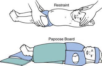
Figure 628-1 Methods of restraining an infant for examination and for procedures such as tympanocentesis or myringotomy.

(From Bluestone CD, Klein JO: Otitis media in infants and children, ed 2, Philadelphia, 1995, WB Saunders, p 91.)

When examining the ear, inspecting the auricle and external auditory meatus for infection can aid in evaluating complications of OM. External otitis can result from acute OM with discharge, or inflammation of the posterior auricular area can indicate a periostitis or subperiosteal abscess extending from the mastoid air cells. The presence of preauricular pits or skin tags also should be noted because affected children have a slightly higher incidence of SNHL; ear pits can develop chronic infection.

Cerumen is a protective, waxy, water-repellent coating in the ear canal that can interfere with examination. Cerumen usually is removed using the surgical head of the otoscope, which allows passage of a wire loop or a blunt curette under direct visualization. Other methods include gentle irrigation of the ear canal with warm water, which should be performed only if the TM is intact, or instillation of a solution such as diluted hydrogen peroxide in the ear canal (with intact TM only) for a few minutes to soften the wax for suction removal or irrigation. Some commercial preparations such as trolamine polypeptide oleate-condensate (Cerumenex) can cause dermatitis of the external canal with chronic use and should be used only under a physician’s supervision.

Inflammation of the ear canal with associated pain often indicates external otitis. Abnormalities of the external auditory canal include stenosis (common in children with trisomy 21), bony exostoses, otorrhea, and the presence of foreign bodies. Cholesteatoma of the middle ear can manifest in the canal as intermittent foul-smelling drainage, sometimes associated with white debris; cholesteatoma of the external canal can appear as a white, pearl-like mass in the canal skin. White or gray debris of the canal suggests fungal external otitis. Newborn ear canals are filled with vernix caseosa, which is soft and pale yellow and should disappear shortly after birth.

The TM and its mobility are best assessed with a pneumatic otoscope. The normal TM is in a neutral position; a bulging TM may be caused by increased middle-ear air pressure, with or without pus or effusion in the middle ear; a bulging drum can obscure visualization of the malleus and annulus. Retraction of the TM usually indicates negative middle-ear pressure, but it also can result from previous middle-ear disease with fixation of the ossicles, ossicular ligaments, or TM. When retraction is present, the bony malleus appears more prominent, and the incus may be more visible posterior to the malleus.

The normal TM has a silvery-gray, “waxed paper” appearance (Fig. 628-2). A white or yellow TM can indicate a middle-ear effusion. A red TM alone might not indicate pathology, because the blood vessels of the membrane may be engorged as a result of crying, sneezing, or nose blowing. A normal TM is translucent, allowing the observer to visualize the middle-ear landmarks: incus, promontory, round window niche, and, often, the chorda tympani nerve. If a middle-ear effusion is present, an air-fluid level or bubbles may be visible (see Fig. 628-2). Inability to visualize the middle-ear structures indicates opacification of the drum, usually caused by thickening of the TM, a middle-ear effusion, or both. Assessment of the light reflex often is not helpful, because a middle ear with effusion reflects light as well as a normal ear.

image

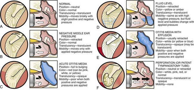
Figure 628-2 A-F, Common conditions of the middle ear, as assessed with the otoscope.

(From Bluestone CD, Klein JO: Otitis media in infants and children, ed 3, Philadelphia, 2001, WB Saunders, p 131.)

TM mobility is helpful in assessing middle ear pressures and the presence or absence of fluid (see Fig. 628-2). To best perform pneumatic otoscopy, a speculum of adequate size is used to obtain a good seal and allow air movement in the canal. A rubber ring around the tip of the speculum can help to obtain a better canal seal. Normal middle-ear pressure is characterized by a neutral TM position and brisk TM movement to both positive and negative pressures.

Eardrum retraction is most common when negative middle-ear pressure is present; with even moderate negative middle-ear pressure there is no visible inward movement with applied positive pressure in the ear canal (see Fig. 628-1). However, negative canal pressure, which is produced by releasing the rubber bulb of the pneumatic otoscope, can cause the TM to bounce out toward the neutral position. The TM can retract in both the presence and absence of middle-ear fluid, and if the middle-ear fluid is mixed with air, the TM might still have some mobility. Outward eardrum movement is less likely in the presence of severe negative middle-ear pressure or middle-ear effusion.

The TM that exhibits fullness (bulging) moves to applied positive pressure but not to applied negative pressure if the pressure within the middle ear is positive. A full TM and positive middle-ear pressure without an effusion may be seen in young infants who are crying during the otoscopic examination, in older infants and children with nasal obstruction, and in the early stage of acute OM. When the middle-ear-mastoid air cell system is filled with an effusion and little or no air is present, the mobility of the TM is severely decreased or absent in response to both applied positive and negative pressures.

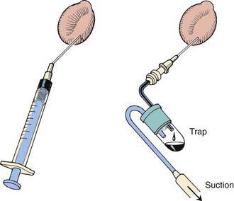
Tympanocentesis, or aspiration of the middle ear, is the definitive method of verifying the presence and type of a middle-ear effusion and is performed by inserting, through the inferior portion of the TM, an 18-gauge spinal needle attached to a syringe or a collection trap (Fig. 628-3). Culturing of the ear canal and alcohol cleansing should precede tympanocentesis and culture of the middle-ear aspirate; a canal culture is taken first to help determine whether organisms cultured from the middle ear are contaminants from the external canal or true middle ear pathogens.

Further diagnostic studies of the ear and hearing include audiometric evaluation, impedance audiometry (tympanometry), acoustic reflectometry, and specialized eustachian tube function studies. Diagnostic imaging studies, including CT and MRI, often provide further information about anatomic abnormalities and the extent of inflammatory processes or neoplasms. Specialized assessment of labyrinthine function should be considered in the evaluation of a child with a suspected vestibular disorder (Chapter 633).