Gastric Volvulus

Published on 13/07/2015 by admin

Filed under Radiology

Last modified 22/04/2025

Print this page

rate 1 star rate 2 star rate 3 star rate 4 star rate 5 star
Your rating: none, Average: 0 (0 votes)

This article have been viewed 3023 times

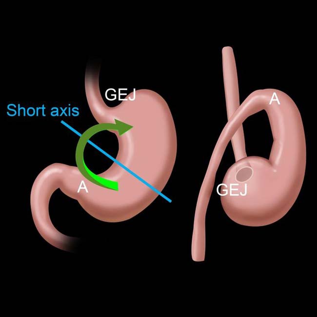
 Most common type; “upside-down stomach”

image Occurs in setting of large paraesophageal hernia
image Stomach rotates upward, with greater curvature lying above lesser curve
• Mesenteroaxial volvulus: Rotation of stomach about its short axis

image More common type in children
• Entire stomach may be herniated (type IV paraesophageal hernia [PEH]) or only part (type III PEH)

image Either can result in volvulus ± obstruction ± ischemia
image Gastric wall pneumatosis indicates ischemia
• Diagnosed with upper GI &/or CT
• CT is better at demonstrating associated hernias and gastric ischemia

TOP DIFFERENTIAL DIAGNOSES

• Hiatal hernia

image Types III and IV PEHs increase risk for gastric volvulus
• Postoperative state, stomach

image Esophagectomy with gastric pull through (conduit may twist and obstruct)
• Epiphrenic diverticulum

CLINICAL ISSUES

• Treatment: Open or laparoscopic detorsion and gastropexy

DIAGNOSTIC CHECKLIST

• Presence or absence of obstruction and ischemia are more important than remembering or reporting whether volvulus is organo- or mesenteroaxial
image
(Left) Graphic illustrates an organoaxial gastric volvulus, in which the stomach twists along its long axis, resulting in the greater curvature (GC) lying above the lesser curvature (LC).

image
(Right) Film from an upper GI series in a 73-year-old woman shows a type IV paraesophageal hernia (PEH) with organoaxial volvulus but little or no obstruction. The greater curvature of the stomach image 
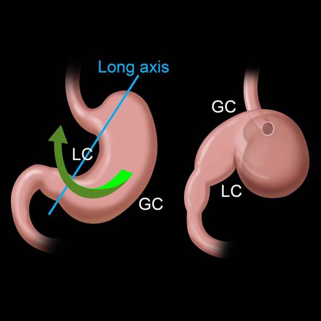
lies above the lesser curvature. The small bowel image is also herniated through a large diaphragmatic defect.
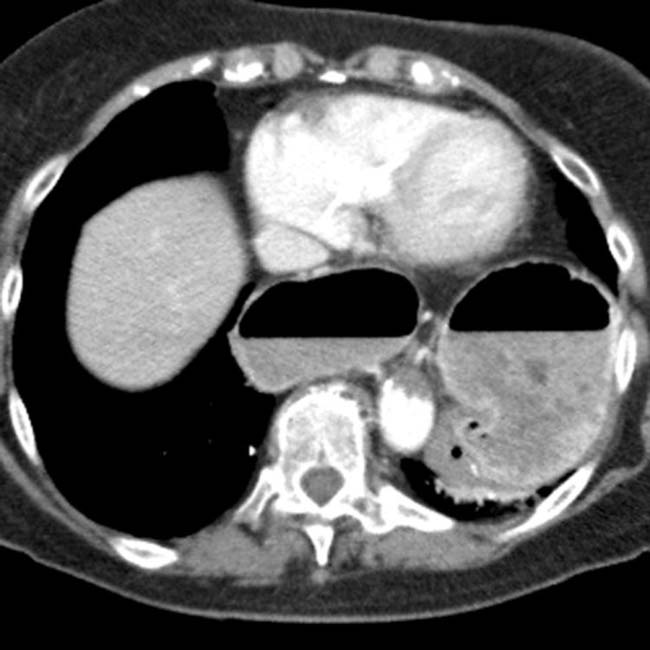
image
(Left) Axial CECT demonstrates an intrathoracic stomach (type IV PEH) in a 81-year-old woman with mild chest pain and a known brain malignancy. The stomach is dilated with 2 air-fluid levels, indicating obstruction.

image
(Right) Coronal CECT in the same patient demonstrates an “upside-down” configuration of the stomach, with reversal of the greater and lesser curvatures, in keeping with an organoaxial volvulus.

TERMINOLOGY

Abbreviations

• Gastric volvulus

Definitions

• Uncommon acquired twist of stomach on itself

IMAGING

General Features

• Morphology

image Abnormal degree of rotation of 1 part of stomach around another part
• Types of volvulus: Organoaxial (most common), mesenteroaxial, mixed
• Organoaxial volvulus: Rotation of stomach around its longitudinal axis

image Around line extending from cardia to pylorus
image Stomach rotates upward, with greater curvature lying above lesser curvature
image Antrum moves from inferior to superior position; fundus rotates superior to inferior
image Usual setting is with a large paraesophageal hernia (PEH)

– Type III PEH = Gastroesophageal (GE) junction and portions of fundus and body herniate into chest
– Type IV PEH = GE junction and almost entire stomach lie within chest
• Mesenteroaxial volvulus: Rotation of stomach about its mesenteric (short) axis

image Axis running transversely across stomach at right angles to lesser and greater curvatures
image Stomach rotates from right to left, or left to right about long axis of gastrohepatic omentum
image Not necessarily in setting of hiatal hernia
image May result from congenital absence or laxity of gastric ligaments
• Mixed volvulus: Combination of organoaxial and mesenteroaxial volvulus

Radiographic Findings

• Radiography

image Abdominal plain films; patient upright

– Double air-fluid level
– Large, distended stomach; seen as air- and fluid-filled spheric viscus displaced upward and to left
– Small bowel collapsed if stomach is obstructed
image Chest film: Intrathoracic; upside-down stomach

– Retrocardiac fluid level; 2 air-fluid interfaces at different heights; suggests intrathoracic gastric volvulus

Fluoroscopic Findings

• Upper GI

image Massively distended stomach in left upper quadrant extending into chest
image Inversion of stomach (upside-down stomach)

– Greater curvature above level of lesser curvature
– Positioning of cardia and pylorus at same level
– Downward pointing of pylorus and duodenum
image Volvulus with > 180° twist causes luminal obstruction
image Incomplete or absent entrance of contrast material into &/or out of stomach; acute obstructive volvulus
image May see “beaking” at point of twist
image Mesenteroaxial: Antrum and pylorus lie above gastric fundus

CT Findings

• CT appearance may be variable

image Depends upon extent of gastric herniation, points of torsion and final positioning of stomach
image May see linear septum within gastric lumen; corresponding to area of torsion
• Entire stomach may be herniated (type IV PEH) or only part (type III PEH)

image Either can result in volvulus ± obstruction ± ischemia
image Ischemia seen as lack of contrast enhancement of gastric wall ± pneumatosis within wall
• CT chest and abdomen; performed preoperatively

image To detect associated malformation or malposition and site, size, level of diaphragmatic defect

MR Findings

• Coronal images demonstrate 2 points of twisting

image Different signal intensities reflect point of torsion

Angiographic Findings

• GV may present as acute upper gastrointestinal hemorrhage

Imaging Recommendations

• Best imaging tool

image Upper GI series

– Demonstrates focus of twist; anatomic detail
image Fluoroscopic guidance may help in advancing nasogastric tube into obstructed stomach

– May allow decompression and stabilization of patient
image CT: Complementary role

– Multiplanar reformations  help depict altered anatomy
– CT is better at diagnosing gastric ischemia

DIFFERENTIAL DIAGNOSIS

Hiatal Hernia

• Stomach entering thorax through esophageal hiatus
• Type I: “Sliding”; GE junction and cardia above diaphragm
• Type II: Herniation of fundus through hiatus; GE junction below diaphragm (rare)
• Type III: Most common type of paraesophageal hernia; GE junction and fundus ± body of stomach in chest
• Type IV: Intrathoracic stomach; GE junction and most of stomach in chest
• Type III and IV paraesophageal hernias predispose to volvulus

Postoperative State, Stomach

• Esophagectomy with gastric pull-through procedure
• Complete mobilization of stomach, resection of lower esophagus, pyloroplasty, transhiatal dissection

image Intrathoracic stomach is the intended result
image Conduit can twist (volvulus) with outlet obstruction

Epiphrenic Diverticulum

• Large pulsion diverticulum above diaphragm
• May have an air-fluid level

PATHOLOGY

General Features

• Etiology

image Stabilizing ligaments are too lax as result of congenital or acquired causes
image Congenital Morgagni or Bochdalek hernia

– May lead to gastric volvulus in children or adults
image Traumatic diaphragmatic rupture with gastric herniation
• Associated abnormalities

image Large paraesophageal hernia

– Permits part or all of stomach to enter chest
image Diaphragmatic eventration or paralysis

– Could result in volvulus of intraabdominal stomach (rare)
image Wandering spleen: Absence of ligamentous connections between stomach, spleen
image Hernia of colonic transverse loop ± other bowel loops

– Invariably through very large diaphragmatic defect

Gross Pathologic & Surgical Features

• Herniated and twisted stomach
• May or may not be ischemic

image Complete obstruction often leads to ischemia and perforation if not corrected

CLINICAL ISSUES

Presentation

• Gastric volvulus can be asymptomatic if there is no outlet obstruction or vascular compromise

image May be an incidental finding on imaging
image Patients usually endorse symptoms of GE reflux, if asked
• Acute volvulus; associated interference of blood supply

image Surgical emergency
image Classic clinical triad (Borchardt triad)

– Violent retching with production of little vomitus
– Constant severe epigastric pain
– Difficulty in advancing nasogastric (NG) tube beyond distal esophagus
• Chronic or intermittent gastric volvulus

image Frequently not recognized early in its presentation
image Vague and nonspecific symptoms suggestive of other abdominal processes, causing delay in diagnosis

– Volvulus may be inadvertently reduced by placement of NG tube prior to imaging, precluding diagnosis
image May be discovered incidentally during clinical work-up for unrelated condition

– CT (or MR) often requested as 1st radiographic study during evaluation
• Symptomatic GV in infancy and childhood may not be as rare as is commonly assumed

Demographics

• Age

image Mesenteroaxial is most common type in children

– Associated anatomic defects are usually found
image Organoaxial accounts for over 60% of adult cases; only 40% of pediatric cases

– More common in elderly
• Epidemiology

image 5 cases of combined organo- and mesenteroaxial GV in children reported in world literature

Natural History & Prognosis

• As hernias enlarge, body and variable portion of antrum come to lie above diaphragm

image Stomach can become entirely intrathoracic organ; prone to volvulus
• Obstruction can occur at points of torsion or at points where stomach reenters abdomen through hiatus

image As much as 180° of twisting may occur without obstruction or strangulation
image Twisting beyond 180° usually produces complete obstruction and clinically acute abdomen
image Organoaxial: Can obstruct; does not usually result in strangulation

– Mesenteroaxial: Can occlude gastric vessels; strangulation
• Upside-down stomach

image Usually constitutes organoaxial volvulus ± obstruction
image Presents with bleeding and anemia; uncommonly induces obstruction or strangulation
• Vascular occlusion leads to necrosis, shock
• Strangulation may lead to mucosal ischemia and perforation

image Areas of focal necrosis; may permit gas to dissect into gastric wall
• Prognosis: gastric volvulus is potentially a catastrophic condition

image Mortality rate: 30% when obstruction is present

Treatment

• Goals: Early recognition and surgical repair

image Detorse stomach
image Repair of associated defects in diaphragm

– Gastropexy; may be prophylactic, prevent recurrence
• Open or laparoscopic detorsion and gastropexy
• Gastric resection for strangulation and necrosis
• Upside-down stomach: Balloon repositioning; fixation by percutaneous endoscopic gastrostomy

DIAGNOSTIC CHECKLIST

Consider

• Anatomical detail of stomach often better delineated on upper gastrointestinal studies

image Identification of gastric volvulus can be incidental finding on CT

– Consider whenever stomach is noted to  be in abnormal anatomic position

Image Interpretation Pearls

• Presence or absence of obstruction and ischemia are more important than remembering or reporting whether volvulus is organo- or mesenteroaxial

image
(Left) Frontal radiograph (not shown) in an elderly woman showed an intrathoracic stomach (type IV PEH). CT showed the esophagogastric junction in the chest. The pylorus image 
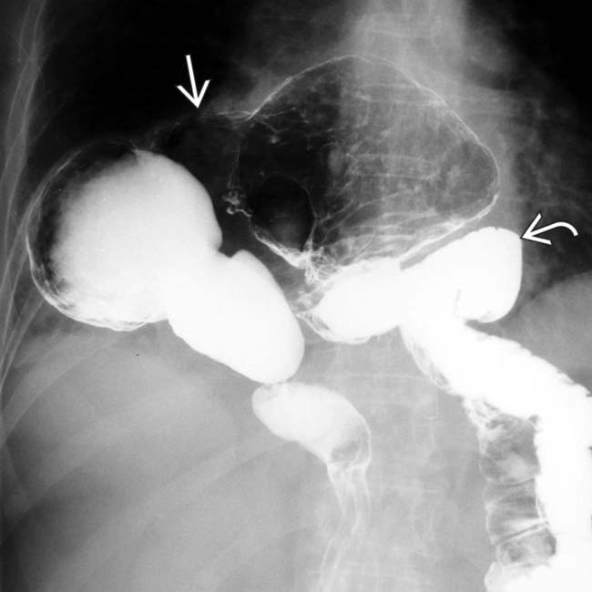
also lies within the chest.
image
(Right) More caudal CT section in the same patient shows a type IV PEH. The fundus image 
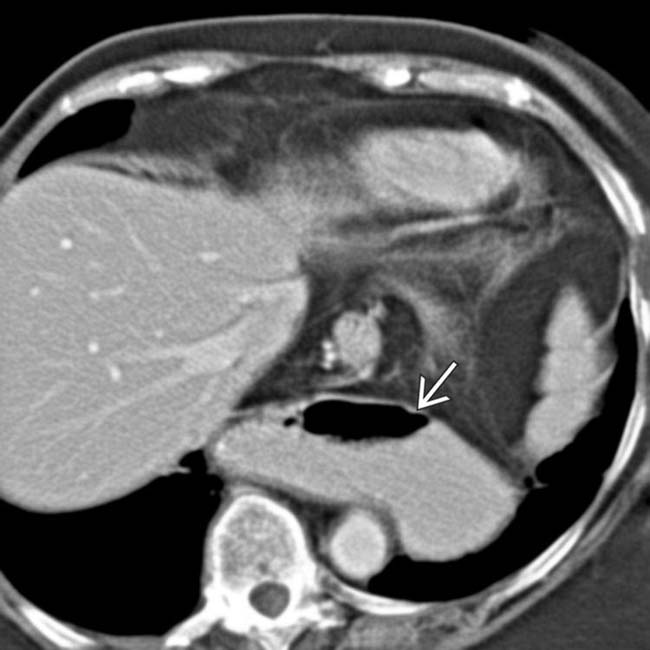
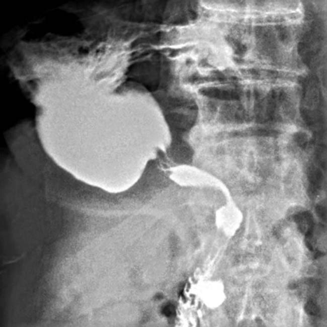
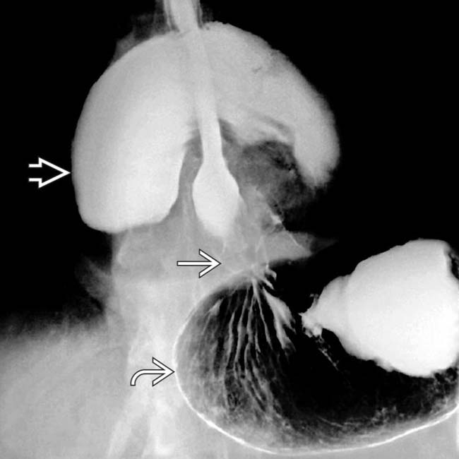
lies lower than the antrum and pylorus. This could be considered organoaxial “position” versus “volvulus,” and no obstruction is present.
image
(Left) Axial NECT in a 63-year-old woman with chronic intermittent chest and abdominal pain demonstrates the entirety of the stomach within the thoracic cavity (type IV PEH). Notice the large amount of contrast retained in the stomach image 
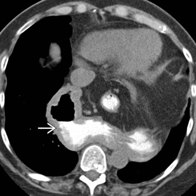
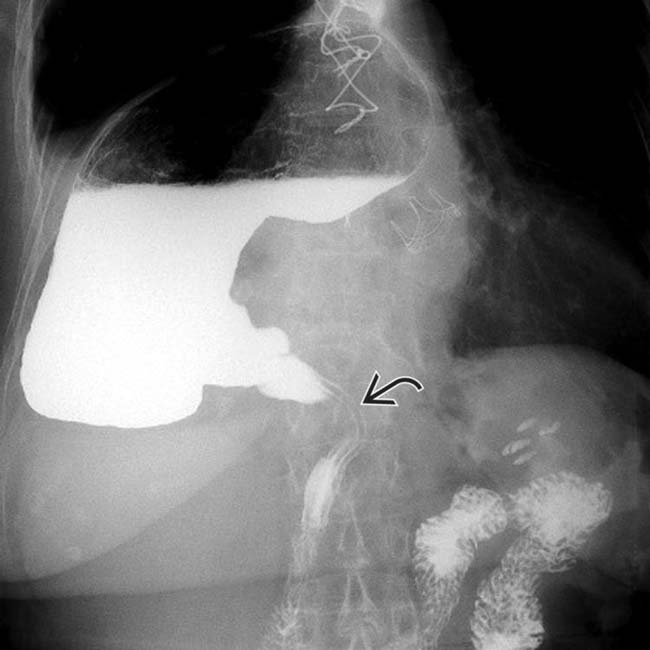
nearly 2 hours after its administration.

image
(Right) Axial NECT in the same patient again demonstrates the intrathoracic stomach image 
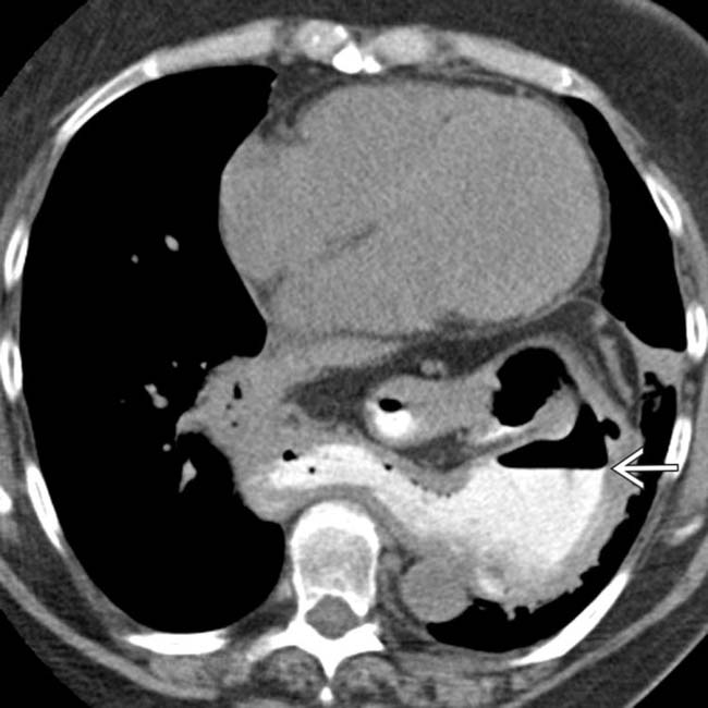
with retained contrast in its lumen.
image
(Left) Coronal NECT in the same patient better demonstrates the twisting of the stomach, with the greater and lesser curvatures having reversed.

image
(Right) Coronal NECT in the same patient demonstrates contrast retained within the midthoracic esophagus image
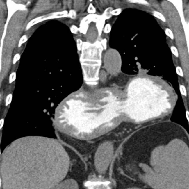
. The patient was felt to be at least partially obstructed, and underwent operative repair, where the organoaxial volvulus was confirmed.

image
(Left) Axial NECT in an 83-year-old woman with chest pain demonstrates the entirety of the stomach located within the thorax, a type IV PEH.
image
(Right) Coronal NECT in the same patient demonstrates the stomach twisted along its long axis, with the greater curvature above the lesser curvature, in keeping with an organoaxial volvulus.
image
(Left) Upright film from an upper GI series in an 80-year-old woman with symptoms only of reflux shows a dilated intrathoracic stomach (type IV PEH) with an organoaxial volvulus. The duodenal lumen image 
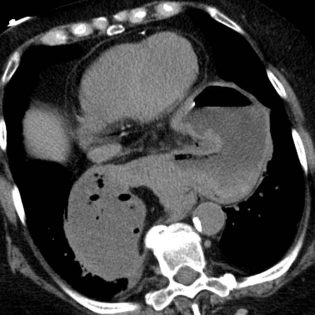
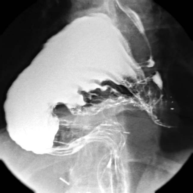
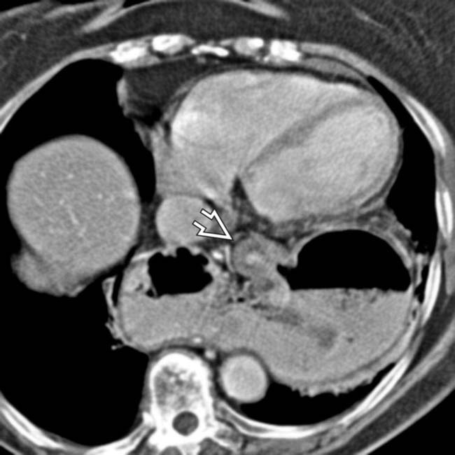
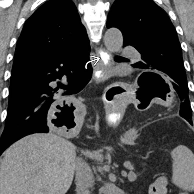
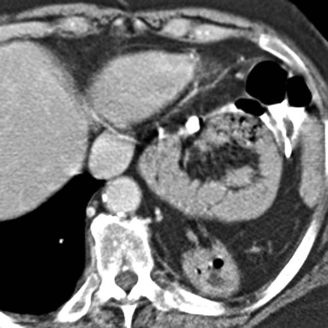
is narrowed as it enters the abdomen.
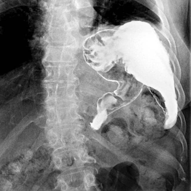
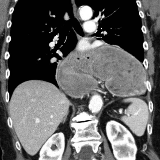
image
(Right) Upright film from an upper GI shows eventration or paralysis of the left diaphragm and an upside-down stomach, another appearance of an organoaxial volvulus. Partial gastric outlet obstruction is evident by delayed passage of barium into the duodenum.
image
(Left) Graphic illustrates a mesenteroaxial volvulus in which the stomach twists along its short axis, resulting in the antrum (A) lying above the fundus and gastroesophageal (GE) junction.

image
(Right) Spot film from an upper GI series shows the GE junction image and fundus image in the abdomen. The body and antrum of the stomach image are in the chest and are twisted and compressed as they traverse the diaphragm, constituting a mesenteroaxial volvulus with obstruction.

image
(Left) Supine film in an elderly woman with severe vomiting, retching, and hematemesis shows gas distention of an abnormal-appearing stomach image 
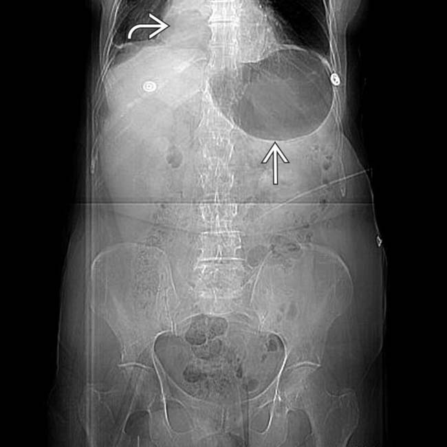
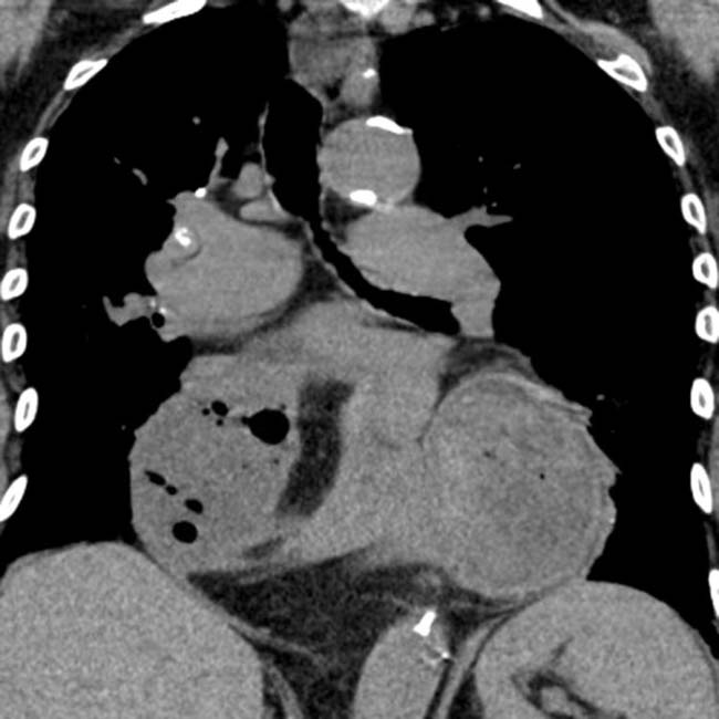
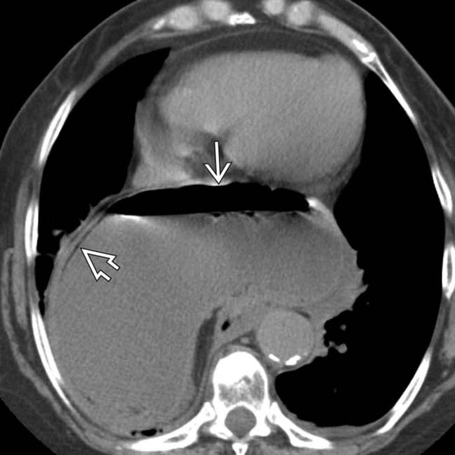
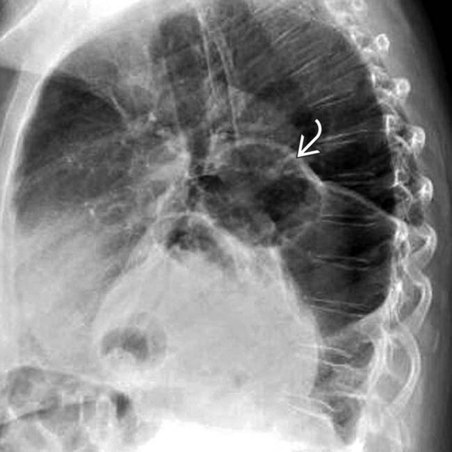
and retrocardiac mass effect image.
image
(Right) Lateral supine scout digital radiograph in the same patient shows 2 air-fluid levels within the stomach image
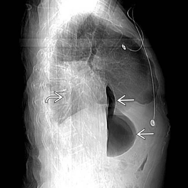
, and the retrocardiac fluid-density mass image.
image
(Left) CECT in the same patient shows a large type III PEH with fluid distention of the herniated stomach image
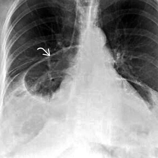
, along with pneumatosis in the gastric wall image, indicating ischemic injury.

image
(Right) Another CT image in the same case shows 2 separate air-fluid levels image 
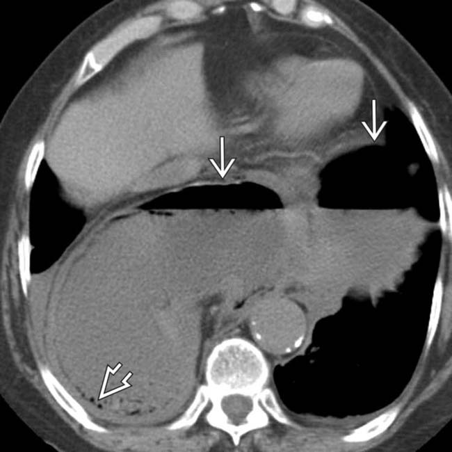
within the distended stomach, and additional evidence of gastric pneumatosis image. The larger collection of gas and fluid is within the gastric body and antrum, which lie in the thorax, while the smaller collection is within the intraabdominal gastric fundus.
image
(Left) More caudal CT section in the same case shows pneumatosis image 
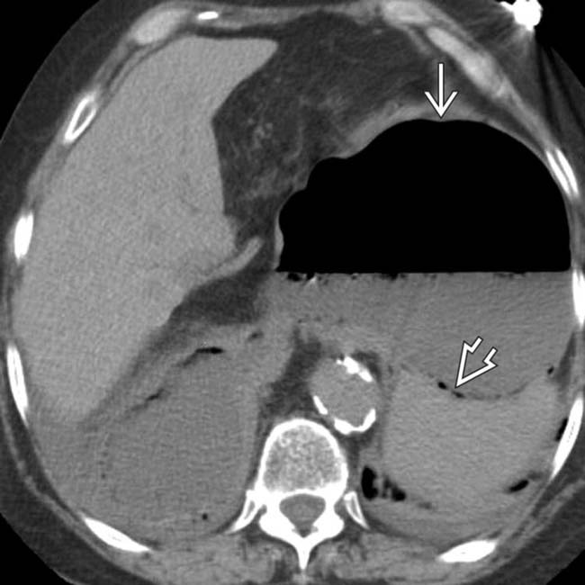
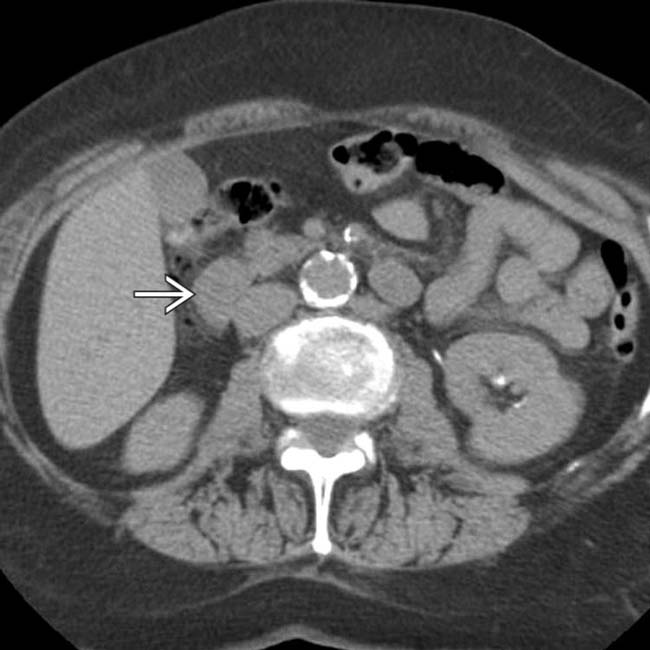
within the gastric fundus image.

image
(Right) More caudal CT section in the same patient shows a gasless duodenum image and small bowel due to the incarcerated gastric volvulus. Volvulus and gastric infarction were confirmed at surgery.
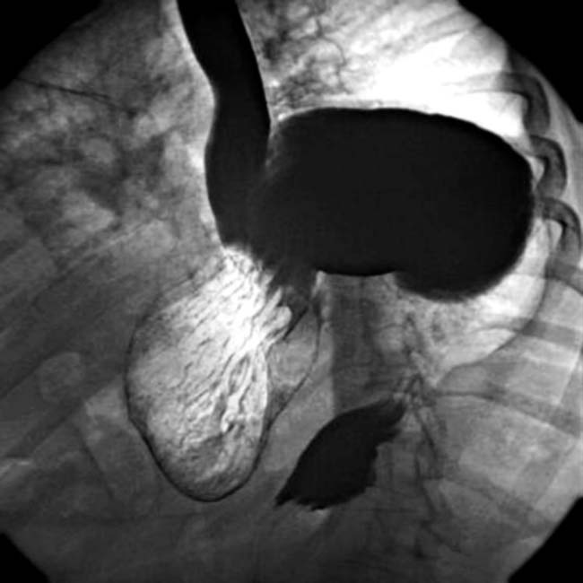
image
Upper GI shows an intrathoracic stomach with organoaxial volvulus and obstruction.

image
Axial CECT shows mesenteroaxial gastric volvulus with the stomach rotated left to right.
image
Lateral chest radiograph in the same patient shows the distended intrathoracic stomach image.
image
PA chest radiograph shows a distended intrathoracic stomach image due to volvulus with acute obstruction.
image
Upper GI series shows intrathoracic stomach with organoaxial volvulus but no obstruction.
image
Upper GI series shows intrathoracic stomach with organoaxial volvulus and partial obstruction.

SELECTED REFERENCES

1. Collet, D, et al. Management of large para-esophageal hiatal hernias. J Visc Surg. 2013; 150(6):395–402.

2. Cross, BN, et al. Roux-en-Y gastric bypass in the setting of congenital malrotation: a report and review of the literature. Surg Obes Relat Dis. 2013; 9(6):e91–e95.

3. Toydemir, T, et al. Laparoscopic management of totally intra-thoracic stomach with chronic volvulus. World J Gastroenterol. 2013; 19(35):5848–5854.

4. Bawahab, M, et al. Management of acute paraesophageal hernia. Surg Endosc. 2009; 23(2):255–259.

5. Chang, CC, et al. A surgical emergency due to an incarcerated paraesophageal hernia. Am J Emerg Med. 2009; 27(1):134.

6. Gerstle, JT, et al. Gastric volvulus in children: lessons learned from delayed diagnoses. Semin Pediatr Surg. 2009; 18(2):98–103.

7. Yano, F, et al. Outcomes of surgical treatment of intrathoracic stomach. Dis Esophagus. 2009; 22(3):284–288.

8. Shivanand, G, et al. Gastric volvulus: acute and chronic presentation. Clin Imaging. 2003; 27(4):265–268.

9. Tabo, T, et al. Balloon repositioning of intrathoracic upside-down stomach and fixation by percutaneous endoscopic gastrostomy. J Am Coll Surg. 2003; 197(5):868–871.

10. Godshall, D, et al. Gastric volvulus: case report and review of the literature. J Emerg Med. 1999; 17(5):837–840.

Schaefer, DC, et al. Gastric volvulus: an old disease process with some new twists. Gastroenterologist. 1997; 5(1):41–45.