


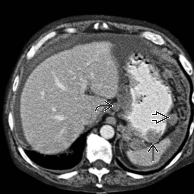
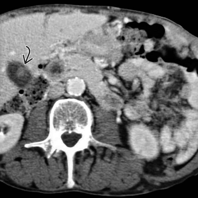
 , lymph nodes
  , and omentum
  .
.
 , lymph nodes
  , and omentum
  , with both nodular and diffuse metastases seen. In addition, the left ureter was obstructed due to a ureteral/retroperitoneal metastasis.
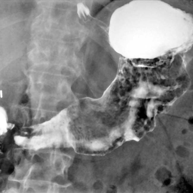
 . These findings are typical of primary gastric lymphoma.
IMAGING
General Features

Fluoroscopic Findings
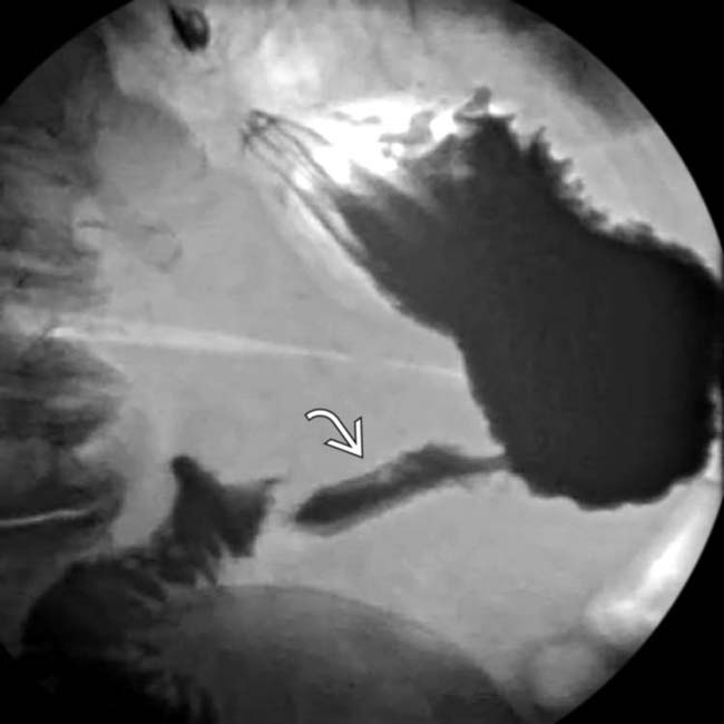
• Fluoroscopic-guided barium study
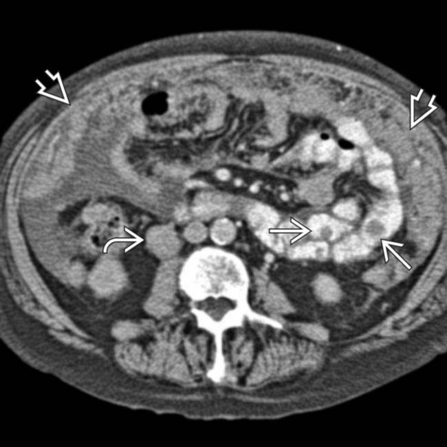
 Breast carcinoma metastases
 Breast carcinoma metastases


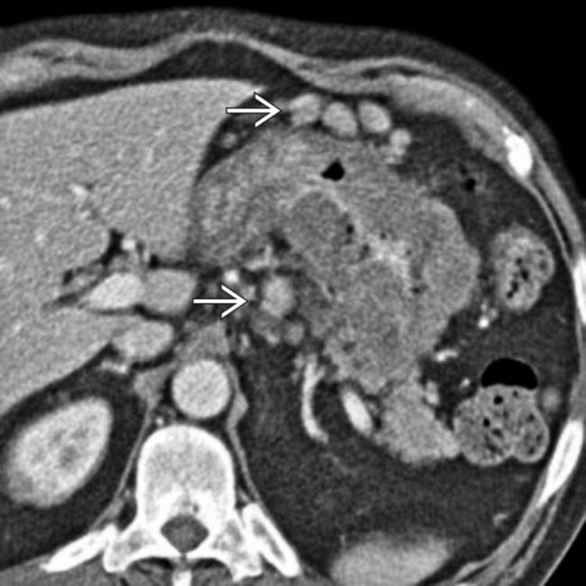
CT Findings
• Multiplanar reformation (MPRs) very useful to judge full extent of disease and relation to adjacent organs, nodes


 . The stomach is not well distended or easily assessed.
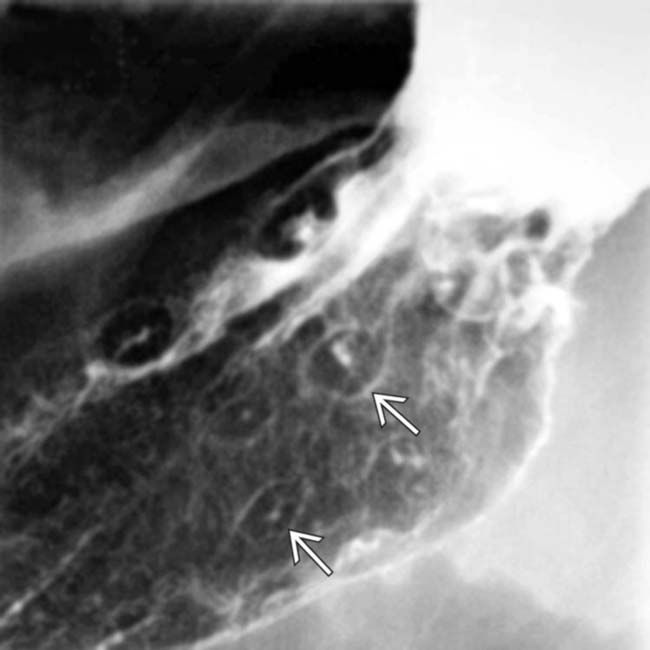
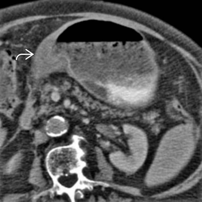
 , consisting of small, intramural masses with a central ulceration.
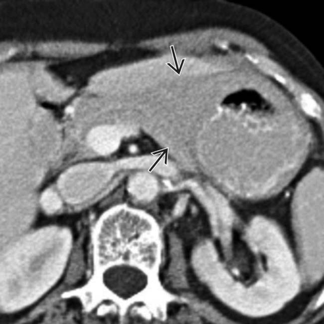
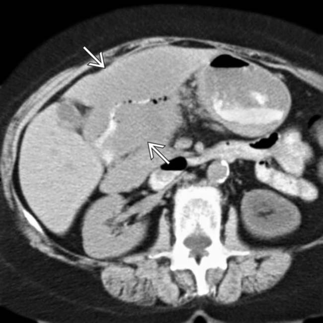
 that diffusely infiltrates the gastric wall. There is no outlet obstruction.
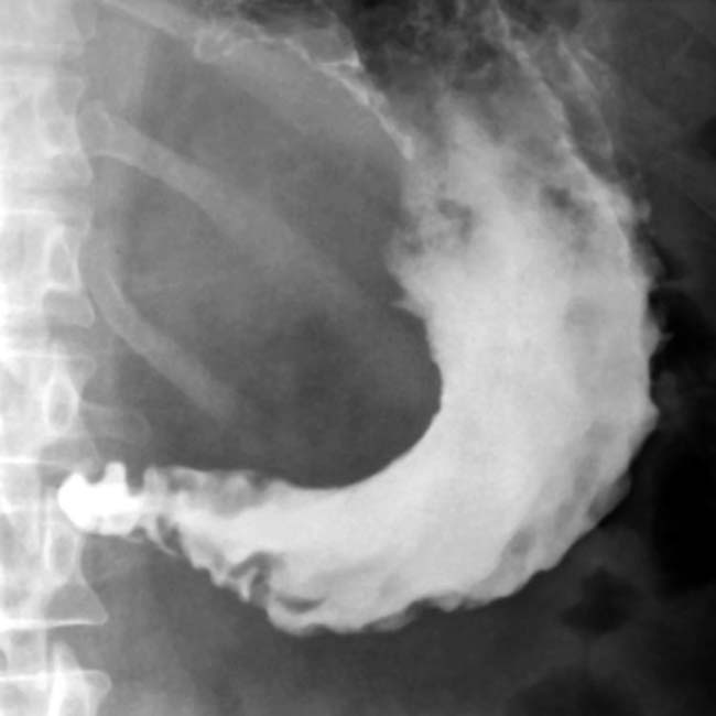
 .
.
 causing delayed gastric emptying. These imaging findings are indistinguishable from primary gastric carcinoma.
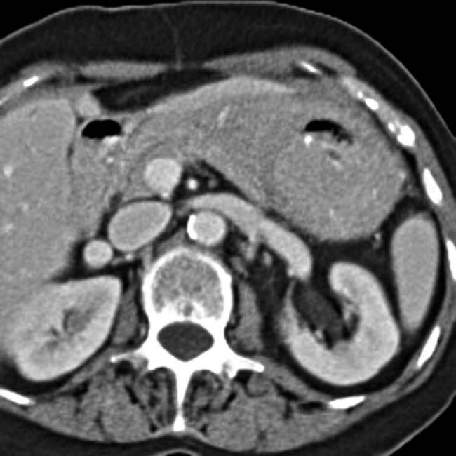
 but no obstruction in this patient with lymphoma.
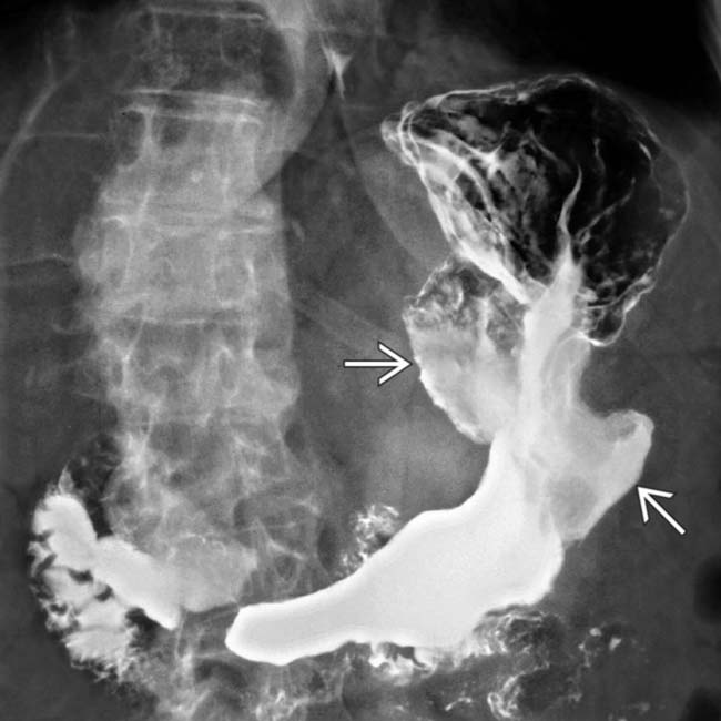
 but no obstruction.
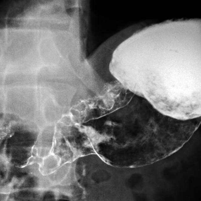
 from metastatic melanoma.
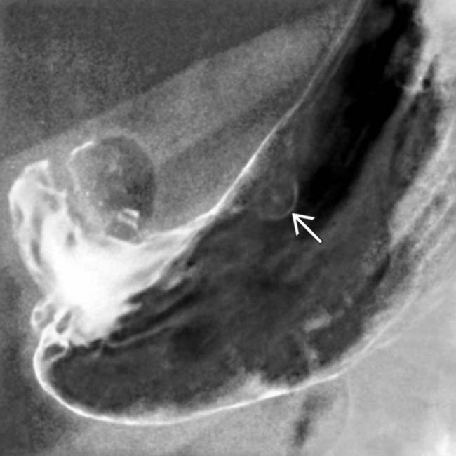
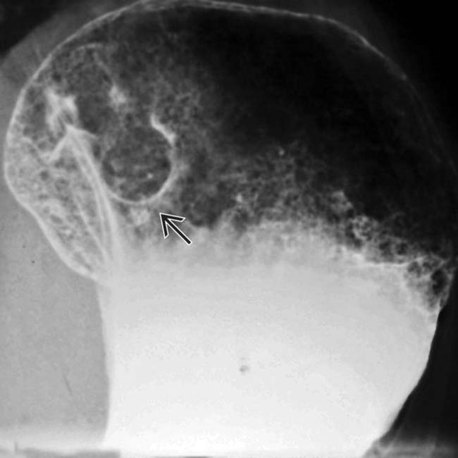
 , a discrete intramural polyp with central ulceration in this patient with metastatic melanoma.
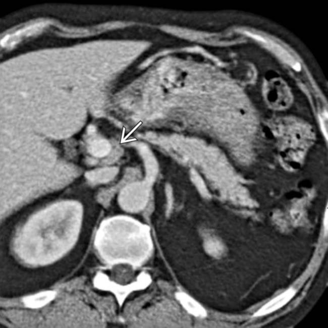
 in this patient with gastric lymphoma.
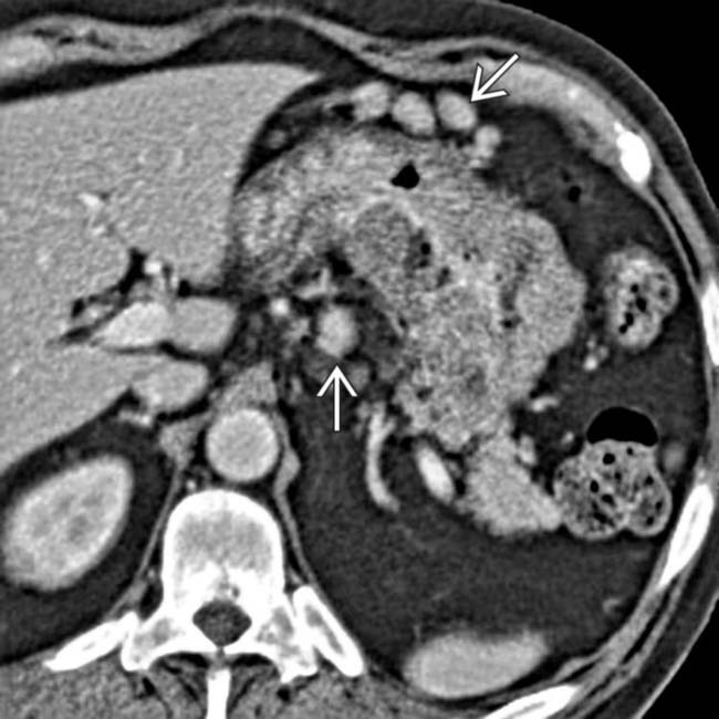
 in this patient with gastric lymphoma.


