Content usually sterile without GB inflammation
• GB empyema: Pus-filled, inflamed, and distended GB secondary to acute cholecystitis with suppurative intraluminal infection


 , findings consistent with gallbladder hydrops.
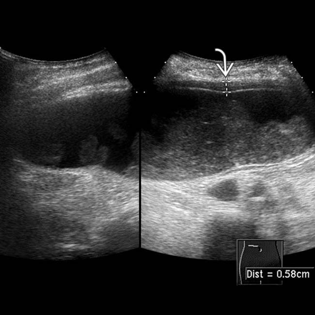
 and rounded but mobile internal echogenic sludge.
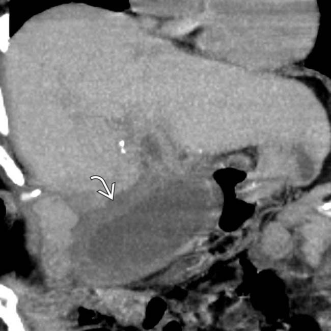
 . Because the patient was considered a poor surgical candidate, she was treated with percutaneous cholecystostomy, which yielded thick bile with a heavy growth of bacteria, consistent with empyema of the gallbladder.


DIFFERENTIAL DIAGNOSIS
Gangrenous Cholecystitis
• GB wall necrosis and gangrene occurs in 1/4 of patients with acute cholecystitis (usually elderly, diabetic, or immunocompromised patients)


 .
.
 . Lack of contrast within the GB in the hepatobiliary phase suggests that this chronic asymptomatic hydrops is due to a cystic duct obstruction.
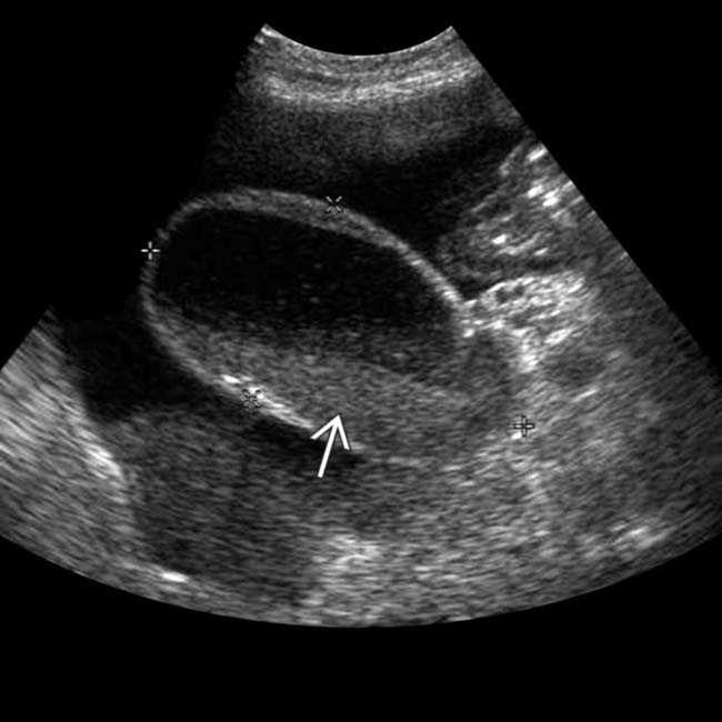
 . The patient had a positive Murphy sign and underwent cholecystectomy, which demonstrated cholecystitis and empyema.
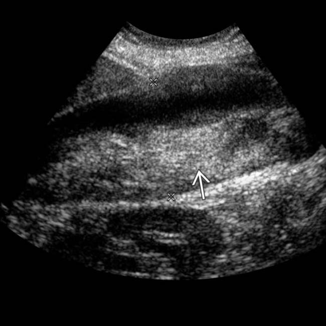
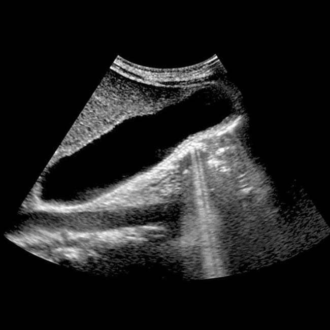
 . Sonographic Murphy sign was positive, compatible with GB empyema.
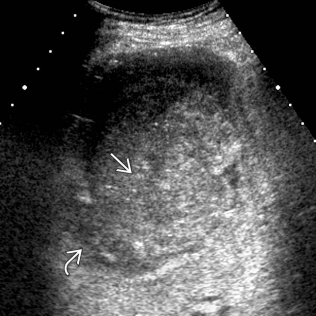
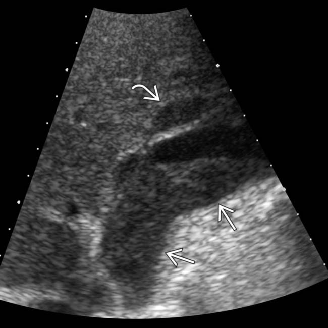
 . The poor definition of the GB wall
  was suspicious for gangrene, confirmed at surgery.
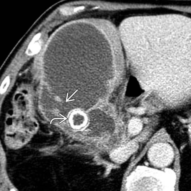
 , and a large gallstone
  , compatible with GB empyema and associated contained perforation.
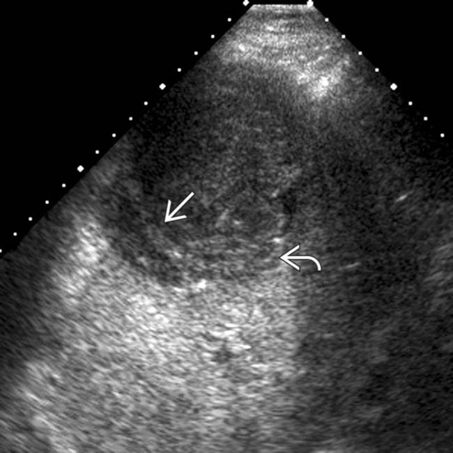
 within the gallbladder due to gangrenous cholecystitis, as well as echogenic pus
  within the gallbladder due to gallbladder empyema.
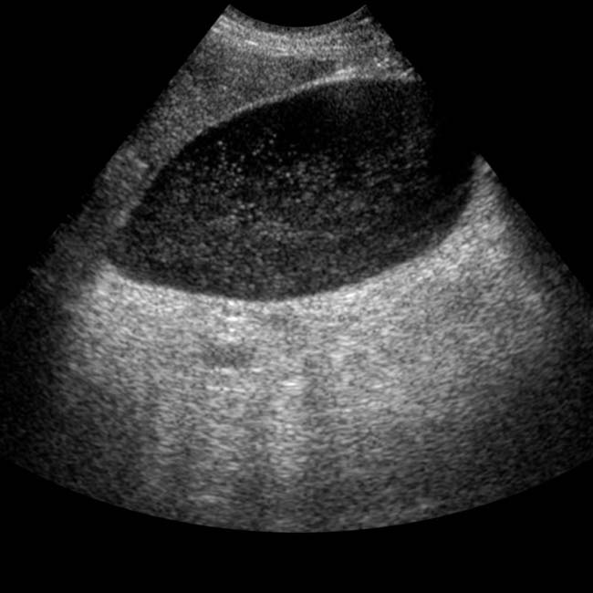
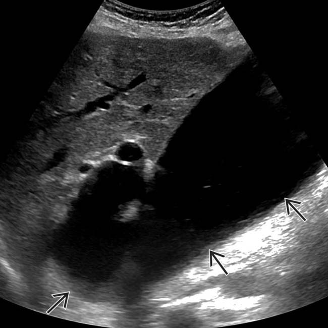
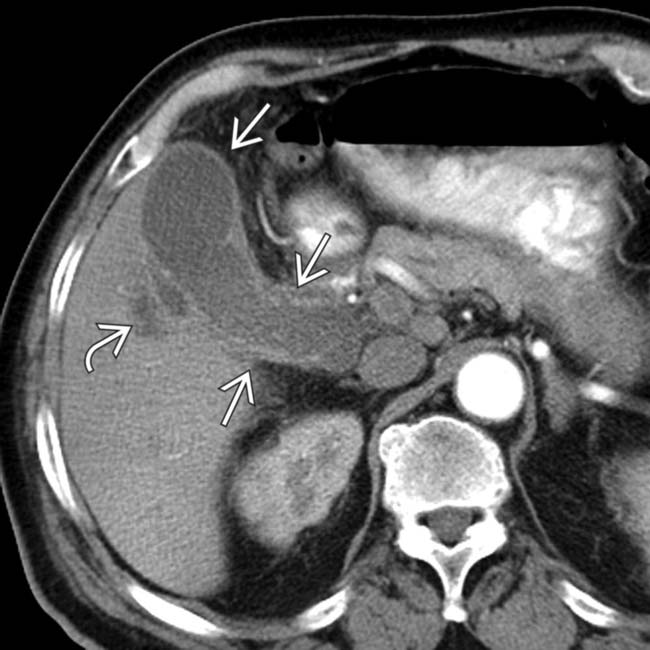
 and a poorly defined pericholecystic fluid collection along adjacent liver
  .
.
 and a pericholecystic fluid collection
  . A perforated, pus-filled GB and an adjacent abscess were identified at laparotomy and cholecystectomy.
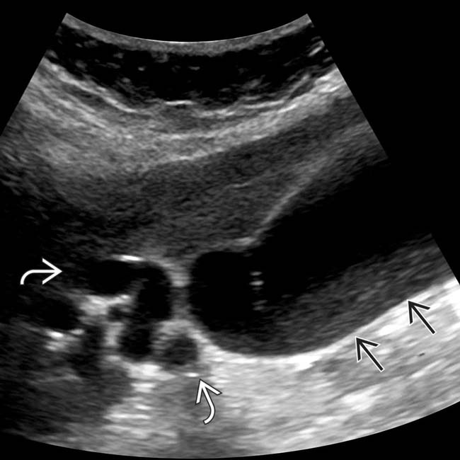
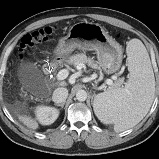
 , and cystic duct dilatation
  . GB distension was the most obvious manifestation of duct obstruction at ultrasound and prompted additional imaging.
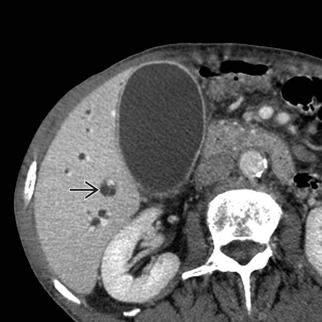
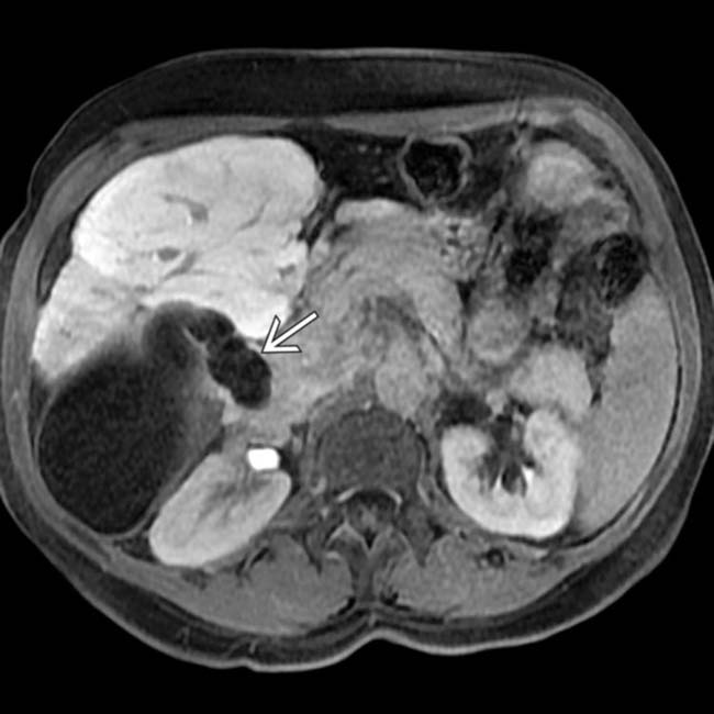
 . GB distension was due to obstruction of the distal common bile duct by a pancreatic head carcinoma (not shown).


