Gallbladder

Published on 10/05/2015 by admin

Filed under Gastroenterology and Hepatology

Last modified 22/04/2025

Print this page

rate 1 star rate 2 star rate 3 star rate 4 star rate 5 star
Your rating: none, Average: 0 (0 votes)

This article have been viewed 1672 times

).

image
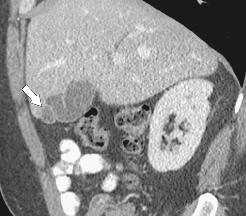
Figure 8-1 Sagittal contrast-enhanced CT in a 47-year-old man with “Phrygian cap” appearance of the gallbladder fundus (arrow).
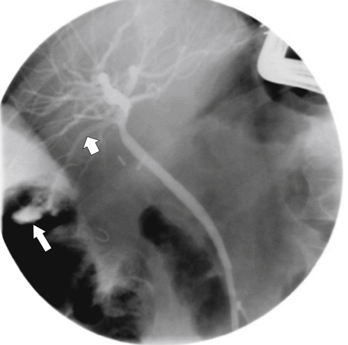
Biliary anatomy has numerous variants, particularly the insertion of the cystic duct into the common hepatic duct. They are a variety of aberrant ducts, perhaps the best known being the duct of Luschka, knowledge of which is critical to prevent surgical transection at cholecystectomy (Fig. 8-2). Less commonly, congenital gallbladder anomalies arise, including duplicated gallbladder or gallbladder agenesis. Gallbladder agenesis is present in approximately 1 per 10,000 individuals. Its only significance is that it is almost always unsuspected and patients with right upper quadrant pain are sometimes inappropriately diagnosed with chronic cholecystitis. They are then referred for cholecystectomy, upon which the gallbladder cannot be identified. This may confound the radiologist and surgeon, who thought they had definitive sonographic finding of a contracted gallbladder full of stones. Rather, the hyperechoic structure in the region of the gallbladder represented gas within the duodenal lumen. Occasionally the gallbladder has a long mesentery and is therefore at risk of torsion with consequent gangrene and perforation.
image
Figure 8-2 ERCP in a 43-year-old woman with recent cholecystectomy and a current bile leak (arrow) from an aberrant duct of Luschka (small arrow).
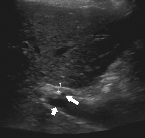
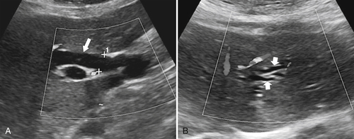
Oral cholecystography, once the mainstay of gallbladder imaging, is no longer used because of its variable gallbladder opacification and because it caused relatively frequent allergic reactions. More recently, however, it has been used in conjunction with multidetector computed tomography (CT) to evaluate biliary tree disease, but its use is not widespread. Ultrasonography (US) has largely replaced oral cholecystograph and is the primary imaging method for the gallbladder given its superficial location and fluid-filled nature. US detects most gallstones in patients who are prepared appropriately and is useful for the evaluation of acute and chronic cholecystitis. It can also detect very small (<5 mm) anomalies, including polyps and cholesterosis. US is also useful for evaluating the intrahepatic and extrahepatic biliary system, particularly if distended. US may detect CBD stones, but more distal stones may prove challenging because of overlying bowel gas that attenuates the sound waves. Subtle biliary dilatation can be observed, and the CBD caliber is readily identified in most patients where the duct crosses the hepatic artery (Fig. 8-3). Normal measurements depend on the age of the patient and any prior cholecystectomy. The CBD typically measures 2 to 3 mm in younger patients, increasing to 4 to 6 mm in the elderly. A CBD greater than 7 mm is considered dilated in the elderly unless there has been prior cholecystectomy, in which case the duct can measure up to 10 mm normally.
image
Figure 8-3 US of a normal common bile duct (between +s) at the level of the hepatic artery (large arrow), which is anterior to the portal vein (small arrow).
CT is less useful than US, although thin-section multidetector CT with multiplanar reformatting can sometimes prove clinically useful. Many gallstones demonstrate variable calcific concentrations, and CT detects only 50% to 70% of gallstones and even fewer CBD stones. Its main use is to evaluate for biliary malignancies (gallbladder carcinoma and intrahepatic cholangiocarcinoma). Common hepatic duct and CBD cholangiocarcinomas are often small and difficult to identify by CT, but their presence can be inferred when they occlude the duct, resulting in biliary obstruction.
Imaging of the biliary system with magnetic resonance cholangiopancreatography (MRCP) has improved dramatically with fast fat-suppressed two- or three-dimensional T2-weighted sequences. Bile is hyperintense on these sequences and is readily differentiated from surrounding anatomy. Images are evaluated either coronally as a composite three-dimensional image or on individual axial maximum intensity projections. This is often useful when evaluating more subtle pancreaticobiliary abnormalities, particularly of the distal pancreatic or bile duct. Although MRCP in its own right is indicated for detection and characterization of pancreaticobiliary disease, the indication for MRCP is often as a replacement for failed endoscopic retrograde cholangiopancreatography (ERCP), either because of absolute or relative contraindications or because of procedure failure. ERCP has better fine-detail resolution, particularly for small intrahepatic or aberrant bile ducts. Furthermore, ERCP offers the opportunity for therapeutic intervention. However, when ERCP is to be avoided for whatever reason, MRCP is an effective screening tool to evaluate for ductal stone disease, choledochal cysts, and intrahepatic ductal anomalies (sclerosing cholangitis, peribiliary cysts).

Choledochal Cyst

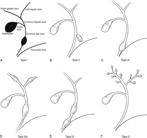
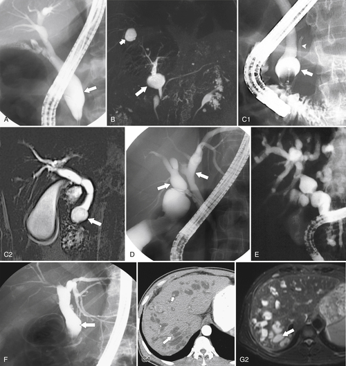
All of the five different forms of choledochal cyst represent variations of congenital cystic dilatation of the bile ducts (Fig. 8-4 and Table 8-1). Their significance is that patients are at increased risk of developing cholangiocarcinoma. Type I is by far the most common (Fig. 8-5), representing approximately 80% to 90% of all forms. Choledochal cysts usually present in childhood, although type IV is just as common in adulthood. There are numerous associated anomalies, including gallbladder agenesis or duplication, biliary atresia, polycystic liver disease and fibrosis (hepatic fibrocystic disease), and annular pancreas. Patients may be asymptomatic, although most present in childhood with secondary signs of cholangitis and jaundice, with or without intraductal stone formation.
image
Figure 8-4 A through F, Schematic representation of types I through V choledochal cysts (choledochoceles).

Table 8-1

Classification of Choledochal Cysts (see Figs. 8-5 and 8-6)

Type I Fusiform dilatation of all or part of CBD (i.e., extrahepatic)
Type II CBD diverticulum (extrahepatic)
Type III Choledochocele (dilatation at junction of CBD and pancreatic duct)
Type IVa Fusiform intrahepatic and extrahepatic duct dilatation
Type IVb Combination of Types I and III
Type V Caroli disease (cystic dilatation of intrahepatic ducts)

CBD, Common bile duct.

image
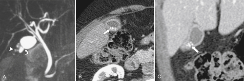
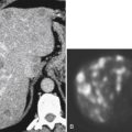
Figure 8-5 Choledochoceles types I through V. A, Type I ERCP demonstrates fusiform dilatation of the distal CBD (arrow). B, MRCP in a 36-year-old man with a type II choledochal cyst (arrow). There is an incidental liver cyst (small arrow). C1 and C2, Type III MRCP and ERCP in a 40-year-old man with choledochocele (arrows). A pancreatic stent (arrowhead) is present. D, ERCP in a 47-year-old woman with fusiform intrahepatic duct dilatation (arrows) caused by a type IVa choledochal cyst. E, ERCP in a 39-year-old woman with a type IVa choledochal cyst. F, Type IVb choledochal cyst. ERCP in a 56-year-old man with fusiform dilatation (arrow) of the CBD and choledochocele of the distal duct. G1 and G2, Type V choledochal cyst. Axial contrast-enhanced CT and T2-weighted MRI in a 40-year-old with Caroli disease with abundant dilated intrahepatic biliary ducts (arrows) and a central dot sign (small arrow) typical of this disease.

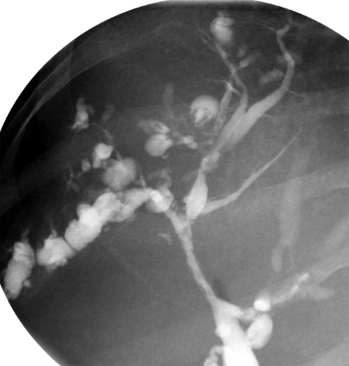
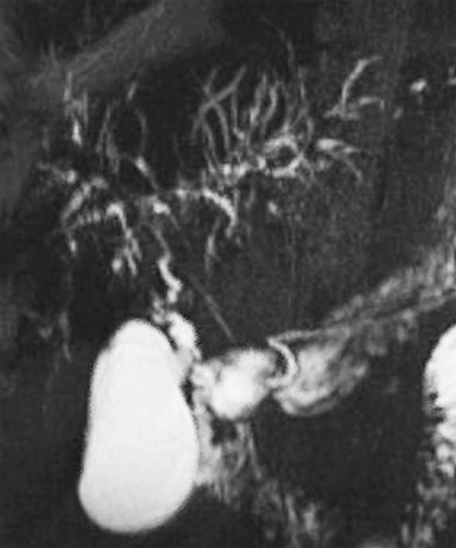
Caroli disease is a congenital disease with two forms, a simple autosomal-dominant type and a complex autosomal-recessive type, but is also classified as a type V choledochal cyst. The simple variety, which usually presents in young adulthood, has isolated ectatic cavernous intrahepatic bile ducts, whereas the complex variety forms part of a Caroli syndrome with ectatic ducts, congenital hepatic fibrosis, and portal hypertension, which usually presents in childhood. The complex form is also associated with autosomal-recessive polycystic kidney disease, medullary sponge kidney, and biliary hamartomas. The defective biliary anatomy is best visualized by ERCP (Fig. 8-6), which shows marked and irregular segmental biliary cavernous ectasia. However, all cross-sectional imaging techniques can identify these findings. A characteristic feature at contrast-enhanced CT is the “central dot” sign, representing enhancing portal tracts within the grossly dilated bile ducts (Fig. 8-5).
image
Figure 8-6 ERCP in a 63-year-old woman with Caroli disease and multiple cystic intrahepatic ducts.

Gallstones and Gallbladder Sludge

Stone formation is secondary to biliary material concretions within the gallbladder, but depending on their size, stones can pass into the cystic or bile ducts, pancreatic duct, or duodenum. Stones may be single (sometimes large) or multiple and when granular are referred to as pseudoliths or, more commonly, gallbladder sludge. Cholesterol makes up approximately 80% of the composition in most stones (75% to 80%). Pigment stones are less common (approximately 20%) and are composed mainly of bilirubin and calcium salts (80%), the remainder being cholesterol. Mixed stones are less common and are composed of between 20% and 80% cholesterol and a mixture of calcium and phosphate salts and bilirubin. A number of predisposing factors are associated with gallstone formation, including obesity, diabetes mellitus, cirrhosis, hemolytic anemias (pigment stones), small bowel malabsorption abnormalities (ulcerative colitis, Crohn disease, and small bowel resection), and hyperthyroidism.
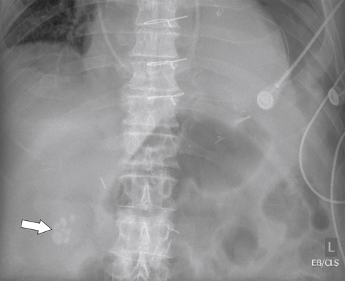
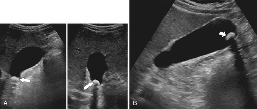
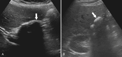
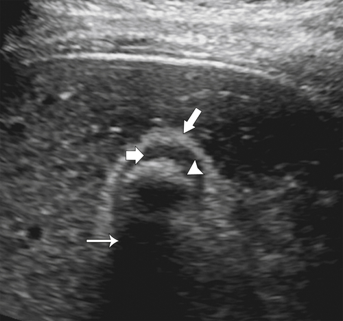
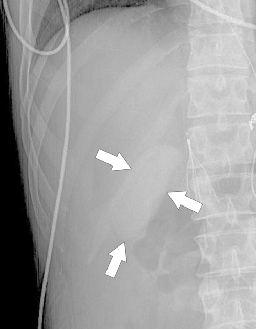
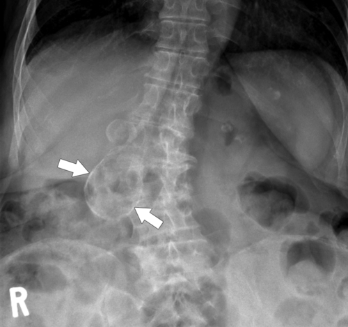
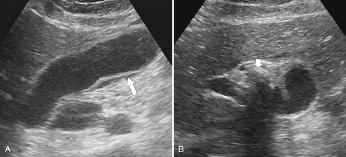
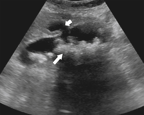
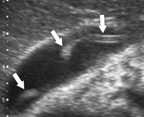
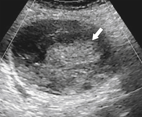
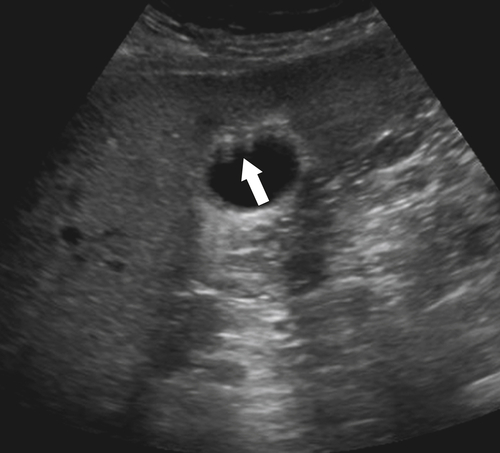
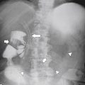
Gallstones are commonly identified at imaging and are usually asymptomatic. Most gallstones are not sufficiently calcified to be visible on plain radiographs, but some mixed (owing to their calcium concentration) stones are visible in the right upper quadrant (Fig. 8-7). US is the investigation of choice but needs adequate patient preparation, which requires the patient to fast for up to 8 hours to ensure gallbladder distention (the gallbladder usually contracts on feeding). Almost all gallstones can then be identified. They are hyperechoic, rounded structures with strong acoustic shadowing (Fig. 8-8). Their presence can be confirmed by placing the patient in the decubitus position, which should cause the stone to drop into the most dependent part of the gallbladder (Fig. 8-8). Stones impacted in the gallbladder neck (or occasionally adherent to the gallbladder wall) may not move, however. Gallstones can be difficult to identify definitively in patients with a contracted gallbladder from chronic cholecystitis. A large stone in a chronically contracted gallbladder can be almost impossible to differentiate from a gas-filled duodenum, which also demonstrates marked hyperechogenicity (Figs. 8-9 and 8-10). Under these circumstances a definitive diagnosis of gallstones can be made only by the presence of the sonographic wall-echo-shadow (WES) sign. In this sign the wall represents the gallbladder wall, the echo the superficial acoustic reflective surface of the gallstone, and the shadow the attenuated sound (or shadow) beyond the gallstone surface (Fig. 8-10). However, between the gallbladder wall and the echogenic stone surface, a sliver of radiolucent bile must be observed to document gallstones definitively and exclude duodenal gas. Without the presence of the WES sign in these circumstances, exclusion of duodenal gas can be made by requesting that the patient swallow water and immediately performing repeat imaging with the patient in the right lateral decubitus position. Swirling ingested gas and water should be observed, confirming the location of the duodenum.
image
Figure 8-7 Plain abdominal radiograph in a 75-year-old man with calcified gallstones in the right upper quadrant (arrow).
image
Figure 8-8 A and B, Sagittal, transverse, and lateral decubitus US in a 47-year-old woman with a hyperechoic gallstone in the gallbladder neck with strong posterior acoustic shadowing (large arrows) that moves in the decubitus position to the gallbladder fundus (small arrow).
image
Figure 8-9 A, Sagittal US in a 55-year-old woman with dense shadowing from gallstones (arrow) in a contracted gallbladder caused by chronic cholecystitis. The US appearance of duodenal gas can be identical. B, Transverse US in a 53-year-old woman with echogenic material in the gallbladder region (arrow). Shadowing is due to duodenal gas and not gallstones.

image
Figure 8-10 Transverse US in a 27-year-old woman with a hyperechoic gallbladder wall (long arrow), sonolucent bile (short arrow), hyperechoic stone (arrowhead), and posterior acoustic shadow (thin arrow), which constitute the WES sign.
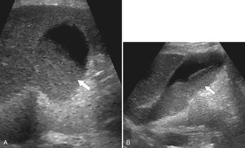
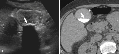
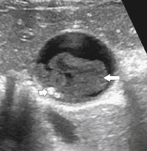
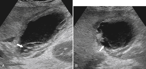
Gallbladder sludge is less commonly observed than gallstones but more commonly recognized in the fasting state, particularly in acutely ill hospitalized patients because fasting and multiple cholestatic medications cause gallbladder stasis, which encourages the deposition of biliary particulate material (calcium bilirubinate or cholesterol crystals). Most patients are asymptomatic. At US the sludge is recognized as a layering, nonshadowing, and slightly hyperechoic material (Fig. 8-11), and at CT it is often slightly hyperdense (Fig. 8-12). Occasionally the sludge coalesces to a more mass-like form, known as tumefactive sludge (Fig. 8-13), which is sometimes mistaken for a gallbladder neoplasm, but the tumefactive mass is often accompanied by mobile sludge. Furthermore, the tumefactive sludge may itself move, and there is no color Doppler flow within the sludge unlike tumors that frequently demonstrate some vascularity.
image
Figure 8-11 A and B, Sagittal and transverse US in a 55-year-old man with layering gallbladder sludge (arrows).
image
Figure 8-12 Transverse US (A) and axial noncontrast (B) in a 64-year-old woman with multiple hyperdense dependent small layering stones (arrows) and sludge, which fill the gallbladder. The sludge is slightly hyperdense at CT (arrowhead).

image
Figure 8-13 Gallstones and sludge. Transverse US images on the gallbladder in a 29-year-old woman with sickle cell disease, a tumefactive sludge ball (large arrow), and pigment stones, which do not shadow (small arrow).
Approximately 80% of gallstones are incidentally detected at CT performed for other reasons. Pure cholesterol stones are hypodense (owing to their lipid content) and isodense to normal bile. The internal architecture can sometimes be appreciated as uniformly dense from predominantly calcified material, laminated from alternating cholesterol and calcified elements (Fig. 8-14), or stellate (also known as the Mercedes-Benz sign), caused mainly by nitrogen gas trapped within the stone matrices (Fig. 8-15). The routine use of CT is not warranted for detection of gallstones but is for some of their complicating features, including cholecystitis, perforation, carcinoma formation, pancreatitis, and ascending cholangitis from cholelithiasis. Gallstones are generally well identified by T2-weighted MRI because of the superior contrast characteristics of high-signal bile and low-signal stones (Fig. 8-16).
image
Figure 8-14 Axial contrast-enhanced CT in a 31-year-old woman with typical CT appearances of cholesterol gallstones (arrow).
image
Figure 8-15 Coronal contrast-enhanced CT in a 61-year-old man with a stellate gallstone with internal nitrogen gas (arrow).
image
Figure 8-16 Axial fat-saturated T2-weighted image in a 44-year-old woman with multiple hypointense gallstones (arrow) surrounded by hyperintense bile.

Choledocholithiasis

Choledocholithiasis is defined as the presence of gallstones within the biliary tree and outside the gallbladder, mostly in the CBD. The stones are usually secondary to the passage of smaller gallstones through the cystic duct into the CBD, where they can move up and down the duct freely or become lodged at the ampulla of Vater. However, primary bile or hepatic duct stone formation is recognized when there is excessive biliary material production (e.g., pigment stone formation in hemolytic anemia) or biliary stasis conditions (congenital ductal anomalies, sphincter of Oddi malfunction), foreign body material (e.g., suture material), or parasitic infections (Ascaris lumbricoides, Clonorchis sinensis). Small stones pass spontaneously, but unlike gallstones, CBD stones are symptomatic and patients may present with obstructive jaundice, fever, pain, or pancreatitis.
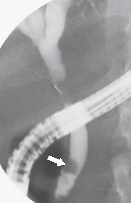
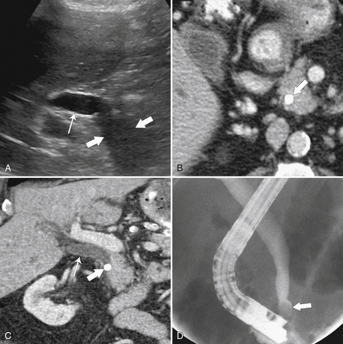
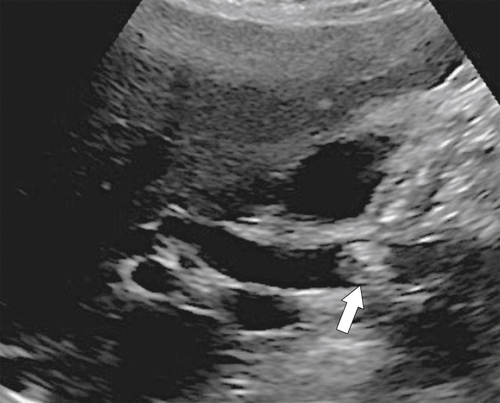
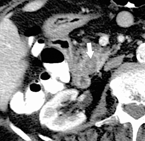
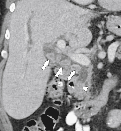
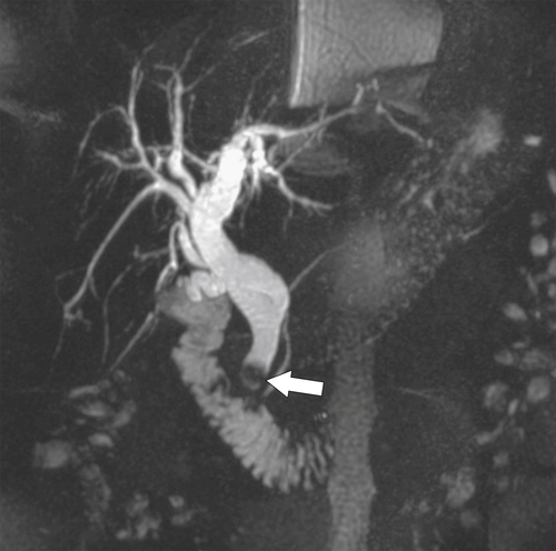
The most sensitive imaging test for CBD stone detection is the injection of water-soluble contrast material into the CBD via either ERCP (Fig. 8-17) or T-tube cholangiography, in which the opacified ducts outline a radiolucent stone. Confusion with an air bubble is usually resolved by placing the patient in the Trendelenburg position (stones are dependent, and gas is nondependent). The stones are less readily identified by US because of normal overlying duodenal bowel gas and other anatomical structures. However, good US technique can identify many stones as hyperechoic shadowing structures within the hypoechoic lumen of the gallbladder. Obstructing stones should also distend the CBD, which then acts as a guide for detection of the offending impacted stone more distally, even if the stone itself is not clearly identified (Fig. 8-18). The absence of posterior acoustic shadowing should not deter the diagnosis, since approximately 10% of CBD stones do not shadow (Fig. 8-19). Most CBD stones are difficult to identify with CT, partly because biliary stones are less conspicuous on CT but also because smaller stones may not be resolved adequately (Fig. 8-20). Denser stones may be identified, particularly if impacting with a distended bile duct above the stone (Figs. 8-18 and 8-21). The absence of a stone at CT does not exclude choledocholithiasis. However, T2-weighted MRI sequences, particularly MRCP, are far more sensitive, detecting approximately 90% of stones because normal bile has intensely bright T2 signal (similar to water), which permits the visualization of even small bile duct stones (Fig. 8-22). This is therefore the noninvasive investigation of choice should US prove unhelpful and is often preferable to ERCP, an invasive procedure.
image
Figure 8-17 ERCP in a 49-year-old man with a radiolucent stone (arrow) in the lower duct.
image
Figure 8-18 Sagittal right upper quadrant US (A), axial (B), and coronal (C) contrast-enhanced CT and ERCP (D) in a 66-year-old man with biliary dilatation due to common duct stone (thin arrows). The stone is not identified on US, but there is strong acoustic shadowing (larger arrows inA) implying a common duct stone. Secondary biliary dilatation and strong acoustic shadows from the impacted stone are identified with CT and ERCP (arrows).
image
Figure 8-19 Sagittal US in a 49-year-old woman with bile duct dilatation caused by a stone in the lower duct (arrow).
image
Figure 8-20 Axial contrast-enhanced CT in a 77-year-old man with gout and a urate stone in the lower bile duct. The stone is only just visible by CT (arrow).
image
Figure 8-21 Coronal contrast-enhanced CT in a 70-year-old man with several CBD stones (arrows), one of which obstructs the lower duct (arrowhead), causing intrahepatic ductal dilatation.
image
Figure 8-22 MRCP in a 59-year-old woman with CBD and intrahepatic duct dilatation caused by an impacted stone at the ampulla of Vater (arrow).
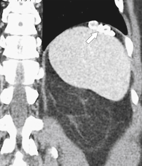
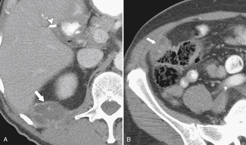
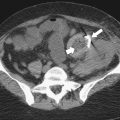
Gallstones may reside outside the gallbladder or biliary tree, either because not all gallstones were removed during cholecystectomy (so-called dropped gallstone) or because of gallbladder perforation resulting from chronic cholecystitis. In the former, the gallstones can be located anywhere in the peritoneal cavity (Fig. 8-23), but their visualization will depend, as in the gallbladder, on their calcium content. Some dropped stones therefore will not be visualized, but often they create a peritoneal or retroperitoneal (depending on where they drop) inflammatory reaction, which should not be confused with a mesenteric neoplasm (Fig. 8-24).
image
Figure 8-23 Coronal contrast-enhanced CT in a 54-year-old man with left subphrenic gallstones (arrow) due to “dropped” stones.
image
Figure 8-24 A and B, Axial contrast-enhanced CT in a 71-year-old man with abdominal pain and two abscesses (arrows) secondary to “dropped” gallstones. Note prior cholecystectomy clips (arrowhead).

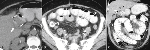
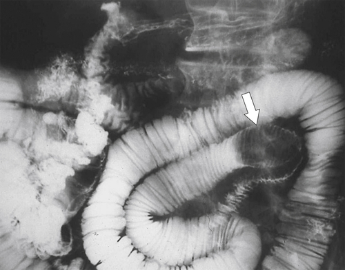
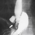
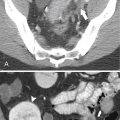
After gallbladder perforation the extruded gallstones can have an appearance similar to dropped gallstones, and the gallbladder itself will invariably appear contracted with signs of previous pericholecystic inflammation. Because of the proximity of the gallbladder to the duodenum, the inflammatory reaction from repeated bouts of cholecystitis may create a biliary enteric duodenal fistula as the gallstones erode into the intestinal lumen, with subsequent retrograde reflux of gas into the biliary system (Fig. 8-25). If small, they may be excreted through the rectum, but larger stones can become lodged in the distal ileum, particularly at its narrowest point, the ileocecal valve. This can cause small bowel obstruction called gallstone ileus (Fig. 8-25). Larger stones may obstruct the small bowel more proximally (Fig. 8-26). Alternatively, the gallstone may erode directly into the colon and be excreted through the rectum, since the colon is more distensible, although in rare cases gallstones are sufficiently large to even cause large bowel obstruction. Erosion directly into the stomach, jejunum, and ileum has also been recognized.
image
Figure 8-25 Axial (A and B) and coronal (C) contrast-enhanced CT in a 77-year-old woman with small bowel obstruction (arrowheads) caused by a gallstone ileus (curved arrow). Small inflammatory mass (large arrow) is due to a choleduodenal fistula with biliary gas (small arrows).
image
Figure 8-26 SBFT in a 56-year-old woman with a large obstructing jejunal gallstone (arrow).

Mirizzi Syndrome

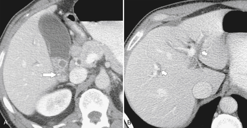
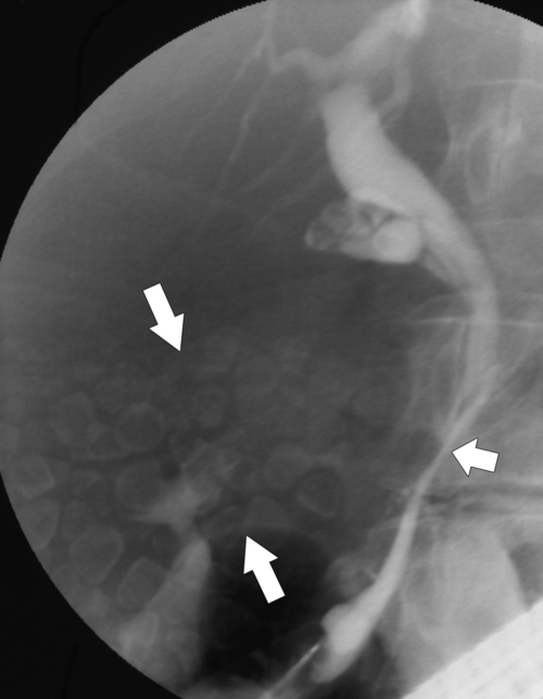
Mirizzi syndrome is a rare complication of gallstone disease in which a large gallstone (usually within a chronically diseased gallbladder) becomes impacted in the cystic duct or gallbladder neck. The stone and the associated inflammatory reaction of the cystic duct then produce mass effect and impinge on the CBD to form a stricture and cause obstructive jaundice. Intrahepatic duct biliary dilatation can be identified on US, CT (Fig. 8-27), or MRI. The impacted stone is more likely to be identified with US or MRI than with CT, although gallbladder distention may or may not be identified because of the often associated chronic cholecystitis. Imaging with ERCP can be helpful, demonstrating a smooth tapering of the CBD, the region of cystic duct insertion, and intrahepatic duct obstruction (Fig. 8-28).
image
Figure 8-27 A and B, Axial contrast-enhanced CT in a 57-year-old man with multiple gallstones in the gallbladder neck (large arrow) causing gallbladder distention and intrahepatic duct dilatation (small arrows) owing to Mirizzi syndrome.
image
Figure 8-28 ERCP in a 49-year-old man with multiple gallstones (large arrows) and bile duct stricture (small arrow) caused by Mirizzi syndrome.

Milk of Calcium Bile

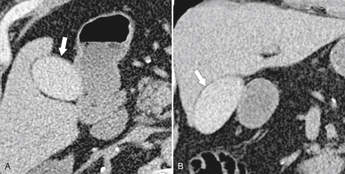
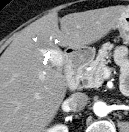
Milk of calcium bile is caused by biliary stasis, which is usually secondary to chronic cystic duct obstruction. Calcium carbonate in static bile increases in concentration and then precipitates, which can then be identified radiographically (Fig. 8-29) or by CT (Fig. 8-30) as either diffuse or layering (Fig. 8-31) radiopacities within the gallbladder. It can be confused with a post-ERCP contrast-filled gallbladder, although this will be unusual (the gallbladder rarely fills with contrast medium injected at ERCP).
image
Figure 8-29 Plain abdominal radiograph of a 66-year-old man with a subtly dense gallbladder (arrows) caused by milk of calcium bile. There is an inferior vena cava filter in situ.
image
Figure 8-30 Axial (A) and coronal (B) contrast-enhanced CT in a 70-year-old man with dense bile caused by milk of calcium bile (arrows).
image
Figure 8-31 Axial contrast-enhanced CT in a 57-year-old woman demonstrates dense layering bile (arrow) within the gallbladder (arrowheads) resulting from milk of calcium bile.

Vicarious Excretion of Contrast Material

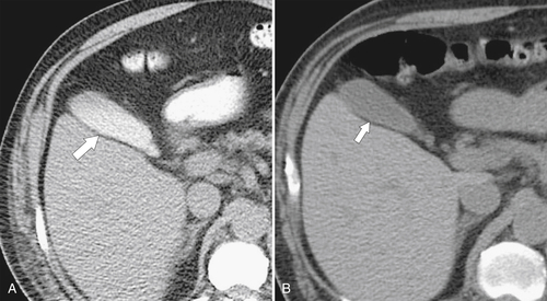
The vicarious excretion of contrast material can have an imaging appearance similar to milk of calcium bile, with the bile uniformly increased in contrast (at plain radiography or CT) because of the biliary excretion of injected or ingested iodinated contrast material. The condition is usually observed in patients with renal failure who fail to excrete the circulating contrast medium; hence, the medium is “vicariously” excreted into bile instead. It is usually observed with CT (Fig. 8-32) because the concentration of contrast medium is generally insufficient to be observed on plain radiography unless the patient received iodinated contrast material in the presence of severe renal failure. Although generally unnecessary, it can be differentiated from milk of calcium bile by repeat imaging a few days later, allowing sufficient time for the biliary contrast medium to have cleared (Fig. 8-32).
image
Figure 8-32 A and B, Axial noncontrast CT in a 51-year-old man with renal failure. There is dense bile caused by vicarious excretion of contrast material (large arrow) from a recent contrast angiographic study. Three weeks later (small arrow), the contrast medium has been excreted.

Porcelain Gallbladder

Porcelain gallbladder is one of the sequelae of chronic cholecystitis. The chronically inflamed gallbladder wall steadily calcifies, sometimes sufficiently to be identified on plain radiography (Fig. 8-33), although most will be identified with US or CT, which will demonstrate curvilinear wall calcification (Fig. 8-34). On US the gallbladder is usually obscured by the calcific wall (Fig. 8-34) and may be confused with a gallbladder full of gallstones. Up to 30% of patients with porcelain gallbladder, left untreated by cholecystectomy, can develop gallbladder carcinoma. Consequently, patients are referred for prophylactic cholecystectomy.
image
Figure 8-33 Plain abdominal radiograph in a 77-year-old woman with a porcelain gallbladder (arrows).
image
Figure 8-34 Transverse US (A) and axial (B) and coronal (C) contrast-enhanced CT in a 74-year-old woman with a diffuse wall shadowing at US and wall calcification at CT from a porcelain gallbladder (large arrows) and gallstones (small arrows).

Inflammatory Biliary Diseases

Cholecystitis

Cholecystitis is a common cause of morbidity among the general population because of the prevalence of gallstones. It may present for the first time acutely or with chronic clinical symptoms. Less commonly (approximately 5%), acute cholecystitis is acalculous (see later in the chapter). Gallbladder inflammatory change typically results from obstruction of the gallbladder by a stone lodged within the cystic duct. Biliary stasis follows with continued mucosal biliary secretion, which distends the lumen and then thickens or inspissates and becomes secondarily infected, usually by Escherichia coli and Bacteroides spp. Symptoms may initially be intermittent and self-limiting, but the ensuing infections ultimately produce symptoms and signs of acute bacterial infection. The gallbladder wall becomes hyperemic, inflamed, and thickened. Treatment is usually antibiotics and interval cholecystectomy, although in severe infection, gallbladder perforation and peritonitis occur, requiring immediate surgery.
Imaging is highly sensitive and specific for the detection of acute cholecystitis, and most patients are referred for US. Classic sonographic features include a distended gallbladder, thickened gallbladder wall (>3 mm), intramural gallbladder wall lucencies (representing wall edema), gallstones (often a gallstone impacted in the gallbladder neck), and a Murphy sign (Fig. 8-35). The Murphy sign is usually elicited by US and is maximal tenderness when the abdomen is compressed precisely over the distended gallbladder. If maximal pain is not directly over the gallbladder, it is unlikely to be a Murphy sign and therefore not acute cholecystitis. However, since this test is often poorly performed, the sign is not necessarily elicited when acute cholecystitis is present.
image
Figure 8-35 A and B, Sagittal US in a 29-year-old woman with a distended gallbladder, slight wall thickening, and intramural gallbladder lucencies (long arrow) caused by acute cholecystitis. There is an impacted stone at the gallbladder neck (short arrow). The patient had a positive sonographic Murphy sign.
Less commonly, patients are referred for CT because the clinical symptoms may not localize specifically to the gallbladder or the referring physician may not specifically suspect cholecystitis. Features on contrast-enhanced CT include a distended gallbladder with enhancement of a circumferentially thickened wall and pericholecystic inflammatory changes (“fat stranding”) in the adjacent peritoneal fat (Fig. 8-36). Gallstones may or may not be identified, depending on their calcium content.
image
Figure 8-36 Axial (A) and coronal contrast-enhanced CT (B) and HIDA scan (C) in a 30-year-old man with an inflamed gallbladder. Pericholecystic edema (large white arrows) and wall thickening are due to acute cholecystitis. On HIDA scan there is no gallbladder filling (small black arrow) of Tc at 60 minutes because of an obstructed cystic duct. Isotope passes into the duodenum directly (arrowhead).
Nuclear scintigraphy with 99mTc hepatobiliary iminodiacetic acid (HIDA), now rarely performed because of the accuracy of US, has high specificity for the diagnosis of acute cholecystitis. Injected isotope is readily excreted into the biliary system, and a normally functioning gallbladder is usually identified at 1 hour. If no gallbladder is observed after 4 hours (because of cystic duct obstruction), a diagnosis of acute cholecystitis is made, assuming that there are accompanying clinical symptoms and signs (Fig. 8-36). Early in the injection (i.e., in the arterial phase), the hyperemic gallbladder wall may demonstrate increased activity and a “rim sign” may be observed, representing a crescentic band of isotope activity in the adjacent inflamed hepatic parenchyma, but this sign is less specific and rarely identified. MRI is rarely performed to evaluate for the diagnosis of acute cholecystitis but will demonstrate MR-equivalent findings to contrast-enhanced CT, with a distended gallbladder and a thickened enhancing wall, and most likely the offending gallstone will be identified. Pericholecystic inflammatory change will be manifest as increased T2 signal.
The complications of acute cholecystitis include gallbladder gangrene and perforation with pericholecystic abscess or gas in the gallbladder wall or lumen (Figs. 8-37 and 8-38). Once the gallbladder has been perforated, a positive Murphy sign is less likely because the gallbladder is no longer distended. The features of a perforated gallbladder are best imaged by CT, demonstrating the extent of pericholecystic abscess formation.
image
Figure 8-37 Axial noncontrast CT in a 77-year-old man with gangrenous cholecystitis with gallbladder perforation, pericholecytic abscess (long arrow), and intraluminal gas (short arrow). There is also a single gallstone that precipitated the infection (arrowhead).
image
Figure 8-38 US in a 76-year-old woman with gallbladder perforation from acute cholecystitis. Numerous stones (long arrow) and a perforation are identified on US (short arrow).
Chronic cholecystitis results from repetitive episodes of subacute cholecystitis caused by the presence of stones but usually is not sufficiently symptomatic to make the patient seek urgent medical care. The symptoms are mild to moderate intermittent pain in the right upper quadrant. There is sometimes biliary colic when the gallstone becomes temporarily impacted in the gallbladder neck, but not long enough to cause the sequelae of full-blown acute cholecystitis. The repetitive milder inflammatory episodes ultimately cause fibrosis, thickening, and contraction of the gallbladder, such that it becomes shrunken and compacted with stones (Figs. 8-9 and 8-10). At that point an increasing number of more painful episodes may develop, precipitating medical attention and subsequent cholecystectomy.

Acalculous Cholecystitis

Acalculous cholecystitis, a relatively unusual form of cholecystitis, demonstrates all the clinical and imaging features of cholecystitis except the presence of gallstones (Fig. 8-39). It is classically identified in patients after a prolonged stay in an intensive care unit (ICU) and less commonly in patients with diabetes mellitus, AIDS, vascular injury or insufficiency to the gallbladder, colitis, or postpartum state. The resulting gallbladder stasis (particularly after hyperalimentation or cholestatic drugs) can ultimately become secondarily infected, leading to cholecystitis. The diagnosis is often challenging because patients who stay in the ICU may not manifest classic clinical symptoms (pain is masked by narcotics, which in themselves are cholestatic). The diagnosis may be suspected only because of clinical features of sepsis and the knowledge by ICU physicians that these patients are at risk for acalculous cholecystitis. It has been demonstrated, however, that many of the sonographic features of cholecystitis in this group of patients can resolve spontaneously. Conversely, if acalculous cholecystitis is identified in the appropriate clinical setting, the gallbladder is often drained via percutaneous cholecystostomy, with culture of bile (typically milky white because of prolonged stasis) to exclude the gallbladder as the infectious source in a patient with a fever of unknown origin.
image
Figure 8-39 Sagittal US (A) and axial contrast-enhanced CT (B) in a 23-year-old man in the ICU with a distended gallbladder (short arrow) and pericholecystic edema (arrowheads) resulting from acute acalculous cholecystitis. The gallbladder wall (long arrows) measures 6 mm.

HIV-Induced Cholecystitis

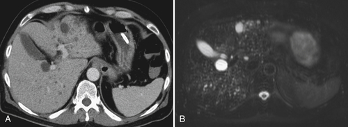
Biliary abnormalities are relatively common in patients with AIDS. Cholestatic and cholecystitis-like findings on US include gallbladder wall thickening, pericholecystic lucencies, gallbladder dilatation, and sludge (Fig. 8-40).
image
Figure 8-40 Sagittal US (A) and transverse US (B) in a 51-year-old man with HIV-associated cholecystitis, gallbladder wall thickening and intramural lucencies (arrows), and gallbladder distention.

Emphysematous Cholecystitis

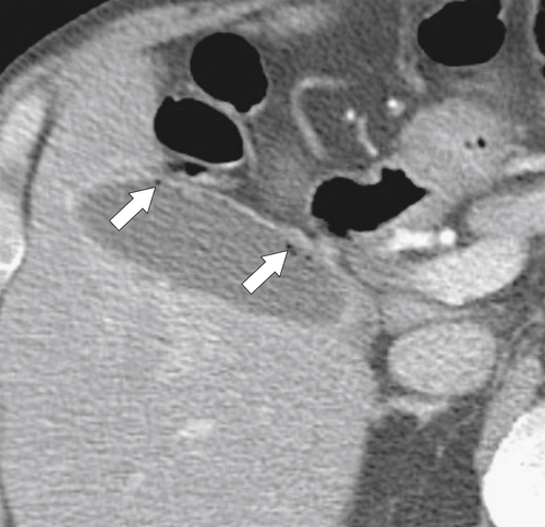
Emphysematous cholecystitis is a rare presentation of acute cholecystitis and is caused by infection with gas-forming organisms, particularly E. coli and Clostridium welchii. It has similarities to emphysematous infections in other organs (kidney and bladder) in that it is much more commonly identified in patients with diabetes, who are particularly susceptible to these infections. It has a relatively high death rate. Patients with widespread atherosclerosis are also at risk (also common to patients with diabetes mellitus) because of relative ischemia. If severe, the gas may be identified on plain radiography, but the gas may obscure visualization of the gallbladder at US. CT is therefore the investigation of choice, readily identifying the inflamed gas-filled gallbladder wall (Fig. 8-41). Emphysematous cholecystitis should be differentiated from ascending cholangitis, which also is caused by gas-forming organisms that affect the gallbladder wall. In ascending cholangitis there is usually gas elsewhere in the biliary tree, whereas the gas in emphysematous cholecystitis is confined to the gallbladder.
image
Figure 8-41 Axial contrast-enhanced CT in a 74-year-old man with diabetes mellitus with subtle gas in the gallbladder wall (arrows) from early emphysematous cholecystitis.

Xanthogranulomatous Cholecystitis

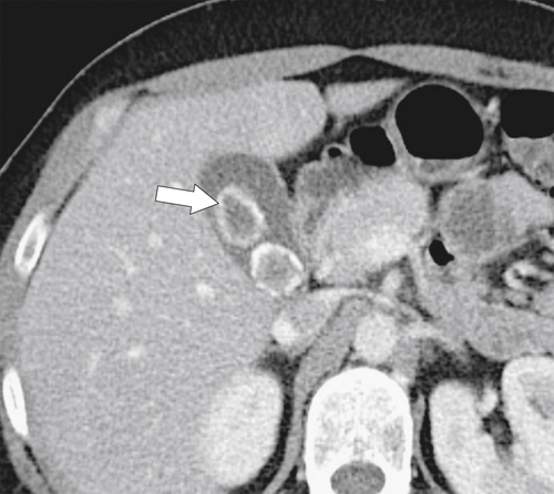
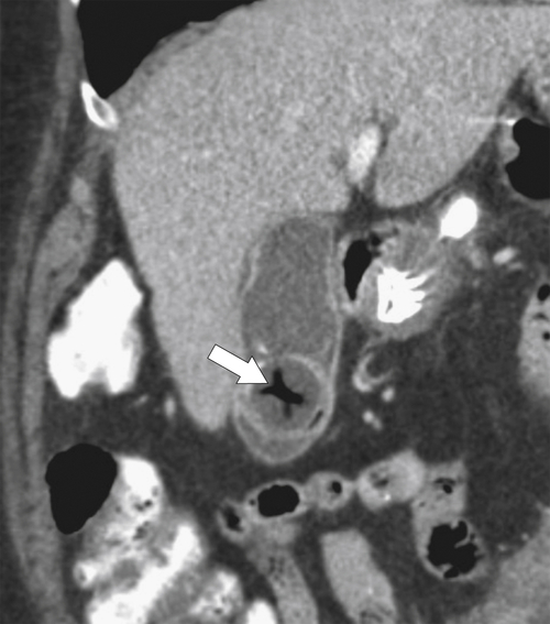
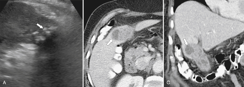
Xanthogranulomatous cholecystitis, a rare form of chronic cholecystitis, is named for the yellow-gray material that results from its lipid-laden macrophages in the gallbladder wall. Most patients (usually those in the fifth to sixth decades) have associated gallstones, and the disease may be secondary to ulceration of the mucosa and extravasation of bile into the gallbladder wall, setting up an inflammatory response. The imaging findings are generally nonspecific and common to chronic cholecystitis in general, with gallstones or sludge and wall thickening, although the wall thickening may be asymmetrical. More characteristic features include hyperechoic nodules and bands within the gallbladder wall (Fig. 8-42), representing the lipid-laden xanthomatous material. On CT, there is mucosal enhancement and mural thickening, often asymmetrical, and the CT equivalent of lipid-laden macrophages within the wall, represented by hypodense intramural nodules (Fig. 8-42). The differentiation from cholangiocarcinoma can be difficult, and usually the diagnosis is made only after cholecystectomy.
image
Figure 8-42 Sagittal US (A), axial (B), and coronal (C) contrast-enhanced CT in a 59-year-old woman with a heterogeneous material in the gallbladder and hypoechoic and hypodense lipid deposits in the gallbladder wall due to xanthogranulomatous cholecystitis (arrows).

Other Benign Gallbladder Diseases

Gallbladder Hydrops

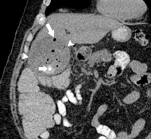
Gallbladder hydrops is also known as a mucocele of the gallbladder because of mucous or watery distention of the gallbladder. It results from obstruction, which sometimes is caused by a gallstone in the cystic duct or gallbladder neck but may also be from neoplastic disease. The gallbladder can distend markedly but remains sterile, although later infection and development of gallbladder empyema are common. Once empyema develops, the patient usually presents with pain and fever and a right upper quadrant mass may be palpated. At US, the gallbladder is markedly distended with echogenic material, usually with a markedly thickened wall (Fig. 8-43). When empyema ensues, gas formation can be identified with CT and US. A number of patients do not have a sonographic Murphy sign, which should not deter the diagnosis of gallbladder empyema. Patients are at significant risk of gallbladder perforation.
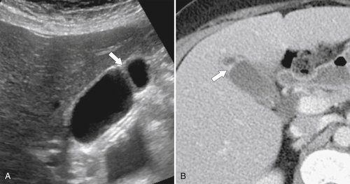
image
Figure 8-43 Coronal contrast-enhanced CT in a 57-year-old man with secondary infection (empyema) of a gallbladder hydrops (arrowheads) with intracystic gas (long arrow). There is an associated gallstone (short arrow).

Hyperplastic Cholecystosis

Hyperplastic cholecystosis is a spectrum of benign inflammatory gallbladder changes, the cause of which is poorly understood. The disease is relatively common (approximately 3% to 5% of the population). Patients are frequently asymptomatic (although associated gallstones can occur in approximately 50% of patients), and usually hyperplastic cholecystosis is diagnosed only after cholecystectomy. However, it can present with intermittent right upper quadrant pain, usually in patients in the fifth decade. Hyperplastic cholecystosis has two predominant forms, adenomyomatosis and hyperplastic cholesterosis.
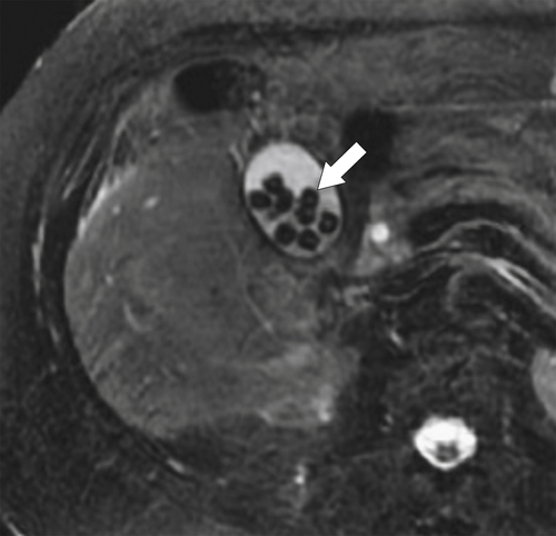
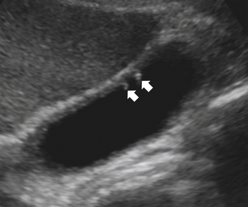
Adenomyomatosis, the more common form, is due to mucosal proliferation and hypertrophy of the muscularis in the gallbladder wall, which is sometimes marked. The gallbladder mucosa then invaginates the hypertrophied wall, forming Rokitansky-Aschoff sinuses. These sinuses can be identified by US, especially in nonobese patients who have been appropriately fasted. The sinuses appear hypoechoic if filled with bile or hyperechoic if filled with sludge or small stones. A characteristic sonographic feature is a so-called comet tail ring-down artifact representing cholesterol crystals within the sinuses (Fig. 8-44), which may also be identified by CT or MRI. On T2-weighted images the Rokitansky-Aschoff sinuses are seen as small multiple cystic structures (sometimes referred to as a “pearl necklace” sign) within the gallbladder wall that may be filled with multiple small stones (a “rosary sign”) at CT (Fig. 8-45). These findings can be diffuse, segmental, or localized. The diffuse form involves the whole gallbladder, the segmental form involves the proximal, middle, or distal gallbladder circumferentially, and the most common form, localized, involves only the fundus. The segmental form typically has waist-like narrowing of the affected gallbladder wall (Fig. 8-46). The fundal or localized form can present as simple mural thickening, but sometimes it is sufficient to appear mass-like and can be difficult to differentiate from gallbladder carcinoma, particularly because with both diseases the affected region may show increased activity on positron emission tomography.
image
Figure 8-44 Sagittal US in a 50-year-old woman with two small filling defects (arrows) with ring-down artifacts caused by cholesterosis.
image
Figure 8-45 MRCP (A) and axial (B) and coronal (C) contrast-enhanced CT in a 58-year-old woman with adenomyomatosis and a CT “rosary sign” with several tiny stones (arrows) lodged within the Rokitansky-Aschoff sinuses and the “string of pearls” sign (arrowhead) and fundal constriction (curved arrow) at MRCP.
image
Figure 8-46 Sagittal US (A) and axial contrast-enhanced CT (B) in a 44-year-old woman with a fundal gallbladder constriction (arrows) caused by adenomyomatosis.
Hyperplastic cholesterosis, sometimes referred to as a strawberry gallbladder because of multiple, small, bright-yellow fatty deposits, is caused by diffuse deposition of submucosal cholesterol/triglyceride-laden histiocytes. The accumulation of these fatty deposits causes mucosal polypoid enlargement, which sometimes can be recognized at US (Fig. 8-47).
image
Figure 8-47 Transverse US in a 48-year-old woman with ring-down artifacts (arrow) in a “strawberry” gallbladder.

Primary Sclerosing Cholangitis

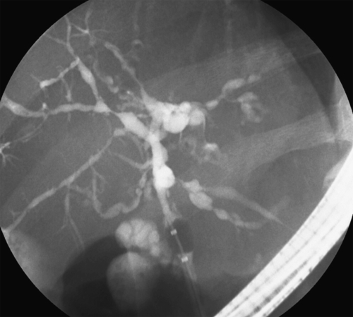
Primary sclerosing cholangitis (PSC) is a chronic disease of the extrahepatic and intrahepatic bile ducts that causes progressive and diffuse biliary duct inflammation and stricture formation, often termed either arborized (or “pruned tree”) or bead like. Cholestasis ensues with the formation of multiple small segmental ectatic bile ducts. PSC is most likely autoimmune in origin because of the presence of numerous circulating autoantibodies and because up to 80% of patients also have ulcerative colitis. It is also, however, associated with Crohn disease and other autoimmune diseases, including autoimmune pancreatitis and thyroiditis. In most patients it appears before the age of 45 years, and the disease is usually unrelenting with progression through fibrosis and biliary cirrhosis, which may ultimately require liver transplantation, even though PSC is known to recur in the allograft. There is also an approximate 10% chance that cholangiocarcinoma will develop.
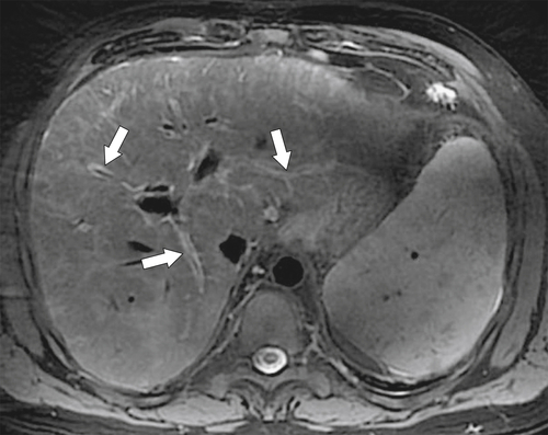
The diagnosis is best made with good biliary ductal opacification, and therefore ERCP (or transhepatic cholangiography) is the imaging method of choice (Fig. 8-48). Segmental strictures are found in the extrahepatic ducts but are most commonly observed in the right and left intrahepatic ducts as multiple short segmental strictures with intermittent focal ductal dilatation (Fig. 8-48). The fine, bead-like ductal abnormalities are generally not identified by CT, although focal bile lakes of ectatic ducts might be visualized. There is often evidence of periductal fibrosis, which is recognized by edematous tracking along the ducts that becomes broader and more confluent as fibrosis progresses and is observed particularly centrally (Fig. 8-49). Finally, as diffuse cirrhosis ensues, the liver has the typical cirrhotic appearances of a shrunken liver with distorted contour. As in other forms of cirrhosis, the caudate lobe is usually hypertrophied, but whereas in other forms the cause is preservation of vascular drainage, in PSC it is relative preservation of the caudate bile ducts. US is superior to CT for evaluating the ductal abnormalities, demonstrating wall thickening (caused by progressive inflammatory change), hypoechoic focal segmental strictures, and biliary ectasia. Echogenic portal triads correspond to fibrotic change. The gallbladder is not spared in this disease, so US may demonstrate segmental and asymmetrical wall thickening. The evolution of the disease is often monitored with MRI and MRCP, which clearly demonstrate the biliary ductal irregularity (Fig. 8-50), and high periductal T2-weighted signal implies edema and fibrosis. Identification of cholangiocarcinoma is suggested by the development of a mass (especially if it demonstrates delayed enhancement on contrast-enhanced CT or MRI) and upstream biliary dilatation.
image
Figure 8-48 ERCP in a 73-year-old woman demonstrating diffuse intrahepatic duct irregularities with a bead-like appearance resulting from primary sclerosing cholangitis.
image
Figure 8-49 T2-weighted fat-saturated MRI in a 35-year-old man with primary biliary cirrhosis and high periductal T2 signal due to edema (arrows). There is associated splenomegaly.
image
Figure 8-50 MRCP in a 33-year-old woman with diffusely irregular intrahepatic bile ducts caused by primary sclerosing cholangitis.

Infectious Cholangitis

Infectious cholangitis usually refers to an infection of the intrahepatic or extrahepatic biliary system, whereas gallbladder infection is termed cholecystitis. It is mostly bacterial in nature (often E. coli) and termed ascending cholangitis. Parasites (Clonorchis sinensis and Ascaris lumbricoides) are common infectious pathogens in many parts of the world. Other AIDS-related infections are responsible for AIDS-related cholangiopathy.

Ascending Cholangitis

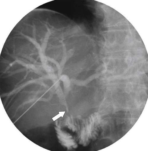
Pyogenic cholangitis is usually associated with gallstones, and patients are particularly susceptible if these are associated with biliary obstruction (as can occur with other benign or malignant strictures). The infection can be devastating if not treated early. Patients present with pain, fever, and jaundice (Charcot triad), and the infection can develop rapidly into bacteremia and overwhelming septicemia. In the correct clinical setting, US is often the imaging method of choice because it is rapidly available and is more sensitive than CT for the demonstration of intrahepatic and extrahepatic biliary duct dilatation, especially if mild (Fig. 8-51). Color Doppler is useful to differentiate normal portal venous strictures from dilated ducts, which are sometimes difficult to identify (Fig. 8-51). Once biliary dilatation has been confirmed, ERCP is usually performed to further define the anatomical site of obstruction and also to remove stones and perform sphincterotomy with or without stent placement, particularly for neoplastic forms of obstruction. Less commonly, transhepatic cholangiography and catheter drainage are performed, mainly for patients in whom ERCP is contraindicated or who have not responded to treatment (e.g., those who underwent prior upper gastrointestinal surgery) (Fig. 8-52).
image
Figure 8-51 A and B, US in a 70-year-old man with ascending cholangitis caused by obstructive jaundice from pancreatic adenocarcinoma and a dilated CBD (16 mm) (long arrow) and intrahepatic ducts (short arrows).
image
Figure 8-52 Transhepatic cholangiogram in a 46-year-old man with prior choledochojejunostomy that is strictured (arrow) with dilated intrahepatic ducts and ascending cholangitis.

AIDS-Related Cholangiopathy

Patients with AIDS-related immunosuppression are susceptible to multiple opportunistic infections, which can lead to repeated biliary inflammatory changes and ultimately strictures and obstruction. The most common opportunistic organism is Cryptosporidium parvum, but others include cytomegalovirus, microsporidia, and Cyclospora. Patients present with a Charcot triad of fever, pain, and jaundice, and most already have a known diagnosis of AIDS. At imaging, both intrahepatic and extrahepatic duct dilatation is usually present, interspersed with multiple stricture formation not dissimilar to that in sclerosing cholangitis. The gallbladder is commonly thickened and often demonstrates irregular linear wall lucencies (Fig. 8-40). Evidence of opportunistic infection elsewhere in the abdomen (retroperitoneal adenopathy) or enteritis is often present.

Parasitic Cholangitis

Ascaris Species Infection

Ascaris lumbricoides is a parasitic nematode worm that typically grows to 20 to 30 inches long. These worms are highly prevalent in most developing countries, and patients are often infested with numerous worms. Given their thin tubular nature, they have a propensity to burrow into the lower CBD through the ampulla of Vater and may cause biliary and pancreatic duct obstruction, the latter leading to pancreatitis. Occasionally a worm manages to navigate up the bile duct and even into the gallbladder, where it can be identified by US or ERCP (Fig. 8-53).
image
Figure 8-53 Sagittal US in a 43-year-old man with a tubular gallbladder filling defect (arrows) caused by Ascaris species worm.

Asiatic or Oriental Cholangitis

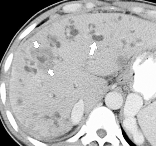
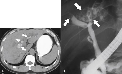
Oriental cholangiohepatitis, caused by Clonorchis sinensis (or Chinese liver fluke), is endemic in Southeast Asia and is responsible for Asiatic or oriental cholangitis, also known as recurrent pyogenic cholangitis. The fluke uses the fresh-water snail as an intermediate host in which cysts mature into cercariae, burrow their way out of the snail, and penetrate fish bodies. Humans who consume the fish become infected. The cercariae resist degradation in the small intestine and migrate to the biliary system, where they sexually reproduce and set up a biliary inflammatory response. The repeated infections lead to intrahepatic and extrahepatic duct inflammation (compounded by fluke volume and eggs), strictures, and stone formation, which are characteristic of this disease. The CT findings are characteristic in the appropriate clinical setting, with irregular and dilated ducts (Fig. 8-54), many of which are filled with stone material (Fig. 8-55). As with gallstones, the overall stone burden may not be evident on CT. Given that the stones are predominantly intrahepatic and many are in distal locations, they can prove almost impossible to remove and repeated cholangitic attacks occur. Cholangiocarcinoma is a complication of the disease and is common in regions where the disease is endemic (Fig. 8-54).
image
Figure 8-54 Axial contrast-enhanced CT in a 43-year-old woman with multiple dilated ducts caused by oriental cholangitis (long arrow). The patient subsequently developed cholangiocarcinoma (short arrows).
image
Figure 8-55 Axial non-contrast-enhanced CT (A) and ERCP (B) in a 28-year-old woman with multiple intrahepatic stones (arrows) caused by oriental cholangiohepatitis.

Portal Biliopathy

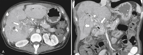
In the presence of extrahepatic portal venous occlusion (which may also extend into the intrahepatic portal veins), multiple hepatopetal portal cavernous veins (or transformation) occur. These act as a means to divert mesenteric blood around the thrombosed portal vein from the peripancreatic region and into the liver. Depending on the acuity of the portal vein obstruction, the portal venous thrombus may or may not be identified, but there will be multiple serpiginous venous collaterals in and around the region of the thrombosed vessel. These dilated collaterals can impinge in the extrahepatic bile duct and cause biliary obstruction, usually mild, within the intrahepatic ducts (Fig. 8-56).
image
Figure 8-56 Axial (A) and coronal (B) contrast-enhanced CT in a 61-year-old woman with cirrhosis, portal vein occlusion (large arrows), and biliary dilatation (small arrows) caused by portal biliopathy (arrowheads).

Gallbladder Wall Thickening

Thickening of the gallbladder wall is defined as a thickness greater than 3 mm. It is important to measure the thickness in transverse diameter; spurious recordings will be made if the wall is measured in oblique or tangential planes. A number of primary gallbladder diseases are responsible for gallbladder wall thickening (Box 8-1). Most causes have already been discussed in this chapter. Gallbladder carcinoma is discussed later in the chapter.
 
Box 8-1   Gallbladder Wall Thickening
Acute and chronic cholecystitis (calculus and acalculous)
Emphysematous cholecystitis
Xanthogranulomatous cholecystitis
Adenomyomatosis
Porcelain gallbladder
Ascites (from any cause)
Hepatitis
Cirrhosis
Pancreatitis
HIV cholangiopathy
Gallbladder carcinoma
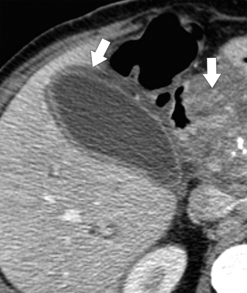
Secondary causes of gallbladder wall thickening are adjacent inflammation, hepatitis or pancreatitis (Fig. 8-57), or ascites from any cause (particularly right-sided heart failure and renal failure). The precise reason is poorly understood but is thought to be elevated portal venous pressure. The gallbladder wall thickening can be surprisingly alarming, particularly in hepatitis, and should not be confused with gallbladder gangrene or necrosis (Fig. 8-58). The wall at US is often multilayered with multiple intramural lucencies (Fig. 8-58). These features all resolve once the immediate inflammatory condition subsides, and cholecystectomy is not warranted.
image
Figure 8-57 Axial contrast-enhanced CT in a 42-year-old man with gallbladder wall thickening (left arrow) caused by adjacent pancreatitis (right arrow).
image
Figure 8-58 Transverse (A) and sagittal US (B) and axial CT (C) in a 61-year-old woman with a 7-mm gallbladder wall thickening and multiple wall lucencies (arrows) caused by hepatitis.

Cystic Biliary Masses

Peribiliary Cysts

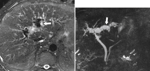
Peribiliary cysts are uncommon but are associated with cirrhosis and portal vein thrombosis. They represent focal dilatation of periductal glands that become obstructed owing to an inflammatory process. They are recognized at imaging as single or multiple small CT-hypodense or MRI-hyperintense cystic lesions along the length of the biliary tree and are sometimes confused with small simple cysts. They are better appreciated with T2-weighted MRI as multiple high-signal cysts (Fig. 8-59).
image
Figure 8-59 Axial fat-saturated T2-weighted MRI (A) and MRCP (B) in a 44-year-old man with multiple small T2 hyperintense peribiliary cysts (arrows).

Biliary Cystadenoma

Biliary cystadenomas are rare cystic tumors most commonly occurring in middle-aged women. They are derived from biliary endothelium and are usually solitary, although occasionally multiple, and often septated and large. They frequently contain ovarian stroma, a diagnostic feature at histological examination. They are usually benign, but some have a malignant potential with a risk of degeneration into biliary cystadenocarcinoma. The cysts are usually complex, which makes them difficult to differentiate from cystic metastases, infected cysts (hydatid or Echinococcus), resolving hematomas, or bilomas. The clinical history is therefore important because most biliary cystadenomas are detected incidentally.
On US a large multiseptated cyst is observed (Fig. 8-60), sometimes with wall calcification. The diagnosis is best suggested with contrast-enhanced CT or MRI, which demonstrates a large multiseptated cyst with a well-defined wall that can calcify. Malignant degeneration demonstrates enhancing septa and larger solid masses within the cyst. The cyst may be of variable signal on both T1- and T2-weighted MRI, depending on the serous or mucoid nature of the cyst contents.
image
Figure 8-60 Sagittal US (A) and axial (B) and coronal (C) contrast-enhanced CT in a 36-year-old woman with a complex 9-cm intrahepatic cyst representing a biliary cystadenoma. Note internal septa (arrows).

Bile Duct Hamartomas

Bile duct hamartomas, also known as von Meyenburg complexes, are rare benign hepatic tumors. A congenital anomaly with malformation of the bile ducts results in multiple biliary cystic lesions. Biliary hamartomas are either incidental or associated with polycystic liver disease, in which they are relatively common. They are usually asymptomatic and of no concern, being detected incidentally on US, CT, or MRI as multiple small (usually <1.5 cm) cystic lesions (Fig. 8-61). US demonstrates multiple, diffuse, small anechoic lesions, although some may display mixed echogenicity because they have solid components (fibrous stroma). On CT, the density similarly depends on the proportion of cystic versus solid components and therefore ranges from water to soft tissue density. A hallmark, however, is the large number and small size of cyst-like lesions, unlike simple hepatic cysts, which are generally not so numerous. On contrast-enhanced CT, the solid elements may enhance but cystic areas do not. MRI can demonstrate similar features, with low signal on T1-weighted imaging, bright T2 signal for cystic lesions, and intermediate signal for the more solid components.
image
Figure 8-61 Axial contrast-enhanced CT (A) and T2-weighted MRI (B) in a 53-year-old man with multiple biliary hamartomas or von Meyenburg complexes that are low density at CT and high signal at MRI.

Gallbladder Masses

Gallbladder Polyps

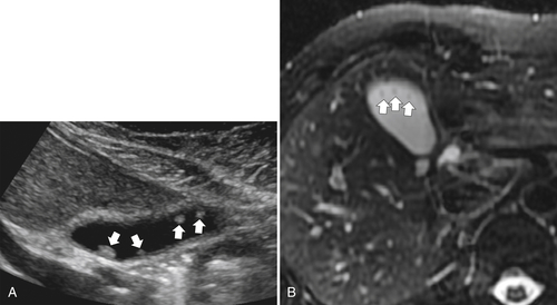
Gallbladder polyps are relatively common and are small polypoid masses that project into the gallbladder lumen. They are most frequently adenomatous. When multiple they are associated with cholesterol deposits and therefore are part of a spectrum of disease with hyperplastic cholecystosis and particularly cholesterolosis. Gallbladder polyps have also been recognized in Peutz-Jeghers (hamartomatous polyps) (see Chapter 4) and familial adenomatous polyposis (adenomatous polyps) (see Chapters 4 and 5). At US, they are recognized by a small intraluminal nonshadowing sessile mass, which helps to differentiate them from gallstones (Fig. 8-62). The presence of cholesterol is indicated by echogenicity within the polyp (without shadowing). Differentiation from tumefactive sludge is usually possible because sludge is typically mobile, larger, and irregular in contour. Furthermore, color Doppler flow can be observed in some larger polyps. If the polyp is sufficiently large (>5 mm), it can be identified with CT. Smaller polyps can be identified with T2-weighted MRI because of the superior contrast resolution between the high-signal bile and low-signal polyp.
image
Figure 8-62 Sagittal US (A) and axial T2-weighted fat-saturated MRI (B) in a 49-year-old woman with several gallbladder polyps (arrows).
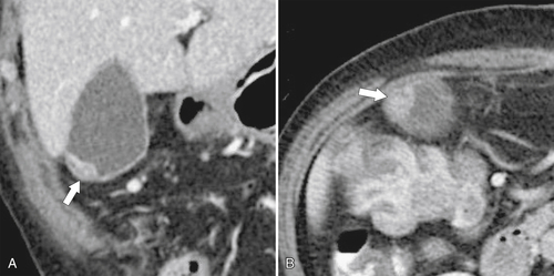
Most polyps are asymptomatic and benign, but they are considered to have premalignant potential, although this is not fully substantiated. Polyps up to 1 cm are serially imaged to determine any increase in size that might indicate malignant transformation to cholangiocarcinoma. When polyps are larger than 1 cm (and especially if >1.5 cm), cholecystectomy is usually indicated to exclude cholangiocarcinoma (Fig. 8-63). Some surgeons remove polyps smaller than 1 cm because of this risk.
image
Figure 8-63 Coronal (A) and axial (B) contrast-enhanced CT in a 56-year-old woman with a small fundal polypoid mass (arrows) that was early gallbladder carcinoma at cholecystectomy.

Biliary Intraductal Papillary Mucinous Neoplasm

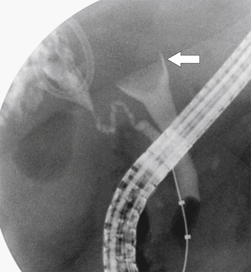
Biliary intraductal papillary mucinous neoplasm (IPMN) causes both intrahepatic and extrahepatic biliary duct dilatation when mucin is secreted from papillary mucinous epithelial cells lining the ducts. Therefore a discrete mass may not be present. Biliary IPMN is far less commonly observed than IPMN of the pancreatic duct. Patients present with intermittent pain, fever, and jaundice. The diagnosis is suggested when ERCP demonstrates multiple intraluminal mucinous filling defects, with or without an associated mass. US, CT, and MRI usually show diffuse (and sometimes grossly enlarged) bile ducts filled with hypodense mucinous material (Fig. 8-64).
image
Figure 8-64 ERCP in a 56-year-old woman with a globular intraluminal CBD filling defect (arrow) caused by intraductal IPMN.

Cholangiocarcinoma

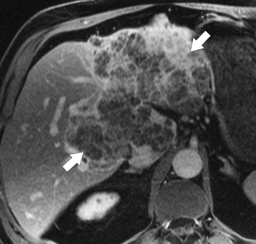
Cholangiocarcinomas are rare tumors with an increased incidence in patients with primary sclerosing cholangitis, oriental cholangiohepatitis, chronic liver disease (especially cirrhosis), Caroli syndrome and other choledochal cysts, Thorotrast exposure, and Lynch syndrome. Tumors occur with equal frequency intrahepatically and extraheptically. Those originating at the biliary confluence are known as Klatskin tumors. Tumors may be exophytic with a larger eccentric mass, polypoid with a smaller intraluminal mass, or infiltrative along the length of the duct. They are almost always ductal adenocarcinomas and invade the periductal tissues with a desmoplastic reaction, and this fibrotic reaction can make it difficult to diagnose the underlying malignancy.
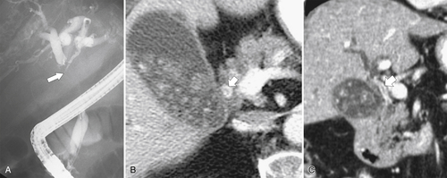
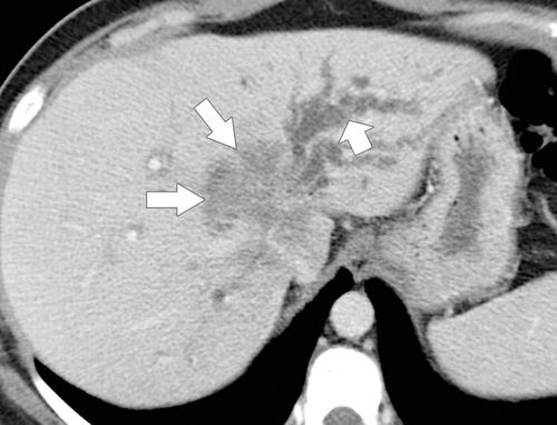
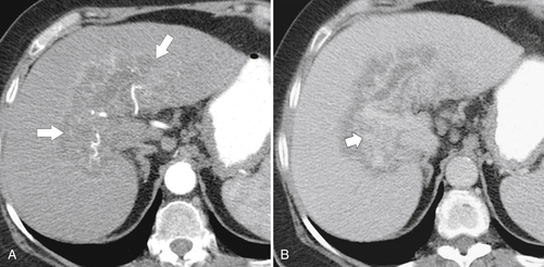
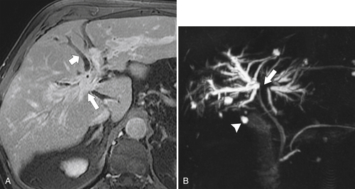
Patients usually present in their sixth and seventh decades with pain and jaundice. The imaging method of choice is ERCP, particularly because many lesions produce little mass effect and are missed by CT. Furthermore, ERCP provides the opportunity to relieve any strictures, at least temporarily. After ductal injection, an irregular stricture should be identified with proximal or upstream biliary dilatation (Fig. 8-65). There may be a small (2- to 5-mm) intraductal papillary mass or a relatively long stricture as seen in the infiltrating type. On contrast-enhanced CT there is usually biliary dilatation but the mass is often not identified, although occasionally the tumor can be observed as subtle ductal enhancement along the affected duct (Fig. 8-66). A small mass in the region of the biliary bifurcation with converging obstructing ducts leading into the mass is highly suggestive of the diagnosis. Larger masses are easier to identify (Fig. 8-67). The mass is generally hypodense on noncontrast CT but often demonstrates peripheral enhancement in the arterial phase that gradually enhances toward the center, which remains hyperdense on delayed imaging, a characteristic feature (Fig. 8-67). This is caused by fibrous desmoplastic tissue that retains the contrast material for variable lengths of time. Imaging with MRCP can provide useful information about the stricture location and intrahepatic duct dilatation (Fig. 8-68). On contrast-enhanced MRI the tumor enhances similarly to that on CT (Fig. 8-69), with peripheral arterial enhancement that is retained on delayed imaging.
image
Figure 8-65 ERCP (A) and axial (B) and coronal (C) contrast-enhanced CT (B and C) in a 71-year-old man with a Klatskin tumor (large arrow) and intrahepatic duct dilatation. The tumor is just visible as an enhancing infiltrating mass along the length of the common hepatic duct (small arrows). Gallstones are present.
image
Figure 8-66 Axial contrast-enhanced CT in a 46-year-old man with a hilar cholangiocarcinoma (long arrows) that mainly obstructs the left-sided ducts (short arrow).
image
Figure 8-67 Axial arterial (A) and delayed (B) contrast-enhanced CT in a 77-year-old man with cholangiocarcinoma. A large hilar heterogeneous mass (arrows) demonstrates delayed enhancement (short arrow).
image
Figure 8-68 Axial postcontrast fat-saturated MRI (A) and MRCP (B) in a 63-year-old man with a Klatskin tumor (arrows) and dilated intrahepatic ducts (small arrow). There are several hepatic cysts (arrowhead).
image
Figure 8-69 Axial postcontrast fat-saturated MRI in a 73-year-old woman with a large infiltrating heterogeneous cholangiocarcinoma (arrows).

Gallbladder Carcinoma

Gallbladder carcinoma (usually adenocarcinoma) is a rare malignancy. Patients present with pain, jaundice, and generalized symptoms of malignancy, particularly if it has metastasized, which it often has done by the time of presentation, primarily by local invasion of the liver. It has a strong association with gallstones, perhaps reflecting a chronic inflammatory contribution to development of neoplastic change. It is also recognized with increased frequency with porcelain gallbladder (Fig. 8-70) and is associated with gallbladder polyps, although this link is controversial.
image
Figure 8-70 Contrast-enhanced CT in an 86-year-old woman with a partially calcified porcelain gallbladder (large arrow) and a large gallbladder carcinoma (small arrows).
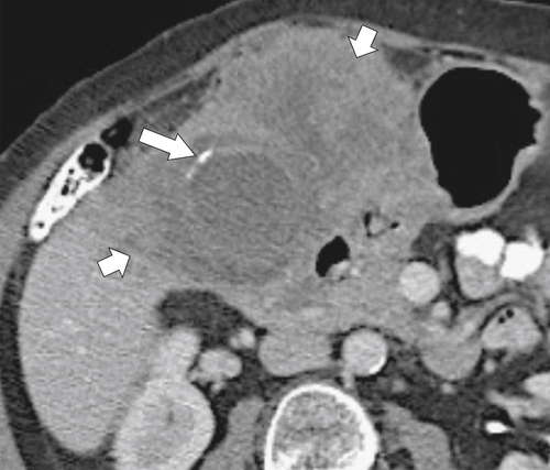
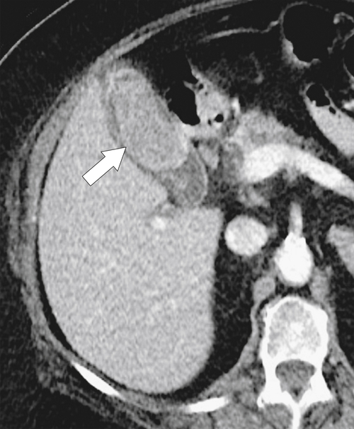
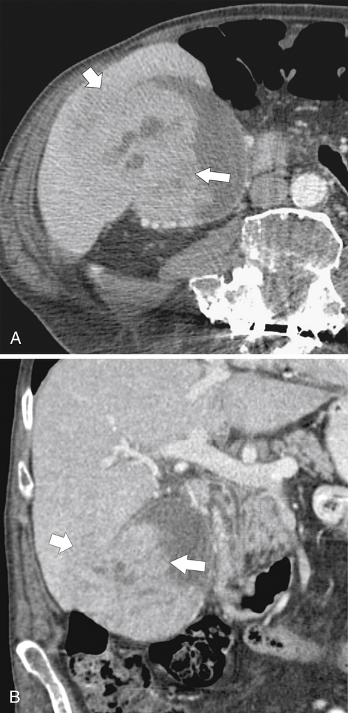
Early in the course of disease a focal mass is confined to the gallbladder wall (Fig. 8-71), but at presentation the mass has often extended beyond the confines of the gallbladder, with an irregular soft-tissue mass infiltrating the liver and porta hepatis with or without associated adenopathy. The mass is usually hypovascular on contrast-enhanced CT and infiltrates the immediate region around the gallbladder fossa (Fig. 8-72).
image
Figure 8-71 Axial contrast-enhanced CT in a 77-year-old man with a 4-cm gallbladder mass (large arrow) that represents a gallbladder carcinoma.
image
Figure 8-72 Axial (A) and coronal (B) contrast-enhanced CT in a 74-year-old man with gallbladder cholangiocarcinoma. A gallbladder-centered mass (long arrows) extends into segment VI of the liver (short arrows).

Ampullary Carcinoma

Ampullary carcinoma is an adenocarcinoma arising from ductal epithelium in the region of the ampulla of Vater. The cause is unknown, but it is associated with polyposis and Gardner syndrome. The tumor is lobulated or infiltrating like other cholangiocarcinomas. Given its location, it leads to early bile and pancreatic duct dilatation (“double-duct” sign) and therefore mostly presents with jaundice. Its prognosis is also better than that of cholangiocarcinoma elsewhere, but the tumor may have metastasized to regional lymph nodes at the time of presentation. It is often not identified on cross-sectional imaging because of its small size but can appear hypodense on CT and similarly hypointense on contrast MRI. Evidence of an underlying mass is inferred from pancreatic and bile duct dilatation (Fig. 8-73). However, a dedicated contrast-enhanced CT using duodenal distention has a better chance than conventional CT of identifying the mass water (the patient drinks 500 mL immediately before imaging). Images are then reformatted into the coronal plane to precisely define the location of the stricture and mass. In practice, once the patient presents with jaundice, ERCP is usually performed and directly visualizes a soft tissue mass at the ampulla.
image
Figure 8-73 Coronal (A) and axial (B) contrast-enhanced CT in a 72-year-old woman with an ampullary tumor. The tumor cannot be visualized by CT but can be inferred from pancreatic (small arrow) and CBD dilatation (large arrows) proximal to the major papilla.

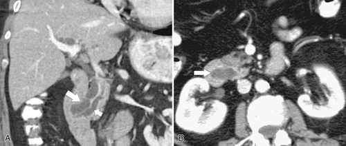
Metastatic Disease

Gallbladder metastases are uncommon but are well recognized in melanoma (15% of patients with disseminated melanoma have gallbladder metastases). These patients can present with acute cholecystitis if an enlarging mass causes cystic duct obstruction. Other metastases are far less common and usually from lung cancer. The metastases are identified as an enhancing polypoid mass on contrast-enhanced CT or MRI. They also demonstrate increased color Doppler flow at US.
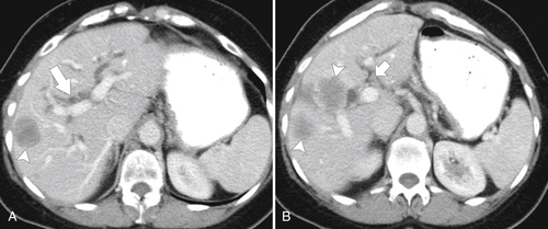
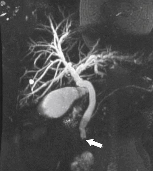
Common bile or hepatic duct obstruction occurs from either direct malignant extension or distant metastatic spread, usually lymphatic. Pancreatic head and uncinate adenocarcinomas often obstruct the pancreatic and CBD early, producing the “double-duct” sign (Fig. 8-74). Lymphatic metastatic disease is usually to lymph nodes at the porta hepatis, particularly from intestinal malignancies. Given the caliber of the bile duct at this point, even small nodes can impinge on and obstruct the duct. Because of their small size, they may not be identified with cross-sectional imaging but can be inferred from upstream biliary dilatation. They typically produce a smooth extrinsic tapering impression on the bile duct rather than the abrupt irregular strictures seen in cholangiocarcinoma (Fig. 8-75).
image
Figure 8-74 Axial (A) and coronal (B) contrast-enhanced CT and MRCP (C) in a 66-year-old woman with pancreatic adenocarcinoma (large arrow) obstructing both the pancreatic duct and the CBD (double-duct sign) (small arrows) that also results in intrahepatic duct dilatation (arrowheads).
image
Figure 8-75 A and B, Contrast-enhanced CT in a 48-year-old woman with colon cancer and hepatic metastases (arrowheads). Biliary dilatation (large arrow) is due to small porta hepatic nodes (small arrow in B) obstructing the common hepatic duct.

Gallbladder Lymphoma

Most lymphomatous biliary involvement is from metastatic spread from lymphoma elsewhere or as part of diffuse nodal disease throughout the abdomen. Primary gallbladder lymphoma is extremely rare and of the non-Hodgkin type. The gallbladder initially produces eccentric wall thickening, which develops into a larger, generally homogeneous mass. Before cholecystectomy is performed, it is generally not possible to distinguish lymphoma from other gallbladder malignancies, especially gallbladder adenocarcinoma.

Ampullary Dysfunction

Ampullary dysfunction, also known as sphincter of Oddi dysfunction, is a difficult disease to diagnose and treat. It is seen in approximately 15% of patients after cholecystectomy, and patients report recurrent episodic abdominal pain. The cause is failure of the sphincter of Oddi to relax normally, causing temporary biliary dilatation until the spasm subsides. The diagnosis is usually one of exclusion, although it can be made using sphincter of Oddi manometry. MRCP performed at the time of sphincter spasm may show ductal distention (Fig. 8-76), but because the disease is transitory, the diagnosis may be missed. Further diagnostic steps involve challenging the sphincter after a fatty meal and imaging shortly afterward with US or preferably MRCP. A better noninvasive test is a secretin stimulation test (see Chapter 9). Injection of secretin causes excessive production of pancreatic exocrine digestive juices (primarily bicarbonate), which challenges the sphincter. This is best monitored by MCRP before and 1, 2, 5, and 10 minutes after secretin injection. The normal pancreatic and bile duct should demonstrate minimal ductal increase, which rapidly returns to normal. Sphincter dysfunction causes pancreatic and biliary sustained dilatation at the time of examination.
image
Figure 8-76 MRCP in a 74-year-old man. Dilatation of the CBD and intrahepatic ducts is due to ampullary stenosis (arrow).

Postoperative Anomalies

Intrahepatic Biliary Gas

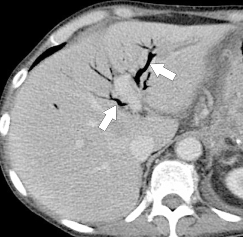
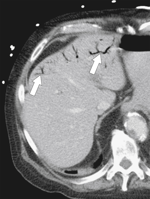
Intrahepatic biliary gas is commonly observed after upper gastrointestinal surgery with choledochoenteric anastomoses. Intestinal gas (and sometimes fluid) passes into a patent bile duct and into intrahepatic ducts. This also commonly occurs after biliary sphincterotomy procedures. It can be differentiated from portal venous gas by being predominantly central and radiating from the hilum (Fig. 8-77). In contradistinction, portal venous gas is identified at the liver margins (Fig. 8-78). Far less commonly, intrahepatic biliary gas is caused by gas-forming organisms, usually secondary to ascending cholangitis. The patient will almost certainly show signs of overwhelming sepsis.
image
Figure 8-77 Axial contrast-enhanced CT in a 59-year-old woman after a Whipple procedure. Intrahepatic biliary gas (arrows) that radiates peripherally from the hepatic hilum is a normal finding.
image
Figure 8-78 Axial contrast-enhanced CT in a 72-year-old man with peripherally located nondependent intrahepatic gas (arrows) that is portal venous in origin.

Biloma

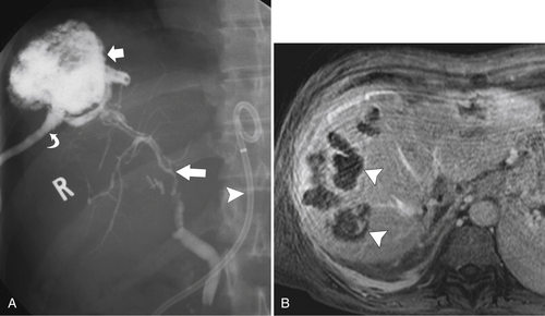
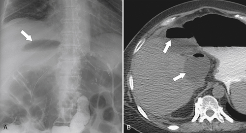
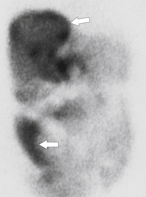
Bile leakage, either intrahepatically or extrahepatically, accumulates in loculated collections known as bilomas, with an attenuation on noncontrast CT similar to bile and therefore hypodense, close to water density. Leakage is usually iatrogenic from biliary surgery (cholecystectomy), percutaneous biopsy or catheter placement, perforation during ERCP, or radiation biliary necrosis after radiation treatment for cholangiocarcinoma (Fig. 8-79). Bilomas are also common after rupture from blunt or penetrating trauma. They usually are easily recognized on US, CT, and MRI as fluid-filled collections arising from the affected biliary structure (Fig. 8-80). Sometimes the cause of perihepatic fluid collections is unclear, and imaging 99mTc HIDA can confirm the biliary nature of the fluid (Fig. 8-81). Leakage into the peritoneum causes severe peritonitis. Treatment is usually by percutaneous catheter drainage.
image
Figure 8-79 ERCP (A) and contrast-enhanced MRI (B) in a 57-year-old man with irregular intrahepatic ducts (large arrow) and a large biloma (small arrow) that drains externally via a biliary cutaneous fistula induced by previous percutaneous catheter drainage (curved arrow). There is a biliary drain in situ (arrowhead). Several other bilomas are also identified at MRI (arrowheads).
image
Figure 8-80 Plain abdominal radiograph (A) and axial noncontrast CT (B) in a 70-year-old man with a partially gas-filled biloma (arrows) as a result of a leak from recent biliary surgery.
image
Figure 8-81 Technetium 99m HIDA imaging in a 67-year-old with fever and perihepatic and peritoneal fluid confirmed as biliary in origin (arrows).

Biliary Traumatic Complications

Most biliary trauma is iatrogenic from bile duct or gallbladder surgery (open or laparoscopic and especially from liver transplantation), percutaneous catheter placement (Fig. 8-82) and biopsy, or complications from ERCP. Bile ducts can also become ischemic and undergo necrosis from radiation treatment, chemoembolization (TACE), or injury to the hepatic artery from hepatobiliary surgery (Fig. 8-79). Other injuries result from complete or partial rupture from blunt or penetrating trauma. These injuries may cause bile leakage (biloma), fistula (biliary-enteric fistula), and stricture formation, usually as a result of ischemia and healing fibrosis, or occasionally because of occlusion from inadvertent surgical suturing. Treatment for most complications is usually by temporizing biliary drainage and, if necessary, balloon dilatation or surgical repair of the offending stricture or defect. Cross-sectional imaging or nuclear scintigraphy may reveal evidence of biloma. Biliary strictures are best evaluated with ERCP but also may sometimes be observed on CT. Hemobilia, the presence of bloody bile, is usually caused by vascular injury, most commonly iatrogenic (e.g., percutaneous biliary catheter drainage that extravasates into the biliary tree). It is usually self-limiting but may require selective arterial catheterization and embolization if severe.
image
Figure 8-82 Transverse US in a 55-year-old man with recent percutaneous gallbladder placement. Irregular hyperechoic intraluminal mass (arrow) is due to gallbladder hemorrhage.

Suggested Readings
Ahualli J. The double duct signRadiology 2007;244(1):314315.
Altun E. et al. Acute cholecystitis: MR findings and differentiation from chronic cholecystitisRadiology 2007;244(1):174183. doi:10.1148/radiol.2441060920.
Anderson S.W. et al. Accuracy of MDCT in the diagnosis of choledocholithiasisAJR 2006;187(1):174180.
Anderson S.W. et al. Detection of biliary duct narrowing and choledocholithiasis: accuracy of portal venous phase multidetector CTRadiology 2008;247(2):418427.
Arai K. et al. Dynamic CT of acute cholangitis: early inhomogeneous enhancement of the liverAJR 2003;181(1):115118.
Bader T.R. et al. MR imaging features of primary sclerosing cholangitis: patterns of cirrhosis in relationship to clinical severity of diseaseRadiology 2003;226(3):675685.
Bennett G.L. et al. CT findings in acute gangrenous cholecystitisAJR 2002;178:275281.
Bennett G.L. et al. Ultrasound and CT evaluation of emergent gallbladder pathologyRadiol Clin North Am 2003;41(6):12031216.
Berk R.N. et al. Carcinoma in the porcelain gallbladderRadiology 1973;106:2931.
Bilgin M. et al. Hepatobiliary and pancreatic MRI and MRCP findings in patients with HIV infectionAJR 2008;191(1):228232.
Brancatelli G. et al. Fibropolycystic liver disease: CT and MR imaging findingsRadiographics 2005;25(3):659670.
Bucceri A.M. et al. Common bile duct caliber following cholecystectomy: two-year sonographic surveyAbdom Imaging 1994;19:251258.
Catalano O. et al. Complications of biliary and gastrointestinal stents: MDCT of the cancer patientAJR 2012;199:W187W196. doi:10.2214/AJR.11.7145.
Catalano O.A. et al. MR imaging of the gallbladder: a pictorial essayRadiographics 2008;28(1):135155:  quiz 324.
Chan W.C. et al. Gallstone detection at CT in vitro: effect of peak voltage settingRadiology 2006;241(2):546553.
Chan Y.L. et al. Choledocholithiasis: comparison of MR cholangiography and endoscopic retrograde cholangiographyRadiology 1996;200:8591.
Chavhan G.B. et al. Pediatric MR cholangiopancreatography: principles, techniques, and clinical applicationsRadiographics 2008;28(7):19511962.
Chen R.C. et al. Value of ultrasound measurement of gallbladder wall thickness in predicting laparoscopic operability prior to cholecystectomyClin Radiol 1995;50:570578.
Choi E. et al. Duplication of the extrahepatic bile duct with anomalous union of the pancreaticobiliary ductal system revealed by MR cholangiopancreatographyBr J Radiol 2007;80(955):e150154.
Choi J.Y. et al. Hilar cholangiocarcinoma: role of preoperative imaging with sonography, MDCT, MRI, and direct cholangiographyAJR 2008;191(5):14481457.
Chung Y.E. et al. Varying appearances of cholangiocarcinoma: radiologic-pathologic correlationRadiographics 2009;29(3):683700. doi:10.1148/rg.293085729.
Corwin M.T. et al. Incidentally detected gallbladder polyps: is follow-up necessary? Long-term clinical and US analysis of 346 patientsRadiology 2011;258(1):277282:  Published online August 9, 2010, doi: 10.1148/radiol.10100273.
Darge K. et al. MR imaging of the abdomen and pelvis in infants, children, and adolescentsRadiology 2011;261(1):1229. doi:10.1148/radiol.11101922.
Dave M. et al. Primary sclerosing cholangitis: meta-analysis of diagnostic performance of MR cholangiopancreatographyRadiology 2010;256:387396.
Dohke M. et al. Anomalies and anatomic variants of the biliary tree revealed by MR cholangiopancreatographyAJR 1999;173(5):12511254.
Elsayes K.M. et al. Gastrointestinal manifestation of diabetes mellitus: spectrum of imaging findingsJ Comput Assist Tomogr 2009;33(1):8689.
Fuks D. et al. Acute cholecystitis: preoperative CT can help the surgeon consider conversion from laparoscopic to open cholecystectomyRadiology 2012;263(1):128138:  Published online February 13, 2012, doi: 10.1148/radiol.12110460.
Furlan A. et al. Gallbladder carcinoma update: multimodality imaging evaluation, staging, and treatment optionsAJR 2008;191(5):14401447.
Ginat D. et al. Incidence of cholangitis and sepsis associated with percutaneous transhepatic biliary drain cholangiography and exchange: a comparison between liver transplant and native liver patientsAJR 2011;196:W73W77. doi:10.2214/AJR.09.3925.
Grand D. et al. CT of the gallbladder: spectrum of diseaseAJR 2003;183:163170.
Gupta R.T. et al. Dynamic MR imaging of the biliary system using hepatocyte-specific contrast agentsAJR 2010;195:405413. doi:10.2214/AJR.09.3641.
Haller J.O. Sonography of the biliary tract in infants and childrenAJR 1991;157:10511058.
Han J.K. et al. Cholangiocarcinoma: pictorial essay of CT and cholangiographic findingsRadiographics 2002;22(1):173187.
Hashimoto M. et al. Evaluation of biliary abnormalities with 64-channel multidetector CTRadiographics 2008;28(1):119134. doi:10.1148/rg.281075058.
Heffernan E.J. et al. Recurrent pyogenic cholangitis: from imaging to interventionAJR 2009;192(1):W2835.
Hoeffel C. et al. Normal and pathologic features of the postoperative biliary tract at 3D MR cholangiopancreatography and MR imagingRadiographics 2006;26(6):16031620. doi:10.1148/rg.266055730.
Jutras J.A. Hyperplastic cholecystosesAJR 1960;83:795827.
Kiewiet J.J.S. et al. A systematic review and meta-analysis of diagnostic performance of imaging in acute cholecystitisRadiology July 12, 2012:  Published online. http://dx.doi.org/10.1148/radiol.12111561.
Kim J.H. et al. MR cholangiography in symptomatic gallstones: diagnostic accuracy according to clinical risk groupRadiology 2002;224(2):410416:  Published online May 17, 2002, doi: 10.1148/radiol.2241011223.
Kim J.H. et al. CT findings of cholangiocarcinoma associated with recurrent pyogenic cholangitisAJR 2006;187:15711577. doi:10.2214/AJR.05.0486.
Kim J.Y. et al. Differentiation between biliary cystic neoplasms and simple cysts of the liver: accuracy of CTAJR 2010;195:11421148. doi:10.2214/AJR.09.4026.
Kim J.Y. et al. Spectrum of biliary and nonbiliary complications after lapatoscopic cholecystectomy: radiologic findingsAJR 2008;191(3):783789.
Kim O.H. et al. Imaging of the choledochal cystRadiographics 1995;15:6988.
Kim S.J. et al. Peripheral mass–forming cholangiocarcinoma in cirrhotic liverAJR 2007;189:14281434. doi:10.2214/AJR.07.2484.
Knowlton J.Q. et al. Imaging of biliary tract inflammation: an updateAJR 2008;190(4):984992.
Kumar A. et al. Carcinoma of the gallbladder: CT findings in 50 patientsAbdom Imaging 1994;19:304310.
Lee W.J. et al. Radiologic spectrum of cholangiocarcinoma: emphasis on unusual manifestations and differential diagnosesRadiographics 2001;21:S97S116:  Spec No.
Levy A.D. et al. Caroli’s disease: radiologic spectrum with pathologic correlationAJR 2002;179(4):10531057.
Lim J.H. Cholangiocarcinoma: morphologic classification according to growth pattern and imaging findingsAJR 2003;181:819827.
Lim J.H. et al. Intraductal papillary mucinous tumor of the bile ductsRadiographics 2004;24(1):5366:  discussion 66-67.
Lim J.H. et al. Parasitic diseases of the biliary tractAJR 2007;188(6):15961603.
Lim J.H. et al. Biliary parasitic diseases including clonorchiasis, opisthorchiasis and fascioliasisAbdom Imaging 2008;33(2):157165.
Lim J.H. et al. Intraductal papillary mucinous tumors of the bile ductsRadiographics 2004;24:5367.
Malet P.F. et al. Gallstone composition in relation to buoyancy at oral cholecystographyRadiology 1990;177:167169.
Mall J.C. et al. Caroli’s disease associated with congenital hepatic fibrosis and renal tubular ectasiaGastroenterology 1974;66:10291035.
Martel J.P. et al. Melanoma of the gallbladderRadiographics 2009;29(1):291296.
Menias C.O. et al. Mimics of cholangiocarcinoma: spectrum of diseaseRadiographics 2008;28(4):11151129. doi:10.1148/rg.284075148.
Miller F.H. et al. Contrast-enhanced helical CT of choledocholithiasisAJR 2002;181:125130.
Miller W.J. et al. Imaging findings in Caroli’s diseaseAJR 1995;165:333340.
Mortelé K.J. et al. Multimodality imaging of pancreatic and biliary congenital anomaliesRadiographics 2006;26(3):715731. doi:10.1148/rg.263055164.
O’Connor O.J. et al. Imaging of cholecystitisAJR 2011;196:W367W374. doi:10.2214/AJR.10.4340.
O’Connor O.J. et al. Structured review: imaging of biliary tract diseaseAJR 2011;197:W551W558. doi:10.2214/AJR.10.4341.
Patel H.T. et al. MR cholangiopancreatography at 3.0 TRadiographics 2009;29:6. doi:10.1148/rg.296095505:  1689-1706.
Reinhold C. et al. MR cholangiopancreatography: potential clinical applicationsRadiographics 1996;16:309331.
Rizzo R.J. et al. Congenital abnormalities of the pancreas and biliary tree in adultsRadiographics 1995;15:4968.
Rybicki F.J. The WES signRadiology 2000;214(3):881882.
Sainani N.I. Cholangiocarcinoma: current and novel imaging techniquesRadiographics 2008;28(5):12631287.
Santiago I. et al. Congenital cystic lesions of the biliary treeAJR 2012;198:825835. doi:10.2214/AJR.11.7294.
Savader S.J. et al. Choledochal cysts: classification and cholangiographic appearanceAJR 1991;156:327331.
Schuster D.M. et al. Magnetic resonance cholangiographyAbdom Imaging 1995;20:353361.
Shakespear J.S. et al. CT findings of acute cholecystitis and its complicationsAJR 2010;194:15231529. doi:10.2214/AJR.09.3640.
Shanbhogue A.K.P. et al. Benign biliary strictures: a current comprehensive clinical and imaging reviewAJR 2011;197:W295W306. doi:10.2214/AJR.10.6002.
Shanmugam V. et al. Is magnetic resonance cholangiopancreatography the new gold standard in biliary imaging? Br J Radiol 2005;78(934):888893.
Sheng R. et al. Cholangiographic features of biliary strictures after liver transplantation for primary sclerosing cholangitis: evidence of recurrent diseaseAJR 1996;166:11091116.
Shin S.M. et al. Biliary abnormalities associated with portal biliopathy: evaluation on MR cholangiographyAJR 2007;188:W341W347. doi:10.2214/AJR.05.1649.
Silva A.C. et al. MR cholangiopancreatography: improved distention with intervenous morphine administrationRadiographics 2004;24:677687.
Silverthorn K. Sonographic follow-up of patients with gallbladder polypsAJR 2001;177:467.
Smith E.A. et al. Cross-sectional imaging of acute and chronic gallbladder inflammatory diseaseAJR 2009;192(1):188196.
Sugita R. et al. Periampullary tumors: high-spatial-resolution MR imaging and histopathologic findings in ampullary region specimensRadiology 2004;231(3):767774.
Taourel P. et al. Anatomic variants of the biliary tree: diagnosis with MR cholangiopancreatographyRadiology 1996;199:521527.
Valls C. et al. Biliary complications after liver transplantation: diagnosis with MR cholangiopancreatographyAJR 2005;184:812820.
Vermani N. et al. MR cholangiopancreatographic demonstration of biliary tract abnormalities in AIDS cholangiopathy: report of two casesClin Radiol 2009;64(3):335338.
Ward J. et al. Bile duct strictures after heptobiliary surgery: assessment with MR cholangiographyRadiology 2004;231:101108.
Weltman D.I., Zeman R.K. Acute diseases of the gallbladder and biliary ductsRadiol Clin North Am 1994;32:933954.
Wiot J.F., Felson B. Gas in the portal venous systemAJR 1961;86:920929.
Yeh B.M. et al. MR imaging and CT of the biliary tractRadiographics 2009;29:6. doi:10.1148/rg.296095514:  1669-1688.
Yu J. et al. Congenital anomalies and normal variants of the pancreaticobiliary tract and the pancreas in adults: part 1, biliary tractAJR 2006;187(6):15361543.
Yun E.J. et al. Gallbladder carcinoma and chronic cholecystitis: differentiation with two-phase spiral CTAbdom Imaging 2003;29(1):102108.
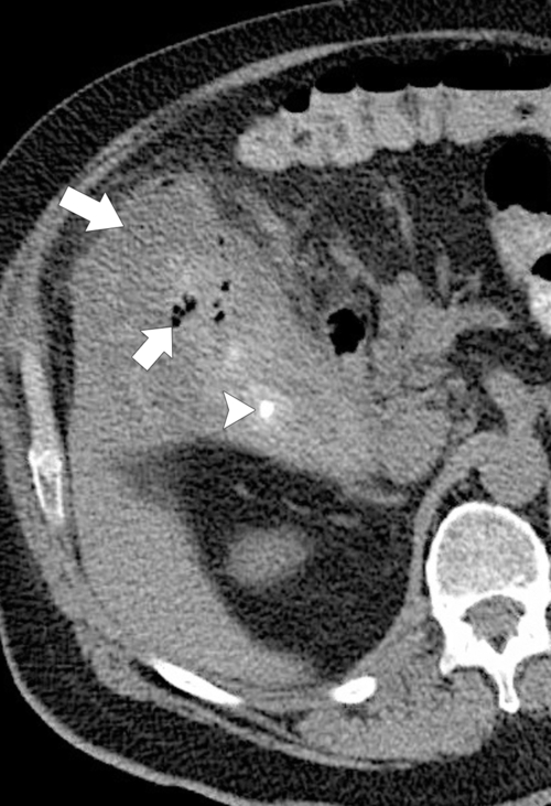
Abraham Vater (1684-1751), German anatomist.
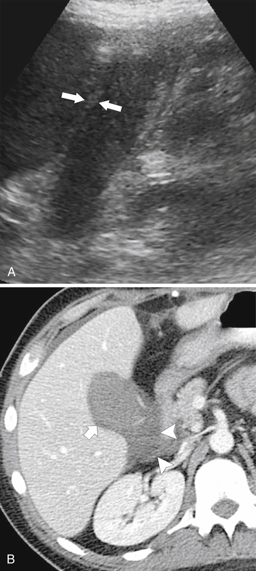
Phrygian cap: refers to the hats worn by residents in the ancient city in central Anatolia.

Hubert von Luschka (1820-1875), German anatomist.

Jacques Caroli (1902-1979), French gastroenterologist.

Christian Doppler (1803-1853), Austrian mathematician and physicist.

Mercédés Adrienne Manuela Ramona Jellinek (1889-1929), daughter of Austrian automobile entrepreneur Emil Jellinek; Karl Freidrich Benz (1844-1929), German engine designer.

Ruggero Oddi (1864-1913), Italian surgeon.

Friedrich Trendelenburg (1802-1872), German surgeon.

Pablo L. Mirizzi (1893-1964), Argentinean physician.

John B. Murphy (1857-1916), American surgeon.

Karl Freiherr von Rokitansky (1804-1878), Austrian pathologist; Karl Albert Ludwig Aschoff (1866-1942), German pathologist.

Jean-Martin Charcot (1825-1893), French neurologist and pathologist.

Jean-Martin Charcot (1825-1893), French neurologist.

Hans von Meyenburg (1887-1971), Swiss pathologist.

Gerald Klatskin (1911-1986), American internal medicine physician.