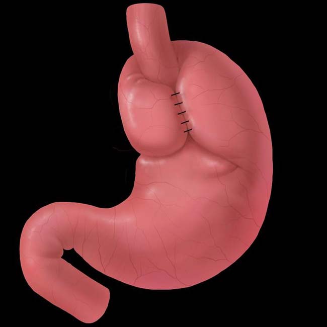
GE junction (at B ring) will be above diaphragm; intact wrap around proximal stomach (neoesophagus) will be below diaphragm
IMAGING


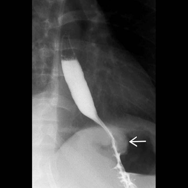
 in its expected subdiaphragmatic location as a filling defect within the air-filled fundus. The distal 3 cm of the esophageal lumen is compressed as it passes through the wrap.
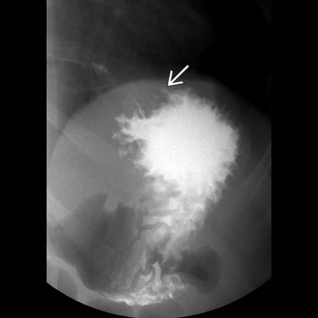
 as a filling defect with the barium pool in the fundus.
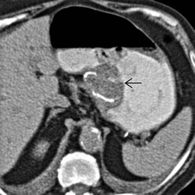
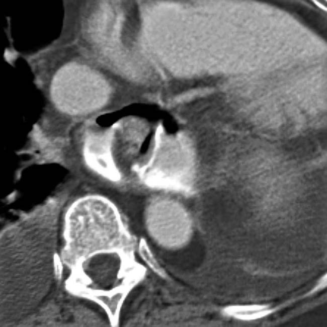
 within the gastric fundus. The metallic staple line is evident within the wrap. The mass effect of the wrap tends to decrease with time following surgery.


IMAGING


Radiographic Findings


• Normal postoperative appearance


CT Findings


• Herniation of intact FDP through diaphragmatic hiatus; seen as mediastinal mass on coronal reformatted images


CLINICAL ISSUES
Presentation


Natural History & Prognosis


 within the gastric fundus.
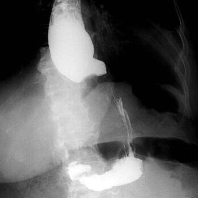
 that constitute the wrap, but the compression of the distal esophagus is maintained.
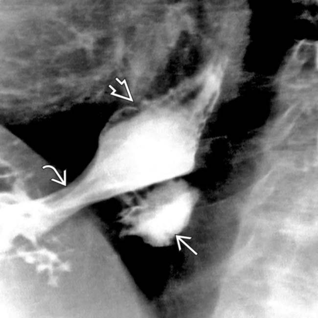
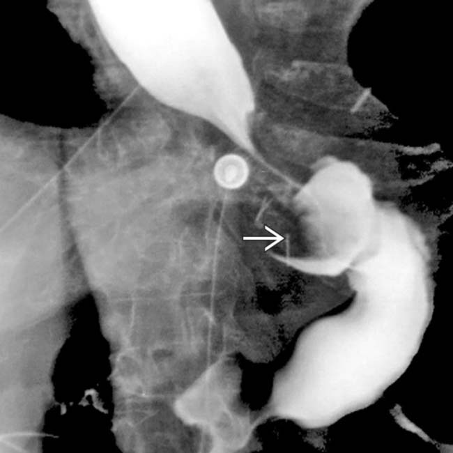
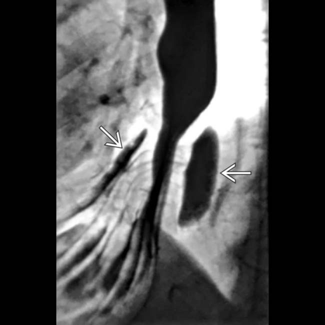
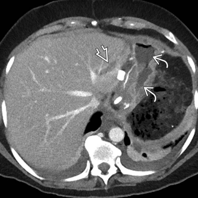
 , which has slipped through the hiatus
  . Suture disruption is implied by the loose compression of the neoesophagus. The B ring
  marks the esophagogastric junction.
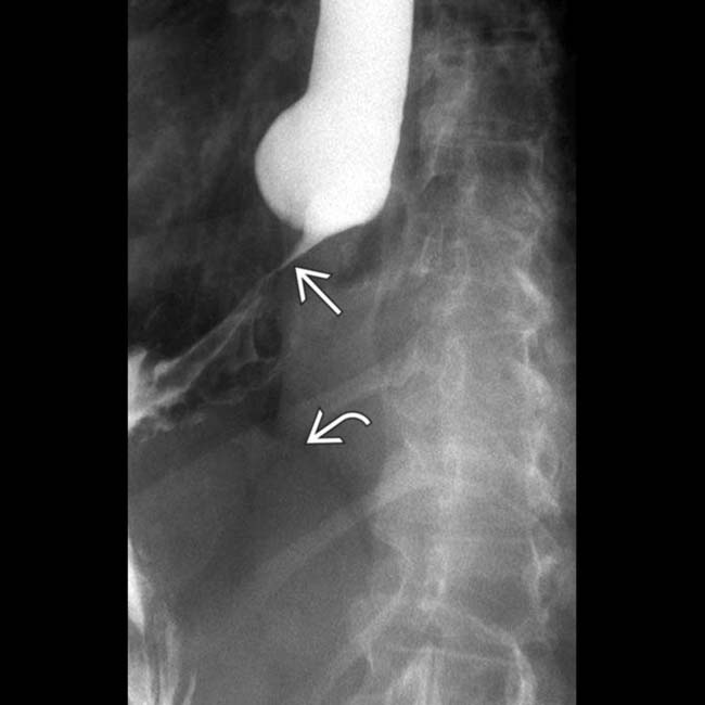
 , but well above the diaphragm
  .
.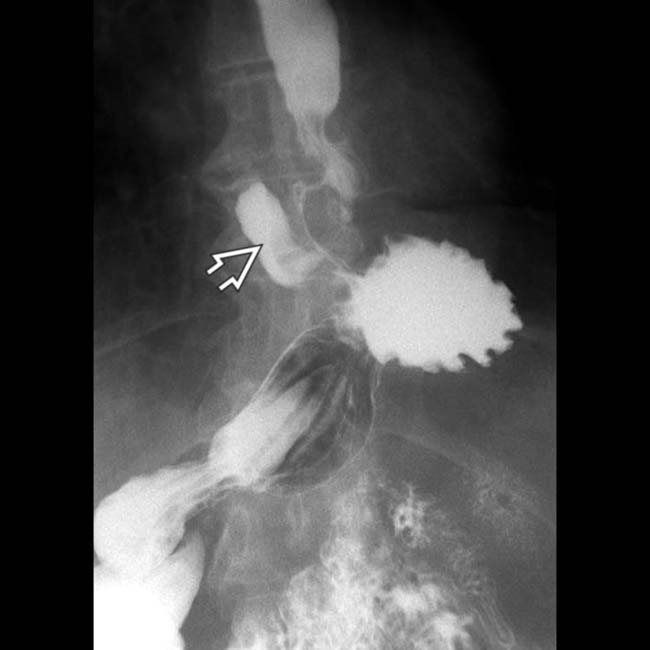
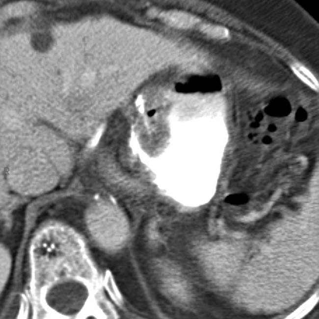
 . This should not be mistaken for a perforation. Note the intact gastric folds within the wrap. The Toupet wrap is often used in older patients with poor esophageal motility, as evident in this patient by slow emptying of the esophagus and tertiary contractions.
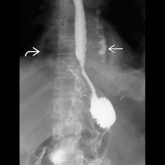
 and gas
  into the mediastinum, indicating perforation of the esophagus or the wrap.
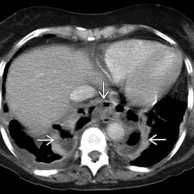
 due to perforation of the Nissen FDP.
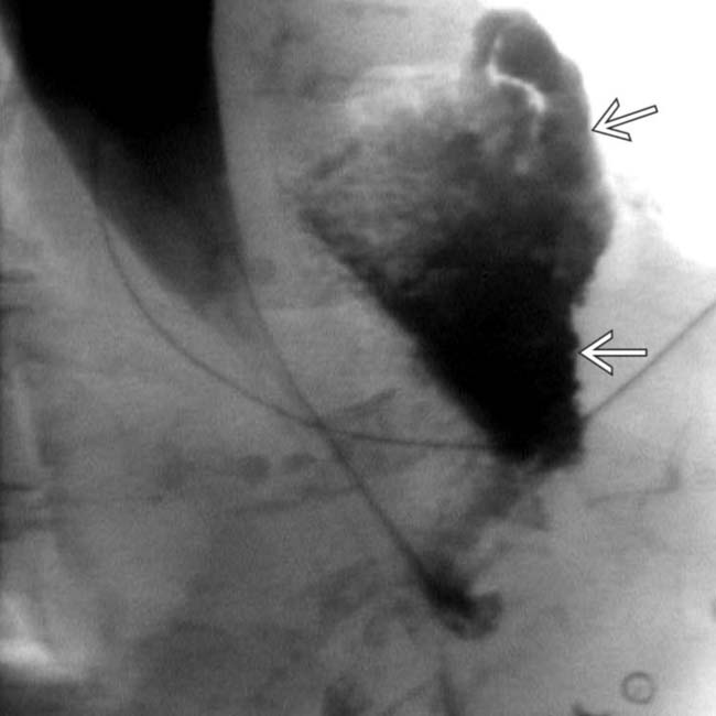
 into the mediastinum, from the distal esophagus. Very little contrast reaches the stomach.
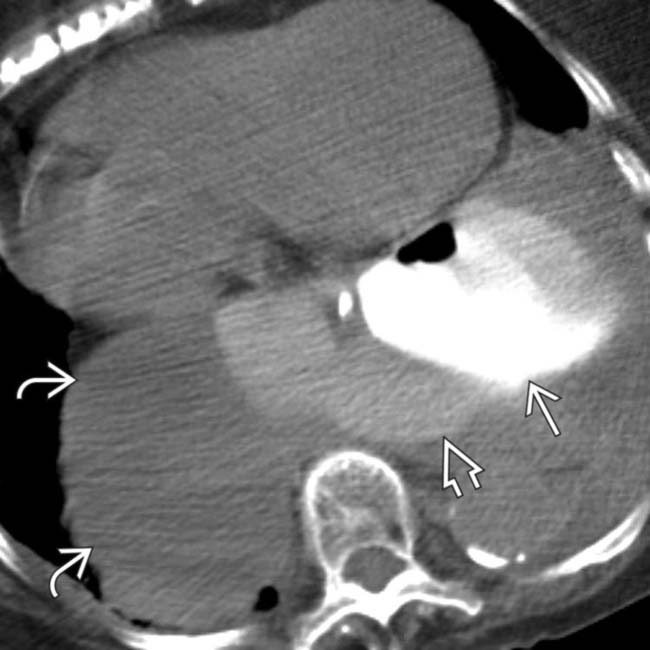
 , and residual herniation of the stomach
  . The dense collection of extraluminal contrast
  indicates a leak from near the operative site.
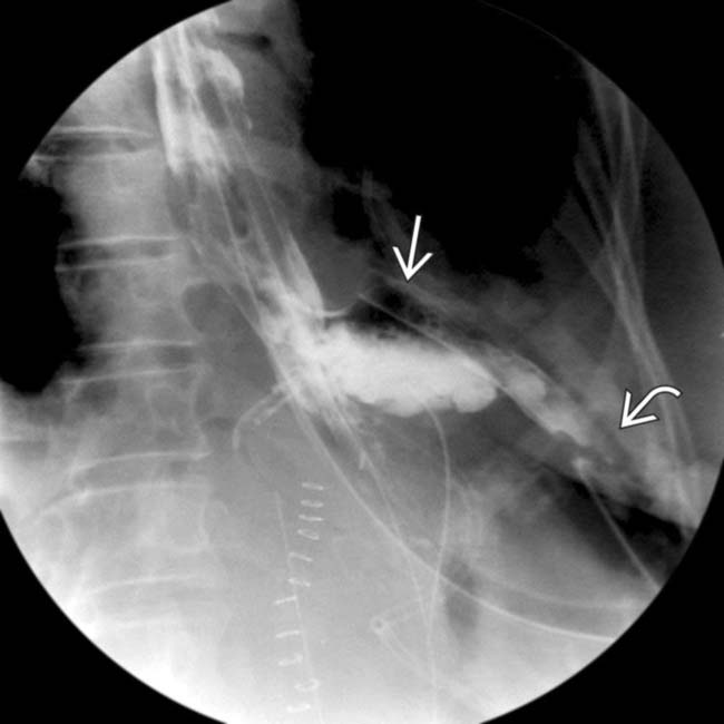
 and left pleural space
  , indicating a leak from the distal esophagus. Very little contrast enters the stomach.
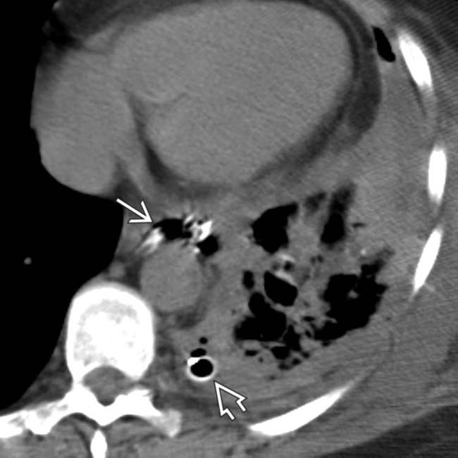
 that drained a loculated left hydropneumothorax (not shown here), left lower lobe pneumonia, and extravasated gas and contrast within the mediastinum
  .
.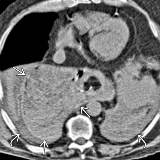
 in the space formerly occupied by the herniated stomach. Hemothorax is also evident
  .
.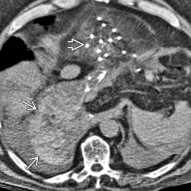
 are present from the mesh repair of the large hernia, and more mediastinal hematoma is seen
  . This hematoma should not be mistaken for leak of contrast medium.
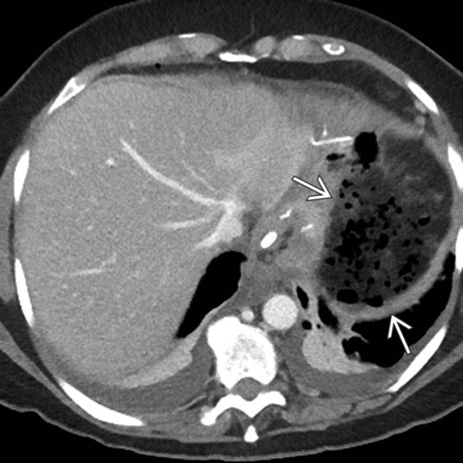
 .
.
 , subsequently confirmed to be due to a leak from the operative site. A small liver laceration
  is also seen.
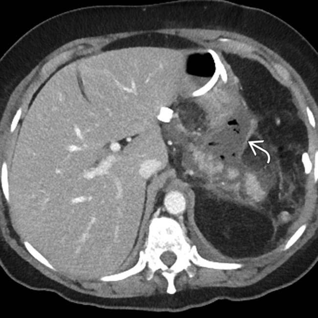
 from the FDP site.
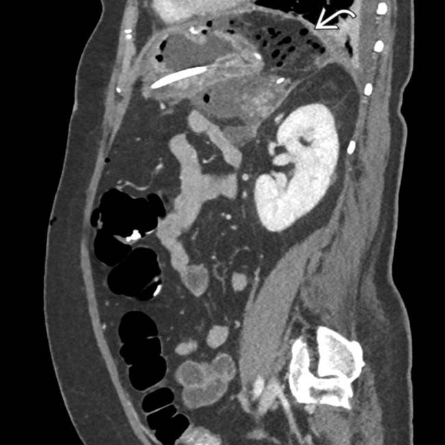
 near the site of the FDP.