Foreign Bodies and Bezoars

Published on 25/03/2015 by admin

Filed under Pediatrics

Last modified 22/04/2025

Print this page

rate 1 star rate 2 star rate 3 star rate 4 star rate 5 star
Your rating: none, Average: 0 (0 votes)

This article have been viewed 1119 times

Chapter 326 Foreign Bodies and Bezoars

326.1 Foreign Bodies in the Stomach and Intestine

Once in the stomach, 95% of all ingested objects pass without difficulty through the remainder of the gastrointestinal tract. Perforation after ingestion of a foreign body is estimated to be <1% of all objects ingested. Perforation tends to occur in areas of physiologic sphincters (pylorus, ileocecal valve), acute angulation (duodenal sweep), congenital gut malformations (webs, diaphragms, diverticula), or areas of previous bowel surgery.

Most patients who ingest foreign bodies are between the ages of 6 mo and 6 yr. Coins are the most commonly ingested foreign body in children, and meat or food impactions are the most common accidental foreign body in adolescents and adults. Patients with nonfood foreign bodies often describe a history of ingestion. Young children might have a witness to ingestion. Approximately 90% of foreign bodies are opaque. Radiologic examination is routinely performed to determine the type, number, and location of the suspected objects. Contrast radiographs may be necessary to demonstrate some objects, such as plastic parts or toys.

Conservative management is indicated for most foreign bodies that have passed through the esophagus and entered the stomach. Most objects pass though the intestine in 4-6 days, although some take as long as 3-4 wk. While waiting for the object to pass, parents are instructed to continue a regular diet and to observe the stools for the appearance of the ingested object. Cathartics should be avoided. Exceptionally long or sharp objects are usually monitored radiologically. Parents or patients should be instructed to report abdominal pain, vomiting, persistent fever, and hematemesis or melena immediately to their physicians. Failure of the object to progress within 3-4 wk seldom implies an impeding perforation but may be associated with a congenital malformation or acquired bowel abnormality.

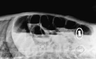
Certain objects pose more risk than others. In cases of sharp foreign bodies, such as straight pins, weekly assessments are required. Surgical removal is necessary if the patient develops symptoms or signs of obstruction or perforation or if the foreign body fails to progress for several weeks. Small magnets used to secure earrings have been associated with bowel perforation. When the multiple magnets disperse after ingestion, they may be attracted to each other across bowel wall, leading to pressure necrosis and perforation (Fig. 326-1). Inexpensive toy medallions containing lead can lead to lead toxicity. Newer coins can also decompose when subjected to prolonged acid exposure. Unless multiple coins are ingested; however, the metals released are unlikely to pose a clinical risk.

image

Figure 326-1 Abdominal radiograph of a boy aged 3 yr, noting 3 attached magnets that resulted in volvulus (i.e., twisting of the bowel) and multiple bowel perforations.

(Courtesy of the US Consumer Product Safety Commission. From Centers for Disease Control and Prevention: Gastrointestinal injuries from magnet ingestion in children—United States, 2003–2006, MMWR 55:1296–1300, 2006.)

Ingestion of batteries rarely leads to problems, but symptoms can arise from leakage of alkali or heavy metal (mercury) from battery degradation in the gastrointestinal tract. Batteries can also generate electrical current and thereby cause low-voltage electrical burns to the intestine. If patients experience symptoms such as vomiting or abdominal pain, if a large-diameter battery (>20 mm in diameter) remains in the stomach for >48 hr, or if a lithium battery is ingested, the battery should be removed. Batteries >15 mm that do not pass the pylorus within 48 hr are less likely to pass spontaneously and generally require removal. In children <6 yr of age, batteries >15 mm are not likely to pass spontaneously and should be removed endoscopically. If the patient develops peritoneal signs, surgical removal is required. The battery should be identified by size and imprint code or by evaluation of a duplicate measurement of the battery compartment. The National Button Battery Ingestion Hotline can be called for help in identification: 202-625-3333. The Poison Control Center can be called as well: 800-222-1222 for ingestion of batteries and caustic materials. Lithium batteries result in more severe injury than a button alkali battery, with damage occurring in minutes.

In older children and adults, oval objects >5 cm in diameter or 2 cm in thickness tend to lodge in the stomach and should be endoscopically retrieved. Thin objects >10 cm in length fail to negotiate the duodenal sweep and should also be removed. In infants and toddlers, objects >3 cm in length or >20 mm in diameter do not usually pass through the pylorus and should be removed. An open safety pin presents a major problem. Razor blades can be managed with a rigid endoscope by pulling the blade into instrument. The endoscopist can alternatively use a rubber hood on the head of the endoscope to protect the esophagus. Open safety pins should also be endoscopically retrieved, but other sharp objects can be managed conservatively. Drugs (aggregated iron pills, cocaine packing) may need to be surgically removed; initial management can include oral polyethylene glycol lavage.

Ingestion of magnets poses a danger to children. The number of magnets is thought to be critical. If a single magnet is ingested, there is the least likelihood of complications. If ≥2 magnets are ingested, the magnetic poles are attracted to each other and create the risk of obstruction, fistula development, and perforation. Endoscopic retrieval is emergent after films are taken when multiple magnets are ingested. Abdominal pain or peritoneal signs require urgent surgical intervention.

Lead-based foreign bodies can cause symptoms from lead intoxication. Early endoscopic removal is indicated of an object suspected to contain lead. A lead level should be obtained.

Children occasionally place objects in their rectum. Small blunt objects usually pass spontaneously, but large or sharp objects typically need to be retrieved. Adequate sedation is essential to relax the anal sphincter before attempted endoscopic or speculum removal. If the object is proximal to the rectum, observation for 12-24 hr usually allows the object to descend into the rectum.

326.2 Bezoars

A bezoar is an accumulation of exogenous matter in the stomach or intestine. They are predominantly composed of food or fiber. Most bezoars have been found in females with underlying personality problems or in neurologically impaired persons. Patients who have undergone abdominal surgery are at higher risk for the development of bezoars. The peak age at onset of symptoms is the 2nd decade of life.

Bezoars are classified on the basis of their composition. Trichobezoars are composed of the patient’s own hair, and phytobezoars are composed of a combination of plant and animal material. Lactobezoars were previously found most often in premature infants and can be attributed to the high casein or calcium content of some premature formulas. Swallowed chewing gum can occasionally lead to a bezoar.

Trichobezoars can become large and form casts of the stomach; they can enter into the proximal duodenum. They manifest as symptoms of gastric outlet or partial intestinal obstruction including vomiting, anorexia, and weight loss. Patients might complain of abdominal pain, distention, and severe halitosis. Physical examination can demonstrate patchy baldness and a firm mass in the left upper quadrant. Patients occasionally have iron-deficiency anemia, hypoproteinemia, or steatorrhea caused by an associated chronic gastritis. Phytobezoars manifest in a similar manner. Detached segments of the bezoar or trichobezoar can migrate to the small intestine as a ““satellite masses”“ and result in small bowel obstruction.

An abdominal plain film can suggest the presence of a bezoar, which can be confirmed on ultrasound or CT examination. On CT a bezoar appears a nonhomogeneous, nonenhancing mass within the lumen of the stomach or intestine. Oral contrast circumscribes the mass.

Bezoars in the stomach can usually be removed endoscopically. If endoscopy is unsuccessful, surgical intervention may be needed. Lactobezoars usually resolve when feedings are withheld for 24-48 hr.