FOREARM, WRIST, AND HAND
Published on 16/03/2015 by admin

Filed under Orthopaedics

Last modified 22/04/2025

Print this page

rate 1 star rate 2 star rate 3 star rate 4 star rate 5 star
Your rating: none, Average: 0 (0 votes)

This article have been viewed 8405 times

CHAPTER 6

FOREARM, WRIST, AND HAND

image

Précis of the Forearm, Wrist, and Hand Assessment*

History (sitting)

Observation (sitting)

Examination (sitting)

Active movements

Passive movements (as in active movements)

Resisted isometric movements (as in active movements, in the neutral position)

Functional testing

Special tests (sitting)

Reflexes and cutaneous distribution (sitting)

Joint play movements (sitting)

Palpation (sitting)

Diagnostic imaging

DIP, Distal interphalangeal; MCP, metacarpophalangeal; PIP, proximal interphalangeal.


*After any examination, the patient should be warned of the possibility of exacerbation of symptoms as a result of the assessment.

SELECTED MOVEMENTS

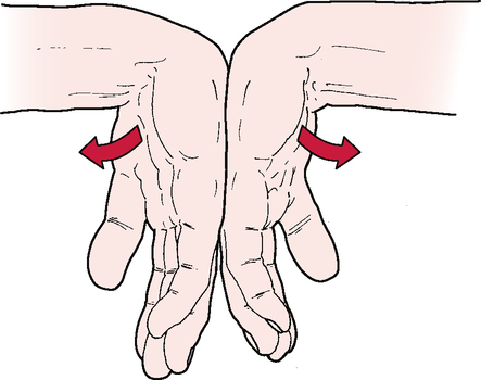
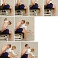
ACTIVE MOVEMENTS image

INDICATIONS OF A POSITIVE TEST

Pronation and supination. Active pronation and supination of the forearm and wrist are approximately 85° to 90°, although this varies from individual to individual. It is more important to compare the movement with that of the normal side. Approximately 75° of supination or pronation occurs in the forearm articulations. The remaining 15° is the result of wrist action.

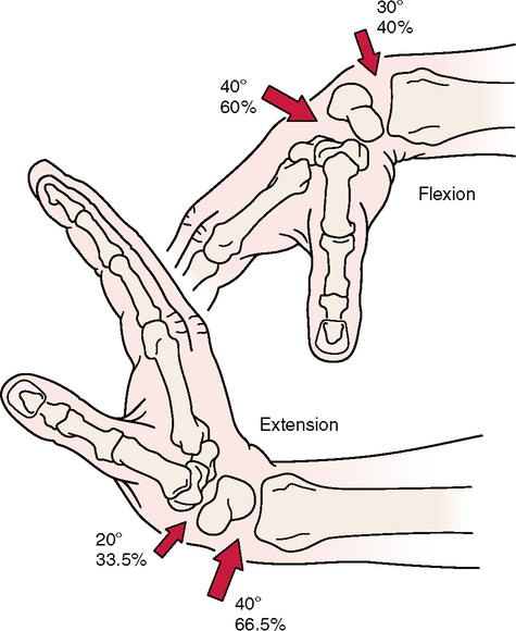
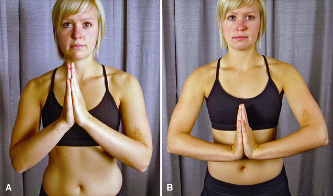
Wrist flexion and extension. Wrist flexion is 80° to 90°; wrist extension is 70° to 90°. The end feel of each movement is tissue stretch.

Radial and ulnar deviation. Radial and ulnar deviations of the wrist are 15° and 30° to 45°, respectively. The normal end feel of these movements is bone to bone.

Finger flexion. Flexion of the fingers occurs at the metacarpophalangeal joints (85° to 90°), followed by the proximal interphalangeal joints (100° to 115°) and the distal interphalangeal joints (80° to 90°).

Finger extension. Extension occurs at the metacarpophalangeal joints (30° to 45°), the proximal interphalangeal joints (0°), and the distal interphalangeal joints (20°). The end feel of finger flexion and extension is tissue stretch.

Finger abduction and adduction. Finger abduction occurs at the metacarpophalangeal joints (20° to 30°); the end feel is tissue stretch. Finger adduction (0°) occurs at the same joint.

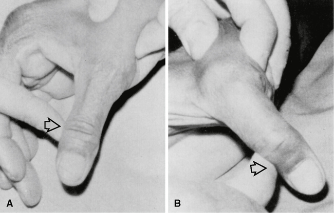
Thumb flexion. Thumb flexion occurs at the carpometacarpal joint (45° to 50°), the metacarpophalangeal joint (50° to 55°), and the interphalangeal joint (80° to 90°). It is associated with medial rotation of the thumb as a result of the saddle shape of the carpometacarpal joint.

Thumb extension. Extension of the thumb occurs at the interphalangeal joint (0° to 5°); it is associated with lateral rotation. Flexion and extension take place in a plane parallel to the palm of the hand.

Thumb abduction and adduction. Thumb abduction is 60° to 70°; thumb adduction is 30°. These movements occur in a plane at right angles to the flexion-extension plane.

CLINICAL NOTES/CAUTIONS

• Pathological conditions in structures other than the joint may restrict ROM (e.g., muscle spasm, tight ligaments/capsules). If the examiner suspects a problem with these structures, passive movement end feels will help differentiate the problem.

• Most functional activities of the hand require the fingers and thumb to open at least 5 cm (2 inches), and the fingers should be able to flex within 1 to 2 cm (0.4 to 0.8 inches) of the distal palmar crease.

• If the patient complains of pain on supination, the examiner can differentiate between the distal radioulnar joint and the radiocarpal joints by passively supinating the ulna on the radius with no stress on the radiocarpal joint. If this passive movement is painful, the problem is in the distal radioulnar joint, not the radiocarpal joints. The normal end feel of both movements is tissue stretch, although in thin patients, the end feel of pronation may be bone to bone.

• Wrist flexion decreases as the fingers are flexed, just as finger flexion decreases as the wrist flexes, and movements of flexion and extension are limited, usually by the antagonistic muscles and ligaments.

• The digits are medially deviated slightly in relation to the metacarpal bones. When the fingers are flexed, they should point toward the scaphoid tubercle. In addition, the metacarpals are at an angle to each other.

SPECIAL TESTS FOR LIGAMENT, CAPSULE, AND JONT INSTABILITY25

Relevant Special Tests

Mechanism of Injury

The most common mechanism of injury is trauma, such as a fall onto the hand (FOOSH) or wrist. Injury also can occur whenever the ligaments are subjected to tensile forces that exceed their physiological capacities. Because the ligaments are damaged, passive stability is lost and active stability is needed. The muscles, tendons, and nerves of the wrist and forearm provide the active stability to the region. However, in the wrist and hand, most joints have no direct muscle or tendon attachment. Instead, the tendons of the muscle overlie the affected joint and have no direct control over the wrist motion or stability. As a result, instability is common after trauma and persists without the neuromuscular system contribution. Although the initial mechanism is different when ligament damage is the result of disease processes, the reason for the lack of stability in the joint is similar.

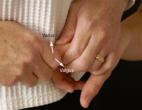
THUMB ULNAR COLLATERAL LIGAMENT LAXITY OR INSTABILITY TEST6,7 image

WATSON (SCAPHOID SHIFT) TEST10,1215 image

TRIANGULAR FIBROCARTILAGE COMPLEX (TFCC) LOAD TEST16 image

SPECIAL TEST FOR MUSCLE OR TENDON PATHOLOGY1719

Relevant Special Test

Finkelstein test

Mechanism of Injury

The pathological condition occurs secondary to overuse or misuse, often as a result of a change in the customary use patterns for the wrist and thumb, leading to inflammation. It may be precipitated by discrete trauma, such as a blow to the wrist, but the onset usually is gradual. Initially, physical irritation results in acute inflammation of the tendon sheath. Over time, chronic degenerative changes develop as a result of the inflammation, resulting in tendinosis. Repeated wrist radial deviation and/or thumb palmar or radial abduction, especially under load, is the most common mechanism of injury.

SPECIAL TESTS FOR NEUROLOGICAL SYMPTOMS2232

Relevant Special Tests

Relevant Signs and Symptoms

Carpal tunnel syndrome commonly presents with progressively worsening pain (classically described as aching and burning) and paraesthesia in the thumb, forefinger, middle finger, hand, and wrist that may extend to the forearm, elbow, shoulder, and neck. Symptoms frequently are worse nocturnally. The patient also may complain of problems with grip and weakness in the wrist and hand. With median nerve involvement, fine motor skills (precision grip) are affected more than power grip.

Variations in the innervation of the hand, particularly the Martin Gruber anastomosis, where the motor nerve crosses over from the median nerve to the ulnar nerve (seen in 10% to 15% of the population), can cause the presentation of entrapment to vary significantly. The condition may involve the flexor pollicis brevis, adductor pollicis, abductor pollicis brevis, lumbricals, and abductor digiti minimi, or even the entire hand.

Mechanism of Injury

In general, nerve injury is classified according to the severity of the injury and the potential for reversibility. Nerve injury may be classified according to the amount of nerve damage and the nerve structure involved. Five categories of nerve injury are frequently used (Sunderland classification).

• First-degree neuropraxia involves distortion of myelin about the nodes of Ranvier caused by ischemia, mechanical compression, or electrolyte imbalance, resulting in temporary loss of nerve conduction.

• Second-degree axonotmesis involves interruption of the axon with secondary wallerian degeneration but preservation of the supporting tissue around the axon. Recovery may be complete but takes longer and depends on the distance between the site of injury and the end structure (denervated muscle).

• Third-degree neurotmesis involves extensive disruption of the nerve and its supporting structures; however, although the endoneurium is disrupted, the perineurium and epineurium remain intact.

• Fourth-degree neurotmesis involves disruption of all neural components except the epineurium.

• Fifth-degree neurotmesis involves complete transection and discontinuity of the nerve, with no capacity for regeneration. Neurotmesis rarely occurs as a result of entrapment. However, when continuity has been disrupted, complete recovery is not possible, even with surgical techniques, and the eventual outcome depends on the individual circumstances.

RELIABILITY/SPECIFICITY/SENSITIVITY COMPARISON3032

  Validity Interrater Reliability Intrarater Reliability Specificity Sensitivity
Tinel’s Sign No association with the severity of carpal tunnel syndrome and test results p > 0.11 0.77 0.80 87% 23%
Phalen’s Test Patients with more severe carpal tunnel syndrome are more likely to have a positive test result p < 0.05 0.65 0.53 76% 51%
Reverse Phalen’s Test Unknown Unknown Unknown Unknown Unknown

image

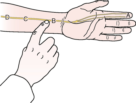
TINEL’S SIGN (AT THE WRIST)20 image

SPECIAL TESTS FOR CIRCULATION AND SWELLING IN THE WRIST AND HAND

Relevant Special Tests

Mechanism of Injury

No single specific mechanism of injury is consistent with every instance of swelling or altered circulation in the upper extremity. Injury may or may not have been a trigger. Both vascular edema and neurological edema can result in altered circulation or the accumulation of fluid and other products of inflammation.
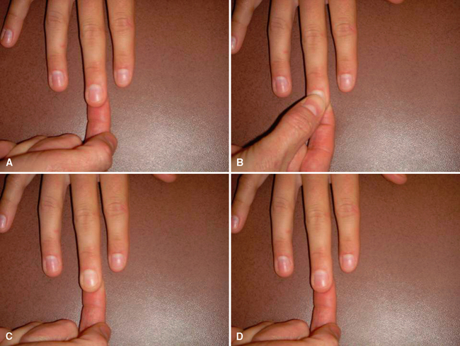
JOINT PLAY MOVEMENTS

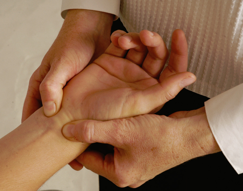
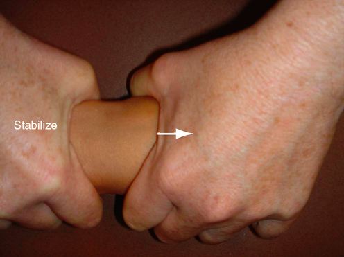
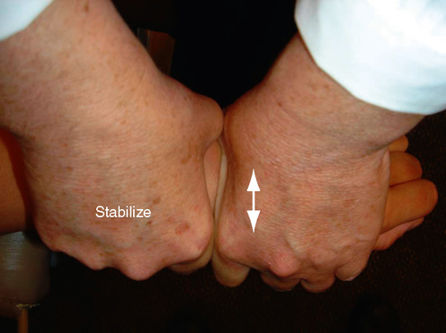
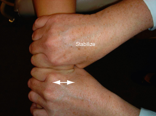
ANTERIOR-POSTERIOR GLIDE OF THE WRIST image

TEST PROCEDURE

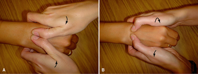
Step 1: Radiocarpal joint. The examiner first places the forearm in a supinated or pronated position. The examiner’s stabilizing hand then is placed around the distal end of the radius and ulna, just proximal to the radiocarpal joint, and the other hand is placed around the proximal row of carpal bones. If the examiner’s hands are positioned properly, they should touch. The examiner applies an anteroposterior gliding movement of the proximal row of carpal bones on the radius and ulna, testing the amount of movement and end feel.

Step 2: Midcarpal joint. The examiner moves the stabilizing hand slightly distally (less than 1 cm [0.4 inch]) so that it is around the proximal row of carpal bones and places the mobilizing hand around the distal row of carpal bones. An anteroposterior gliding movement is applied to the distal row of carpal bones on the proximal row to test the amount of movement and end feel (midcarpal joint).

Step 3: Carpometacarpal joint. The examiner then moves the stabilizing hand slightly distally again (less than 1 cm [0.4 inch]); the hand will be around the distal carpal bones. The mobilizing hand is placed around the metacarpals. An anteroposterior gliding movement is applied to the base of the metacarpals to test the amount of joint play and end feel.

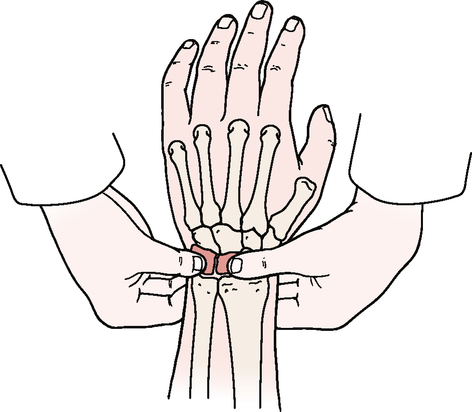
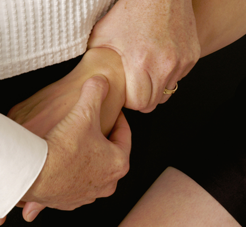
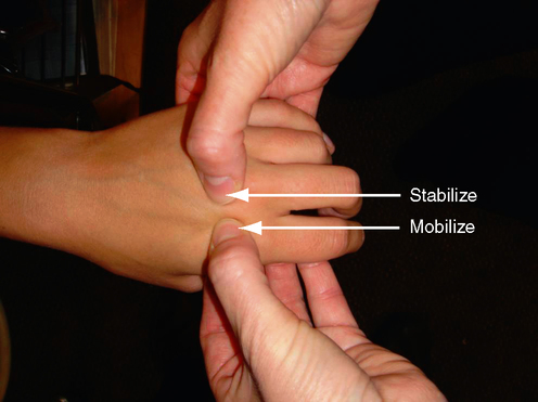
SHEAR TEST OF THE INDIVIDUAL CARPAL BONES image

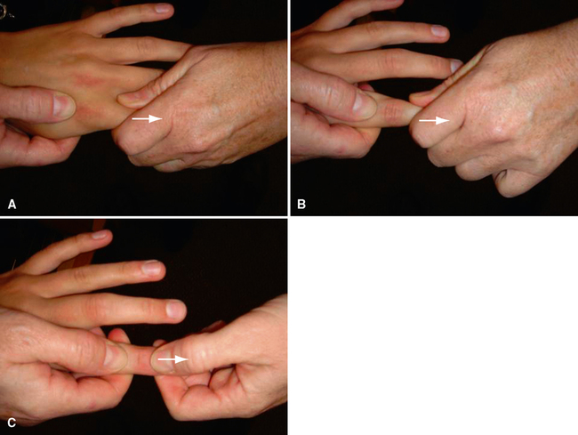
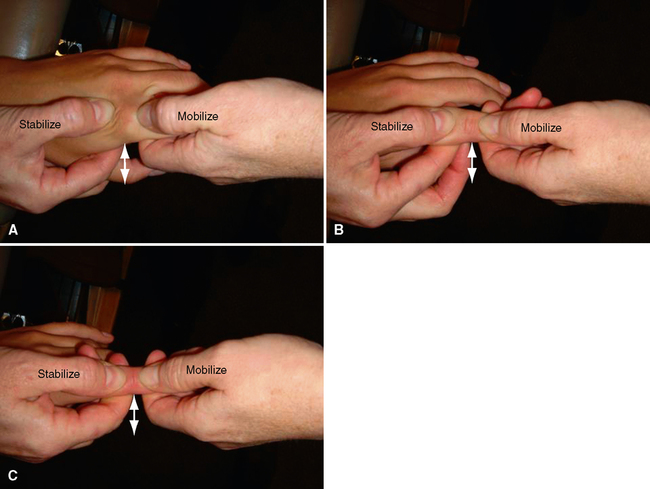
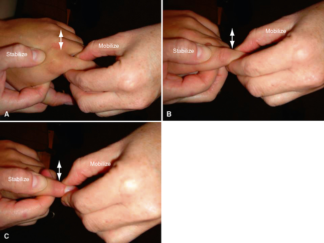
ANTERIOR-POSTERIOR GLIDE OF THE JOINTS OF THE FINGERS image

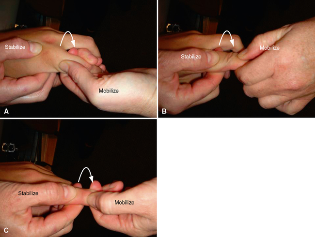
SIDE GLIDE OF THE JOINTS OF THE FINGERS image