Anesthesia for therapeutic and diagnostic procedures
A Brachytherapy
Palladium-103 prostate implants are being used for brachytherapy as an optional treatment for prostate cancer. The sources are permanently implanted directly into the tissue. The seeds are encased in a lead-lined cartridge that attaches to lead-lined placement needles. The seeds are placed by the radiation oncologist through these needles into the prostate through the perineum. Placement is guided by ultrasonography.
2. Preoperative assessment and patient preparation
a) The treatment is performed in the radiation oncology department.
b) Each patient is prescreened and admitted as an outpatient through the short stay unit.
c) The procedure lasts a maximum of 2 hours.
d) Patients may undergo a bowel preparation at home before the procedure.
e) An 18-gauge, 2-inch angiocatheter is placed before the procedure.
f) Each patient will require intravenous (IV) antibiotics before the procedure. One gram of cefazolin (Ancef) is required. If the patient has an allergy to penicillin, then clindamycin (Cleocin), 600 mg, is administered.
Required monitoring equipment, anesthesia, and airway supplies are provided.
a) Anesthesia is usually maintained by spinal anesthesia. A minimal T8 level is desired. If the patient is required to have consecutive treatments, an epidural anesthetic may be placed.
b) Sedation is given as needed.
c) The patient is placed in a lithotomy position for the procedure. A Foley catheter is inserted with Hypaque in the balloon. An ultrasound probe and perineum template are placed before needle placement.
d) The time of treatment is the time of radiation. Everyone must leave the room. Visual contact is always present through monitors. Treatment time is usually 5 to 15 minutes.
(1) Occasionally, seeds can become dislodged from the implanted tissue. Therefore, dressings and linens should not be removed from the room until they have been checked and cleared by the oncology physicist.
(2) If a seed is discovered, do not touch it with your hands. Use long forceps to place it in a lead storage container.
(3) Some seeds may pass in the urine for the first few days. Therefore, urine should be strained before being discarded.
(4) Pregnant personnel are not to provide anesthesia for these patients.
(5) All personnel remaining in the room during the procedure should have proper badges. No person or material should leave the room without being surveyed with a Geiger counter.
B Computed tomography
Computed tomography (CT) uses x-rays generated from a rotating anode x-ray generator. The patient is placed supine on a flat, wheeled platform and moved inside the scanning gantry. X-rays are then projected through the patient at different angles. The X-rays penetrate tissues differently according to the atomic numbers of the atoms within the tissue. The diagnostic quality of a CT scan is enhanced with the injection of IV contrast media (ICM). Contrast media containing iodine may be administered to the patient enterally or parenterally to further attenuate the x-ray beam to enhance the images for CT vascular or gastrointestinal studies.
a) CT scans require that the patient remain as motionless as possible for several minutes to an hour. Patient motion can produce artifacts in the diagnostic images to be read by the radiologist.
b) Patients should lie on a flat, lightly padded wheeled platform, which is rolled into the short bore scanning gantry of the CT scanner.
c) Although the majority of patients are able to cooperate and tolerate CT, others may not be able to cooperate because of extremes in age, concurrent medical conditions, or mental disability.
d) The CT scan is neither physically invasive nor painful. Patients enter the CT scanner without precautions for ferromagnetic objects as for a magnetic resonance imaging (MRI) scan. CT is more rapidly performed than an MRI scan, especially if a spiral CT scanner is used.
e) The patient may require anesthesia anywhere along the continuum from minimal sedation to general anesthesia.
f) Use of ferromagnetic anesthesia equipment and supplies around the CT scanner is not a concern.
g) A standard anesthesia machine, laryngoscope and blades, and IV infusion pumps can be used as if in the operating room.
h) A laryngeal mask airway (LMA) is an appropriate alternative choice as a minimally invasive and secure airway in the patient without contraindications to its use.
i) Attention should be paid to securing the airway, and the anesthesia breathing circuit, the leads for the electrocardiogram (ECG), the noninvasive blood pressure cuff, the IV line, and the pulse oximeter should extend into the scanning gantry. If the use of general anesthesia is used, it is important to verify the presence of a suction canister with tubing that will reach the patient’s mouth in the event of an emergency. An Ambu bag with appropriately sized face mask should also be present.
j) The anesthesia provider should allow for extra anesthesia circuitry length and electrical monitoring leads lengths because of patient movement that will occur because of intermittent repositioning of the mechanized table that positions the patient within the scanning gantry.
k) Sedation can be performed with a variety of agents, including midazolam, chloral hydrate, diazepam, or propofol.
l) General anesthesia can be performed with total IV anesthesia (TIVA), such as with IV propofol, or with inhalation agents.
m) All personnel must be aware of the use of ionizing radiation during the CT scan and should take precautions to be shielded from any exposure to the radiation.
n) Radiation protection can be accomplished with the use of a lead glass barrier, a lead apron, a lead thyroid collar, and lead-glass safety glasses. Radiation dose badges that attach to the clothing are available.
o) ICM can cause an unexpected allergic reaction in some patients, varying from itching with hives to severe, life-threatening anaphylactoid and anaphylactic reactions that have led to patient death.
p) ICM can also cause renal toxicity as well as local tissue sloughing and necrosis if the ICM extravasates from the vein into the surrounding tissue.
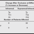
q) The anesthesia provider will be involved with patient care related to ICM extravasation and should be familiar with treatment protocols to minimize patient morbidity, as shown in the following box.
C Gastroenterologic procedures: Colonoscopy, esophagogastroduodenoscopy, and endoscopic retrograde cholangiopancreatography
Endoscopy for gastrointestinal procedures is the use of a flexible fiberoptic endoscope that transmits brilliant, coherent, high-resolution, magnified, direct visual images to the operator. The operator can then examine, biopsy, dilate, or cauterize portions of the gastrointestinal tract. The endoscopist may pass accessory devices down the endoscope such as biopsy forceps, dilation devices, cytology brushes, measuring devices, needles for injection, Doppler probes, ultrasound probes, and probes to measure electrical activity and pH. Even foreign bodies may be removed with the aid of a snare passed through an endoscope.
a) A colonoscopy allows total diagnostic visualization of the mucosa of the tortuous colon from the anus to the cecum.
b) An upper endoscopy, such as an esophagogastroduodenoscopy (EGD), is an accurate way for the operator to evaluate the mucosa of the esophagus, stomach, and duodenum.
c) Endoscopic retrograde cholangiopancreatography (ERCP) is used for the diagnosis of obstructive, neoplastic, or inflammatory pancreatobiliary structures.
d) Endoscopy for gastrointestinal procedures may be performed by a gastroenterologist, a general surgeon, a family practitioner, or a proctologist.
e) The endoscope is passed into the gastrointestinal tract with the aid of lubricant.
f) The endoscope has controls to change the direction of the flexible tip, allow flushing with water, apply suction, or insufflate air or carbon dioxide within the portion of the gastrointestinal tract being observed.
3. Anesthetic technique for colonoscopy
a) Because of the expectations of patients, endoscopically caused discomfort, and the desirability for no patient movement, moderate sedation, deep sedation, and, in some cases, general endotracheal anesthesia are used.
b) A proper preanesthetic assessment of the patient should be performed, focusing on the areas of age, ability to cooperate, level of anxiety, mental disability, allergies, fluid status, laboratory electrolyte values, cardiac history, hypertension, bleeding history, clotting status, respiratory status, obesity, drug and alcohol abuse, gastroesophageal reflux, and pregnancy.
c) Patients should adhere to proper NPO (nothing by mouth) guidelines.
d) Bacteremia is possible as a result of endoscopic procedures.
e) Necessary medications may be given, such as cardiac medications, antihypertensives, and antibiotics.
f) Preemptive analgesia with gargled flavored viscous Xylocaine helps patient acceptance of the procedure.
g) Moderate sedation is usually accomplished with the short-acting sedatives midazolam or propofol and analgesics such as remifentanil, alfentanil, or fentanyl.
h) Deep sedation can be achieved with titration of propofol until effective along with an analgesic medication. Upper endoscopy may necessitate the use of any antisialagogue such as glycopyrrolate.
i) Colonoscopy requires thorough cleansing of the lumen of the colon of fecal material. The colon may be partly prepared with a cleansing enema. Full preparation of the colon is accomplished commonly with orally administered balanced electrolyte solutions in a volume of up to 4 L.
j) After the preparation, abdominal cramping, diarrhea, weakness, and nausea can occur. Patients who arrive for the procedure require reassessment and the insertion of an IV catheter with IV fluid, usually lactated Ringer’s solution or normal saline.
k) Conventional monitors, including pulse oximeter, noninvasive blood pressure monitor, and ECG, are attached.
l) The patient is supplied with oxygen through a disposable nasal cannula or disposable face mask. The procedure is usually performed with the patient positioned in a lateral decubitus position with the body flexed, the head and back bent downward toward the knees, and the legs bent upward toward the abdomen.
m) Patient anxiety, distention because of insufflation, and acute discomfort during the maneuvering of the endoscope usually necessitate the administration of deep sedation or a general anesthetic in some cases.
n) Strong vagal nerve stimulation can occur as a result of distention of the colon. This may cause hypotension, bradydysrhythmia, and ECG changes.
4. Anesthetic technique for EGD
a) EGD requires a general patient assessment with special emphasis on any cardiac history, hypertension, bleeding disorders, postoperative nausea and vomiting (PONV), dysphagia, and gastroesophageal reflux.
b) The patient should be NPO according to guidelines. Most patients are able to have EGD performed with a spray or gargle of topical anesthetic such as Cetacaine, benzocaine, or 4% lidocaine liquid.
c) Rapid absorption of highly concentrated local anesthetics, applied topically over highly vascular and absorptive mucosal tissues, can lead to possible toxicity reactions whose symptoms could be masked while the patient is receiving sedative anesthesia.
d) Topical benzocaine can pose a small risk of methemoglobinemia if overused.
e) An IV catheter is inserted, with fluids such as lactated Ringer’s solution or normal saline attached.
f) The patient is connected to standard monitors. Oxygen can be supplied through a disposable nasal cannula or a disposable face mask. EGD is generally performed with the patient positioned supine.
g) After the patient is adequately sedated, the operator inserts a hollow oral airway gently into the patient’s mouth, and the endoscope is advanced through this airway, allowing direct visualization of the larynx, hypopharynx, esophagus, and stomach, and through the pylorus into the duodenal bulb.
5. Anesthetic technique for ERCP
a) ERCP requires thorough assessment of the patient, including a review of laboratory values of a complete blood count, serum liver chemistries and amylase or lipase levels to evaluate liver function, and clotting studies.
b) Patients should also be evaluated for anticoagulant medications, bleeding history, and prosthetic heart valves.
c) Allergies should be evaluated, especially those to iodinated contrast media.
d) Patients who require ERCP are usually more ill than patients seen routinely for colonoscopy or EGD.
e) The patient should be NPO according to guidelines.
f) IV access is obtained, and fluid is administered.
g) Standard monitors are applied, and oxygen is supplied to the patient via a disposable face mask.
h) The procedure requires that the patient be in a prone or slightly left lateral decubitus position.
i) Deep sedation is generally required, although painful or complex ERCP may require general anesthesia.
j) Pediatric endoscopy has been performed with patients under deep IV sedation with agents such as propofol when the patient will allow placement of the IV catheter and under general endotracheal anesthesia.
k) These procedures can cause bowel rupture or duct rupture. One should be ready with immediate airway and hemodynamic support as necessary along with monitored emergency transport to the operating room for surgical intervention.
a) Postprocedure morbidity differs with each of the described procedures. All patients should be monitored in a postanesthesia care area until they have recovered from the sedation or general anesthetic.
b) Patients having colonoscopy frequently complain of intestinal or abdominal pain immediately after the procedure caused by insufflation during the examination. Rectal bleeding, PONV, and hypotension may also be seen. Administration of a bolus of IV fluids along with an IV antiemetic agent, such as ondansetron, dolasetron, or granisetron, is indicated.
c) EGD morbidity relates to bleeding, PONV, aspiration, dysphagia, and hypotension. Treatments such as those used for colonoscopy may be indicated.
d) ERCP morbidity relates to possible reactions to iodinated contrast media. Patient reactions can be mild (e.g., PONV, pruritus, diaphoresis, flushing, or mild urticaria), moderate (e.g., faintness, severe vomiting, profound urticaria, mild bronchospasm, mild hypotension, mild tachycardia, or bradycardia), or severe (e.g., hypotensive shock, angioedema, respiratory arrest, cardiac arrest, convulsions, or death). PONV can be treated as described previously.
D Interventional radiology, radiotherapy, stereotactic radiosurgery, and interventional neuroradiology
Interventional radiology (IR) involves minimally invasive procedures and therapies performed by radiologists, especially in patients at high medical risk. Major IR therapies include angiography, embolization of blood vessels such as arteriovenous malformations or for epistaxis, delivery of chemical or physical vascular occlusive devices, removal of thrombi, ablation of aneurysms, and angioplasty of blood vessels with stent placement.
Gamma radiation is used for radiotherapy and radiosurgery. The gamma radiation is introduced to the patient by the use of either a Gamma Knife or a CyberKnife. The CyberKnife therapy delivers a sequence of many hundreds of gamma beams to the cancerous tumor from many different directions. Gamma Knife therapy delivers gamma radiation to the cancerous tumor simultaneously in a single dose.
Interventional neuroradiology (INR) is used for diagnosis and treatment of central nervous system (CNS) diseases endovascularly to deliver therapeutic medications or devices. Digital subtraction angiography first uses an original angiograph of the blood vessels to be studied. Then a contrast medium is injected into the same blood vessels, and opaque structures such as bone and tissues can be digitally subtracted or removed from the angiographic image, leaving a clear picture of the blood vessels.
Improvements in vascular access techniques, new thin and flexible catheters and guidewires, and the development of innovative coils and therapeutic medications have made new treatments possible. Conditions that once required extensive surgery with accompanying patient morbidity and mortality can now be performed less invasively. Some major procedures performed with INR are mechanical or chemical removal of emboli or thrombi that cause stroke, the physical occlusion of malformed vascular structures such as an arteriovenous malformations with chemicals or flow-directed balloons, dilation of stenotic blood vessels, and embolization (blocking blood flow) of cerebrovascular aneurysms using catheter-deployed coils.
a) As skills, techniques, and technology progress, more procedures will be performed with radiation or under radiological guidance.
b) These procedures all require the absolute immobility of the patient, with periods of controlled apnea, which assist in the viewing or treatment of the targeted area of the patient, especially during whole-body therapeutic radiation treatment.
c) These procedures are also time consuming, taking up to several hours to complete. Procedures may be necessary in patients within a wide range of ages (infant to geriatric) and with significant coexisting disease.
d) A thorough preanesthetic assessment is imperative.
e) With the exceptions of angiography or radiotherapy, procedures for IR are painful, are physically invasive to the patient, and may need to be accomplished over several treatment sessions.
f) Treatment may be required electively or urgently.
g) Patients may require anesthesia along the continuum from minimal or moderate sedation, local or regional anesthesia, and general anesthesia.
h) Full monitors and IV access are required.
i) Additional catheterization and monitoring of arterial pressure and central venous pressure may be necessary.
j) Certain procedures require monitoring of the patient’s neurophysiologic status for changes. The patient may also need to be assessed awake and then resedated at times during the procedure.
k) Anesthetics that can be used are midazolam, propofol, ketamine, isoflurane, and the other potent inhaled general anesthetics.
l) Rapid recovery from anesthesia at the end of the case is ideal to assess and monitor the patient’s neurologic functioning.
m) It may be necessary to manipulate or manage normal systemic blood flow, normal cerebral blood flow, or other regional blood flow. The anesthesia provider may be called upon to control deliberate hypertension or deliberate hypotension, manage anticoagulation, and manage unexpected procedural complications.
n) Intraoperative radiation therapy (IORT) is the delivery of radiation to the patient via a linear accelerator, at times in conjunction with tumor surgery. If surgery is performed coincidental to the dose of radiation, normal tissues may be able to be moved away from the ionizing radiation beam. Normal tissues and organs can be shielded with lead beforehand.
o) Some facilities use a dedicated IORT suite, and others use an operating room with transport of the patient to the radiation oncology suite. General anesthesia is performed if the surgical and radiation procedures are concurrent.
p) All personnel should leave the room during IORT and stereotactically guided Gamma Knife or CyberKnife surgery so that high-dose radiation can be delivered to the patient and to protect personnel from the scattered radiation. The radiation oncology suite is heavily shielded and has a heavy lead or iron door that can take from 30 to 60 seconds to open.
q) The patient is monitored via closed-circuit video and hands-off anesthesia delivery during treatment.
r) Complications can occur rapidly and can be life threatening. Foremost is the possible complication of hemorrhage. A sedated patient experiencing hemorrhage may show sudden signs of headache, nausea and vomiting, and vascular pain.
s) A patient under general anesthesia may experience sudden bradycardia. The airway should be secured first if necessary followed by support of the cardiovascular system, discontinuation of heparin, and administration of protamine (1 mg/100 units of total heparin dose administered).
t) Other possible complications are radiocontrast reactions, embolization of particles or tissue, perforation of an aneurysm, and obliteration of unintended physiologically necessary arteries.
u) Patient safety necessitates skilled and competent staff assistance in treatment of complications.
v) Complications may necessitate the safe transfer of the patient to the operating room.
E Magnetic resonance imaging
MRI uses the dipole moment (the ability of the atomic nucleus to behave as a magnet) of the hydrogen atom. The patient is placed supine within the scanning gantry or bore of the magnet. The magnet used for MRI can be a permanent magnet or a powerful superconducting electromagnet cooled with liquid helium to 4° Kelvin. The quality of the MRI image is directly related to the strength of the magnetic field. Contrast media are also used in MRI studies to enhance the patient’s tissues and allow the scan to provide further diagnostic information. MRI contrast is most commonly gadopentetate dimeglumine (Magnevist) than CT scans, which contain the element gadolinium, bound as a chelated structure and administered primarily parenterally but rarely enterally.
a) MRI can take up to 1 hour or longer. During this time, the patient should remain extremely still to reduce motion artifacts. These artifacts can cause unfaithful representations of the tissues being studied. The motions of breathing, the heart, blood flow, swallowing, and even cerebrospinal fluid flow produce artifacts in a highly sensitive MRI scan.
b) The patient is exposed to varying magnetic fields of up to 4 tesla (T), along with additional exposure to variable radiofrequency radiation. Blood flow is decreased by strong magnetic fields, and blood pressure compensates by rising. Patients also have reported symptoms of vertigo, nausea, headache, and visual sensations.
c) The MRI machine produces loud vibratory and knocking noises as coils are switched on and off during the course of the study.
d) Most patients are content with an explanation of what to expect during the procedure and with reassurance. Some patients need minimal or moderate sedation. Patients with claustrophobia and those who cannot or will not remain motionless during the study (children) as well as critically ill patients may require deep sedation or general endotracheal anesthesia.
e) MRI is not painful, so opioids are not usually required. Sedation has been performed with oral and IV midazolam, ketamine, pentobarbital, chloral hydrate, and propofol.
f) Minimal or deep sedation or general anesthesia requires IV access and full monitoring.
g) The LMA serves as an excellent, relatively noninvasive airway for MRI. Some anesthesia providers prefer general endotracheal intubation.
h) Because of the intense magnetic field always present in the MRI suite, anesthesia providers should be aware of every item on their persons and every item that is to be used in conjunction with anesthesia administered to the patient.
i) Ferromagnetic (iron-containing) substances are attracted at astonishing rates of speed into the bore of the magnet. Personal items such as pens, certain types of eyeglasses, jewelry, watches, pagers, personal computers, calculators, name badges, coins, audiotapes, videotapes, and credit cards are some of the items that should never enter the MRI suite, as well as any ferromagnetic anesthesia equipment, medication vials, and supplies. If a patient were present within the bore of the MRI, injury or death could be possible from the missile created.
j) Metals that are known to be safe within the proximity of the MRI bore are stainless steel, nonferrous alloys, nickel, and titanium. Materials and equipment constructed of plastic are safe.
k) Patients possessing certain medical therapeutic devices may be prohibited from an MRI scan.
l) Cardiac pacemakers may be affected several ways by the electromagnetic field: reprogramming may occur, the pacemaker may be inhibited, it may revert to an asynchronous mode, it may have the reed switch close, it may become dislodged, or it may become heated by the magnetic field.
m) Any monitor leads and IV tubing should be kept in straight alignment because the intense magnetic fields in the MRI suite can induce current flow in coiled leads or tubing, and severely burn the patient.
n) Flexible LMAs and endotracheal tubes (ETTs) that contain wire windings can also be sources of burns.
o) The American College of Radiology recommends strong attention to and the elimination of induced current that can be large tissue loops, such as the loop created by the hand touching the hip or thigh, or the loop created when the feet or calves of the legs touch.
p) Consideration should be given to the MRI contrast media administered to patients. The dyes used for MRI contrast are nonionic gadolinium chelates and have extremely low allergy rates. Nausea is a common side effect. Urticaria (hives) and anaphylactoid reactions occur in fewer than 1% of patients. The risk of a reaction to MRI dye is increased in patients with a history of asthma and other allergies or drug sensitivities, especially to iodinated contrast dyes.
q) Although MRI does not use ionizing radiation, patients and personnel are exposed to constant levels of magnetic force while in the MRI suite. Acute exposure to magnetic fields less than 2.5 T have not been shown to have adverse effects in humans. All care providers should make their own determinations regarding how much magnetic exposure they will accept during a patient’s MRI scan. Doses both to the patient and to all personnel should be minimized.
r) If the anesthesia provider is away from the patient during the procedure, it should be ensured that all airway circuitry, monitoring leads, and IV connections are secure and tight.
s) Use monitors with both audible and visual alarms. Have a clear and continual view of the patient and the anesthesia monitors, in conjunction with recognized standards of safety. Consideration should be made for safe and rapid access to the patient, should the need exist.
t) Manufacturers have developed a host of MRI-compatible anesthesia equipment and supplies. This host of equipment and supplies allows performance of the anesthetic procedure directly within the MRI suite.
u) The following boxes list the equipment and supplies and the devices to be aware of for patient selection.
F Pediatric anesthesia for therapeutic and diagnostic procedures
Radiation therapy
Radiation therapy uses ionizing photons to destroy lymphomas, pediatric acute leukemias, Wilms tumor, retinoblastomas, and tumors of the CNS. Repeat sessions are typical and require reliable motionlessness and remote monitoring with the child in isolation. As in many off-site locations, the key issue is maintaining an adequate airway because of limited access to the patient.
2. Preoperative assessment and patient preparation
a) The treatment is performed in the radiation oncology department.
b) Standard preoperative assessment is performed.
c) Check the previous anesthetic record for any potential problems and anesthetic requirements. Because several repeat sessions are common, it is likely that the patient will have a recent anesthetic record available for the anesthetist to review preoperatively.
d) Most children will have some type of medication access port. Flush the catheter with 0.9% normal saline using sterile technique. Give a bolus of propofol and start a propofol infusion in the preoperative area. Take the patient to the treatment room and maintain the airway.
a) Use standard monitoring equipment.
b) Bring portable oxygen if it is not in the treatment area.
d) Emergency medications are available.
e) Position the patient supine or prone.
f) Simulations: Use an offsite pediatric anesthesia machine.
a) The case may last up to 60 minutes. General anesthesia with an oral ETT usually is required. Many procedures are done with the patient in the prone position for brainstem or spinal cord tumors. The purpose of this is to have the child motionless, so the clinicians may design a mold to be used to hold the patient in a particular position for the upcoming treatments and for Groshong catheter insertion. Active scavenging through wall suction may be available, but TIVA is frequently used (after an IV line and an oral ETT are established) to expedite emergence and discharge.
b) Treatments: Pediatric patients come for treatment series after the simulation-established mold and Groshong catheter insertion. Treatment may be as short as 10 minutes or as long as 40 minutes. Use a nasal cannula with end-tidal carbon dioxide tubing or two pediatric nasal cannulas.
Diagnostic urology: Voiding cystourethrogram
Children requiring a voiding cystourethrogram (VCU) usually have had a failed previous attempt without anesthesia. The test requires placement of a urinary catheter followed by filling of the bladder and voiding. Children usually object to placement of the catheter.
Mask induction is a very quick procedure (less than 10 minutes), and an IV line is not usually started. The gas machine should be connected to scavenging through the wall suction pin index before induction.
4. Postoperative considerations
After the catheter is placed, start waking up the patient so he or she will be able to go to the bathroom to void. The age of the child is usually about 4 years.
Dimercaptosuccinic acid imaging
G Positron emission tomography scan
Positron emission tomography (PET) scan is used for the imaging and detection of malignant disease, neurologic function, and cardiovascular disease. The isotope fluorodeoxyglucose (FDG) is injected and is then absorbed into metabolically active cells. The absorbed isotope emits minute amounts of positron antimatter that are detected and produce high-resolution images of diseased tissue.
The patient should remain still for about 1 hour after the injection of FDG to minimize the amount of the amount of muscle uptake of this glucose-like molecule. The patient should have fasted to minimize blood glucose levels. Any sedation medications containing sugar should also be avoided.
H Transjugular intrahepatic portosystemic shunt
Transjugular intrahepatic portosystemic shunt (TIPS) is an interventional radiologic procedure that creates a shunt between the hepatic and portal veins, created in the liver parenchyma and maintained by placing metallic stents across the tract. The aim is to decrease the portal venous pressure, thereby directing blood flow away from the portosystemic varices and decreasing the formation of ascitic fluid. Portal hypertension, most commonly from hepatic cirrhosis, leads to development of portosystemic varices and ascites. These varices can develop in a variety of sites, with gastroesophageal the most common. Rupture of gastroesophageal varices leads to massive hemorrhage.
Refer to Section II of Part 2 for anesthetic implications related to hepatic cirrhosis.
a) Pretreat with H2-blockers and nonparticulate antacid.
b) Type and crossmatch 4 units because of the risk of hemorrhage.
d) Check laboratory values, including electrolytes, blood urea nitrogen, creatinine, complete blood count, coagulation studies, and liver function tests.
I Additional information
The following boxes provide additional information relevant to anesthesia for therapeutic and diagnostic procedures.
Remote anesthetic monitoring using telecommunications technology
Communications technology and reliability in conjunction with reliable and accurate electronic monitoring have made it possible to perform anesthetic monitoring (telemonitoring) with the anesthesia provider in one location and the therapeutic or diagnostic procedure in a physically remote, geographically isolated, or environmentally extreme location.
The anesthesia provider may be involved with communication and monitoring involving landline telephone, cellular telephone, wireless walkie-talkies, amateur (ham) radio communications, satellite communications, real-time audio and video, computer and monitor interlinks, the Internet, and videoconferencing software.
The purposes of telemonitoring are the benefits to patients requiring therapeutic or diagnostic procedures with the added safety of available expert care to assist the anesthesia provider in performing anesthesia in a challenging environment. Anesthesia providers can collaborate and use their combined skills during the entire anesthetic procedure from preoperative planning to postprocedure care and eventual discharge. Telemonitoring also provides a tool for mentoring and teaching.


