Femoral Hernia

Published on 19/07/2015 by admin

Filed under Radiology

Last modified 22/04/2025

Print this page

rate 1 star rate 2 star rate 3 star rate 4 star rate 5 star
Your rating: none, Average: 0 (0 votes)

This article have been viewed 2429 times

 Inguinal hernias seen anterior to horizontal plane of pubic tubercle

image Abdominal contents within inguinal canal anteromedial to femoral vessels with extension into scrotum
• Obturator hernia

image Hernia into superolateral aspect of obturator canal
• Lymphadenopathy

CLINICAL ISSUES

• Primarily occur in elderly women, with 36% occurring in patients > 80 years old
• Relatively uncommon, representing only 2-4% of groin hernias in adults

image ∼ 1/10 as common as inguinal hernias
image ∼ 1/3 of groin hernias occur in women
• Highest risk of incarceration/strangulation (25-40%) among all groin hernias

image 8-12x more prone to incarceration/strangulation than inguinal hernias
• Significant risk of mortality, primarily related to incarceration and intestinal obstruction

image Mortality: 1% in 70-79 age group; 5% in 80-90 age group
• Symptomatic hernia (or newly discovered asymptomatic hernia) should undergo immediate surgical repair

DIAGNOSTIC CHECKLIST

• 
image
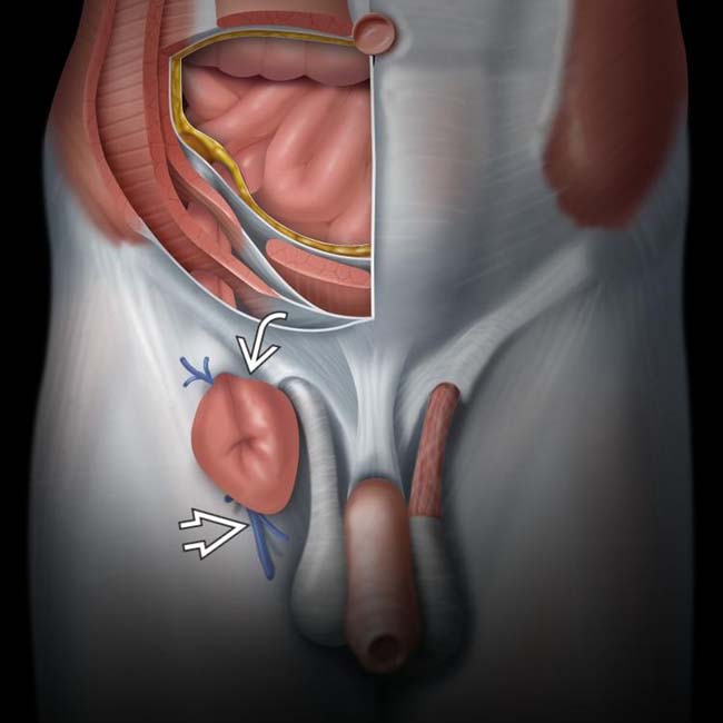
(Left) Illustration of a femoral hernia demonstrates a characteristic “knuckle” of small bowel image closely associated with the femoral vein image. Femoral hernias are usually found medial to the femoral vessels with frequent compression of the femoral vein.
image
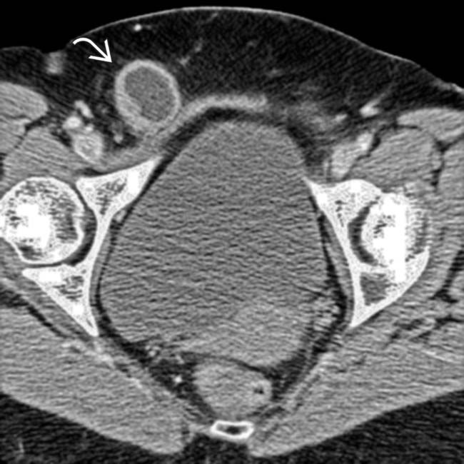
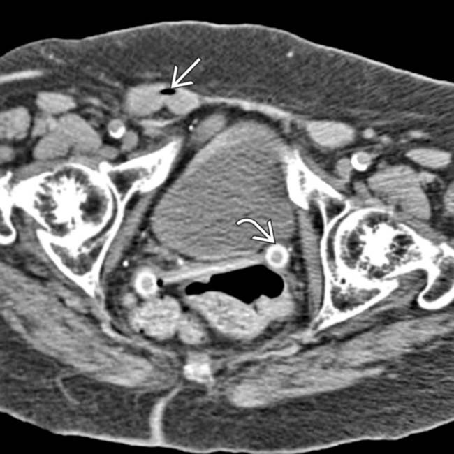
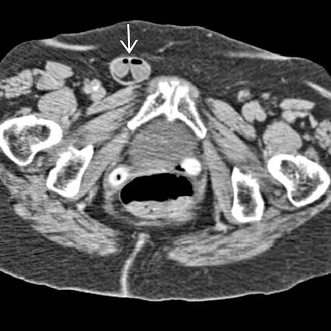
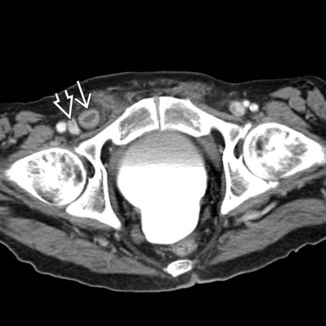
(Right) Axial CECT demonstrates a herniated small bowel loop image lying within the femoral canal, compressing the femoral vessels, compatible with a femoral hernia.
image
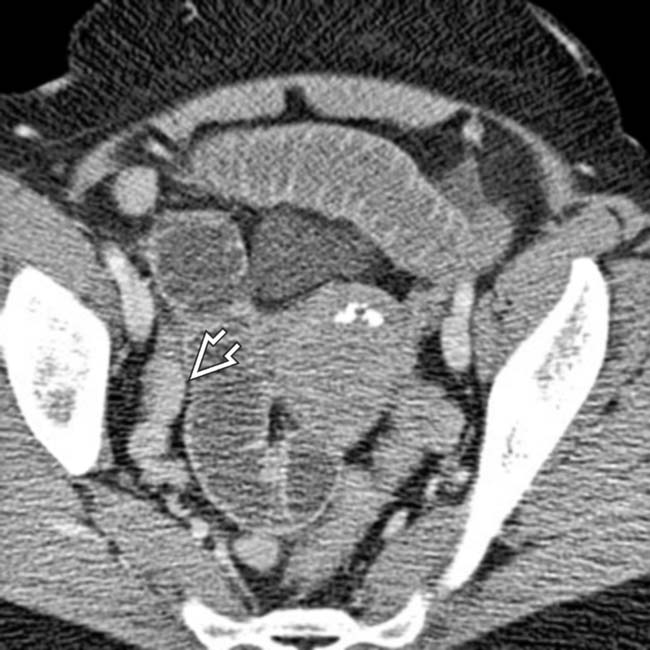
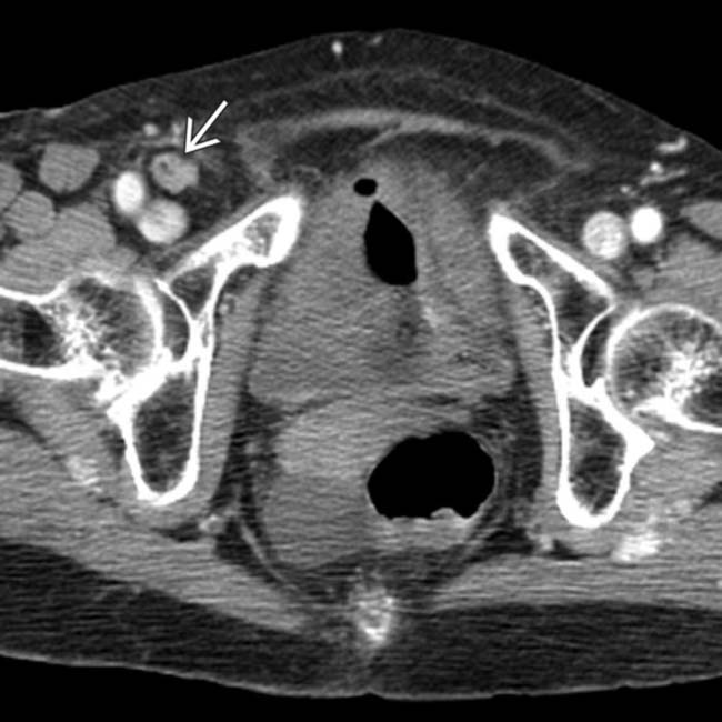
(Left) Axial CECT shows a loop of thickened, hyperemic bowel image herniating into the right groin medial to the femoral vessels. Notice that the femoral vein image is being compressed, and the herniated bowel lies posterolateral to the pubic tubercle.
image
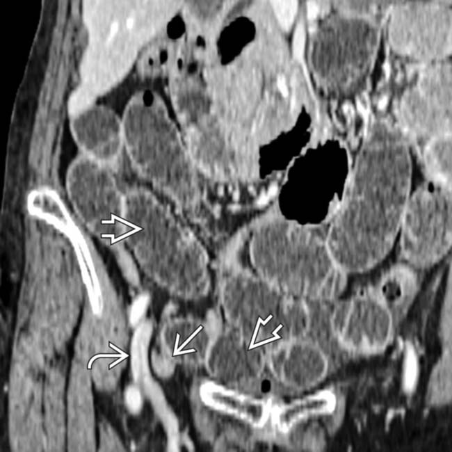
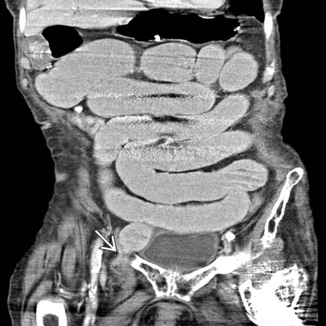
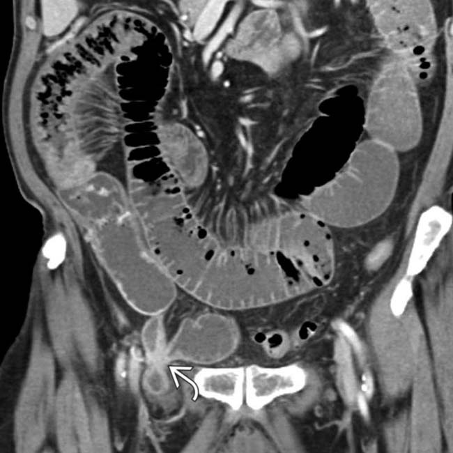
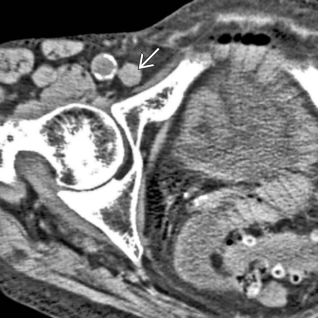
(Right) Coronal CECT in the same patient shows multiple dilated small-bowel loops with a transition point image within the hernia. This thickened, hyperenhancing bowel within the hernia sac was found to be ischemic at surgery. Femoral hernias are at high risk for strangulation and obstruction.
TERMINOLOGY

Abbreviations

• Femoral hernia (FH)

Synonyms

• Crural hernia, enteromerocele, femorocele

Definitions

• Protrusion of abdominal contents through femoral ring into femoral canal

IMAGING

General Features

• Best diagnostic clue
• Location

image Protrusion of hernia sac contents at right angle to inguinal canal through femoral ring into femoral canal

– Posterior to inguinal ligament, anterior to pubic ramus periosteum (Cooper ligament), and medial to femoral vessels
image Inguinal ligament not visible on CT as discrete structure, but horizontal plane connecting pubic tubercles defines plane of inguinal ligament

– Femoral hernia posterior to plane of pubic tubercle
image Twice as common on right side compared to left
• Morphology

image Narrow neck with characteristic pear shape

CT Findings

• Omental fat or bowel herniating into femoral canal medial to femoral vein and inferior to inferior epigastric vessels

image Femoral vein indented/compressed by hernia sac
• Hernia sac located posterior and lateral to pubic tubercle
• Narrow, funnel-shaped, or pear-shaped neck

Ultrasonographic Findings

• Hernia sac visualized extending medial to femoral vein
• Hernia sac may be easier to define with Valsalva maneuver

Radiographic Findings

• Herniography: Hernia curves smoothly over superior pubic ramus on all projections

image Pear-shaped hernia sac with a narrow neck

Imaging Recommendations

• Best imaging tool

image CECT
• Protocol advice

DIFFERENTIAL DIAGNOSIS

Inguinal Hernia

• Abdominal contents within inguinal canal  anteromedial to femoral vessels with extension into scrotum
• Seen anterior to horizontal plane of pubic tubercle
• Does not involve femoral canal or compress femoral vessels

Obturator Hernia

• Hernia into superolateral obturator canal
• Typically occurs in elderly women (80-90%) with a high risk of incarceration

Lymphadenopathy

• When medial to femoral vessels, can theoretically mimic femoral hernia on clinical exam, but distinction easily made with CT

PATHOLOGY

General Features

• Etiology

image May be partially attributable to congenital defect in insertion of transversalis fascia to ileopubic tract
image Femoral ring connective tissues may dilate during pregnancy, placing women at ↑ risk for femoral hernia
image Associated with increased intraabdominal pressure

Gross Pathologic & Surgical Features

CLINICAL ISSUES

Presentation

• Most common signs/symptoms

image Swelling, groin discomfort, vague pelvic discomfort
image Lump usually felt at top of thigh, below groin crease
image Pain is uncommon in absence of strangulation
• Other signs/symptoms

image Nausea, vomiting, severe pain with strangulated hernia
image Difficult to diagnose clinically, especially in obese patients, due to deep location of femoral canal

Demographics

• Age

image 36% occur in patients > 80 years old
image 16% occur in 7th decade
image < 1% of all groin hernias in children
• Gender

image Predominantly women (M:F = 1:4)
• Epidemiology

image ∼ 2-4% of groin hernias in adults
image ∼ 1/10 as common as inguinal hernias
image ∼ 1/3 of groin hernias in women

Natural History & Prognosis

• Complications

image High risk of incarceration &/or strangulation (25-40%), primarily due to narrow neck and unyielding margins of femoral ring

– Highest rate of incarceration of all groin hernias
– 8-12x more prone to incarceration/strangulation than inguinal hernias
– Rarely, inflamed appendix extends into hernia sac
• Morbidity and mortality

image Primarily related to incarceration/bowel obstruction
image Mortality: 1% in 70-79 age group; 5% in 80-90 age group

Treatment

• Symptomatic hernia (or newly discovered asymptomatic hernia) should undergo immediate surgical repair
• Longstanding, asymptomatic hernias may theoretically be treated conservatively with watchful waiting
• No consensus on laparoscopic vs. open surgery

DIAGNOSTIC CHECKLIST

Consider

Image Interpretation Pearls

• Femoral hernias lie medial to femoral vein and inferior to inferior epigastric vessels, often compressing femoral vein
image
Axial CECT shows a femoral hernia with a small bowel obstruction. Note the loop of bowel entrapped in the right femoral canal image.

image
Axial CECT at higher level in the same patient reveals a small bowel obstruction image.
image
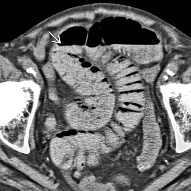
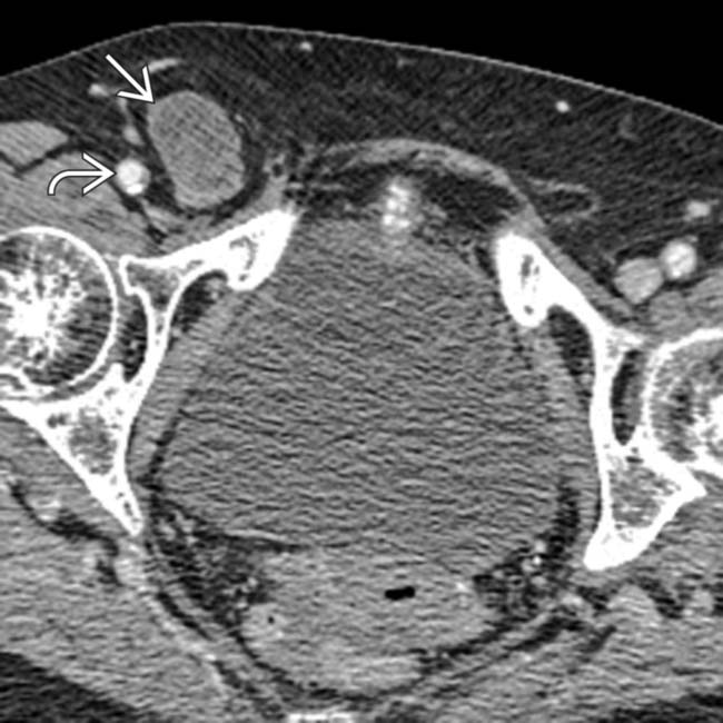
Axial CECT shows an incarcerated femoral hernia in the femoral canal. Note the decompressed distal small bowel, suggesting a small bowel obstruction image.
image
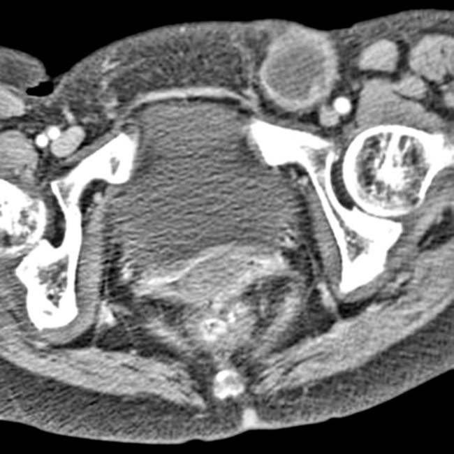
Axial CECT in the same patient at a lower level identifies the source of the obstruction: Small bowel trapped in the femoral canal image.
image
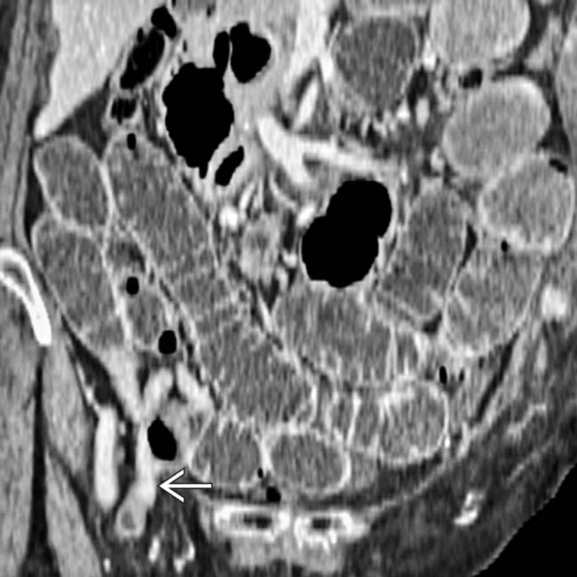
Coronal CECT reformation of a femoral hernia causing small bowel obstruction shows that the neck of the hernia image is medial to the femoral artery image. Note the proximal dilation of the small bowel due to obstruction image.
image
Coronal reformation in the same patient clearly demonstrates a herniated loop of small bowel image.
image
Axial CECT at a lower level demonstrates herniated bowel image medial to the femoral vein image.
image
Axial CECT shows a left femoral hernia containing a “knuckle” of strangulated bowel.
image
Axial CECT in an elderly woman shows a right femoral hernia image and pessary image.
image
Axial CECT shows a right femoral hernia image containing small bowel that caused obstruction.
image
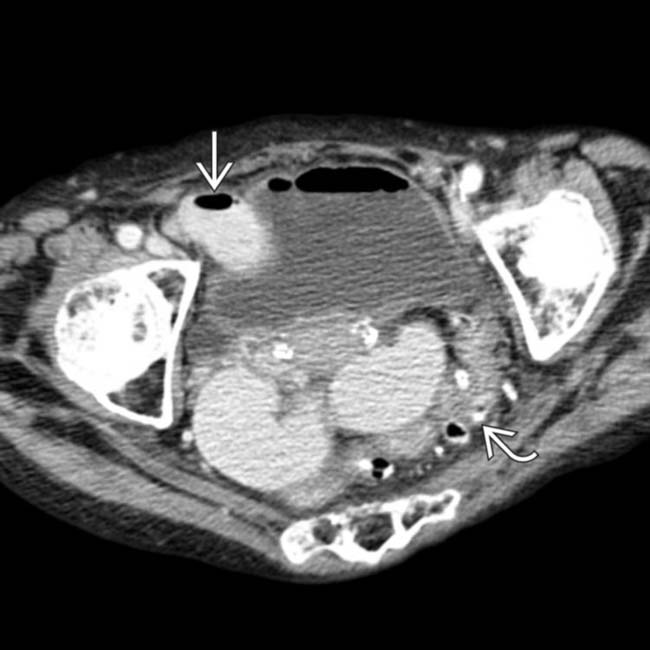
Axial CECT in an 80-year-old woman with abdominal pain, fever, and nausea shows a loop of bowel image coursing into the femoral ring medial to the femoral vessels; the transition point from the dilated to the decompressed bowel, causing a small bowel obstruction. Note the presence of diverticula image within the decompressed sigmoid colon.
image
Coronal CECT reformation image in the same patient demonstrates an incarcerated loop of small bowel image within the femoral ring.

SELECTED REFERENCES

1. Dahlstrand, U, et al. Limited potential for prevention of emergency surgery for femoral hernia. World J Surg. 2014. [ePub].

Alhambra-Rodriguez de Guzmán, C, et al. Improved outcomes of incarcerated femoral hernia: a multivariate analysis of predictive factors of bowel ischemia and potential impact on postoperative complications. Am J Surg. 2013; 205(2):188–193.

Burkhardt, JH, et al. Diagnosis of inguinal region hernias with axial CT: the lateral crescent sign and other key findings. Radiographics. 2011; 31(2):E1–12.

Cherian, PT, et al. The diagnosis and classification of inguinal and femoral hernia on multisection spiral CT. Clin Radiol. 2008; 63(2):184–192.

Cherian, PT, et al. Radiologic anatomy of the inguinofemoral region: insights from MDCT. AJR Am J Roentgenol. 2007; 189(4):W177–W183.

Suzuki, S, et al. Differentiation of femoral versus inguinal hernia: CT findings. AJR Am J Roentgenol. 2007; 189(2):W78–W83.

Akopian, G, et al. De Garengeot hernia: appendicitis within a femoral hernia. Am Surg. 2005; 71(6):526–527.

Bringman, S, et al. Intestinal obstruction after inguinal and femoral hernia repair: a study of 33,275 operations during 1992-2000 in Sweden. Hernia. 2005; 9(2):178–183.

Holzheimer, RG. Inguinal Hernia: classification, diagnosis and treatment—classic, traumatic and Sportsman’s hernia. Eur J Med Res. 2005; 10(3):121–134.

Ikossi, DG, et al. Laparoscopic femoral hernia repair using umbilical ligament as plug. J Laparoendosc Adv Surg Tech A. 2005; 15(2):197–200.

Alvarez, JA, et al. Incarcerated groin hernias in adults: presentation and outcome. Hernia. 2004; 8(2):121–126.

Malek, S, et al. Emergency repair of groin herniae: outcome and implications for elective surgery waiting times. Int J Clin Pract. 2004; 58(2):207–209.

Hachisuka, T. Femoral hernia repair. Surg Clin North Am. 2003; 83(5):1189–1205.

Swarnkar, K, et al. Sutureless mesh-plug femoral hernioplasty. Am J Surg. 2003; 186(2):201–202.

Zollinger, RM, Jr. Classification systems for groin hernias. Surg Clin North Am. 2003; 83(5):1053–1063.

Lau, WY. History of treatment of groin hernia. World J Surg. 2002; 26(6):748–759.

Dieudonne, G. Plug repair of groin hernias: a 10-year experience. Hernia. 2001; 5(4):189–191.

Zhang, GQ, et al. Groin hernias in adults: value of color Doppler sonography in their classification. J Clin Ultrasound. 2001; 29(8):429–434.

Ianora, AA, et al. Abdominal wall hernias: imaging with spiral CT. Eur Radiol. 2000; 10(6):914–919.

Toms, AP, et al. Illustrated review of new imaging techniques in the diagnosis of abdominal wall hernias. Br J Surg. 1999; 86(10):1243–1249.

Loftus, WK, et al. Case report: Femoral hernia causing small bowel obstruction—ultrasound diagnosis. Clin Radiol. 1998; 53(8):618–619.

Radcliffe, G, et al. Reappraisal of femoral hernia in children. Br J Surg. 1997; 84(1):58–60.

Harrison, LA, et al. Abdominal wall hernias: review of herniography and correlation with cross-sectional imaging. Radiographics. 1995; 15(2):315–332.

Chamary, VL. Femoral hernia: intestinal obstruction is an unrecognized source of morbidity and mortality. Br J Surg. 1993; 80(2):230–232.

Lewin, JR. Femoral hernia with upward extension into abdominal wall: CT diagnosis. AJR Am J Roentgenol. 1981; 136(1):206–207.