(Left) CT in an 85-year-old woman with chronic constipation and acute abdominal pain shows free intraperitoneal air .
(Right) Sagittal reformatted CT section in the same case shows massive fecal distention of the rectosigmoid colon with disruption of the anterior wall of the sigmoid colon.
(Left) Axial CECT in an 80-year-old woman presenting with chronic constipation and acute abdominal pain shows free intraperitoneal gas and massive distention of the rectum and left colon with gas and impacted feces .
(Right) Axial CECT in the same patient shows the rectum massively distended with high density feces . Infiltration of the perirectal fat suggests stercoral ulceration, confirmed at surgery. The rectal perforation by stercoral colitis was fatal in this case.


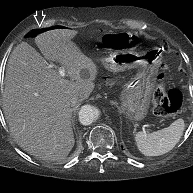
 .
.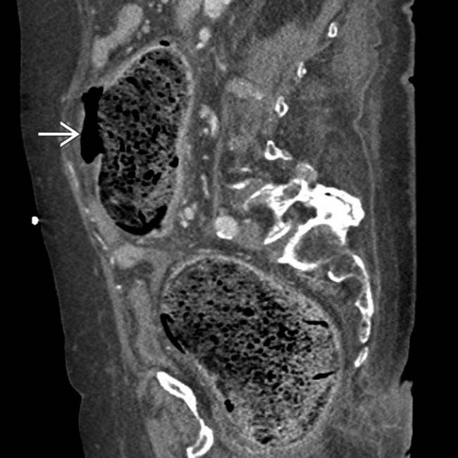
 of the anterior wall of the sigmoid colon.
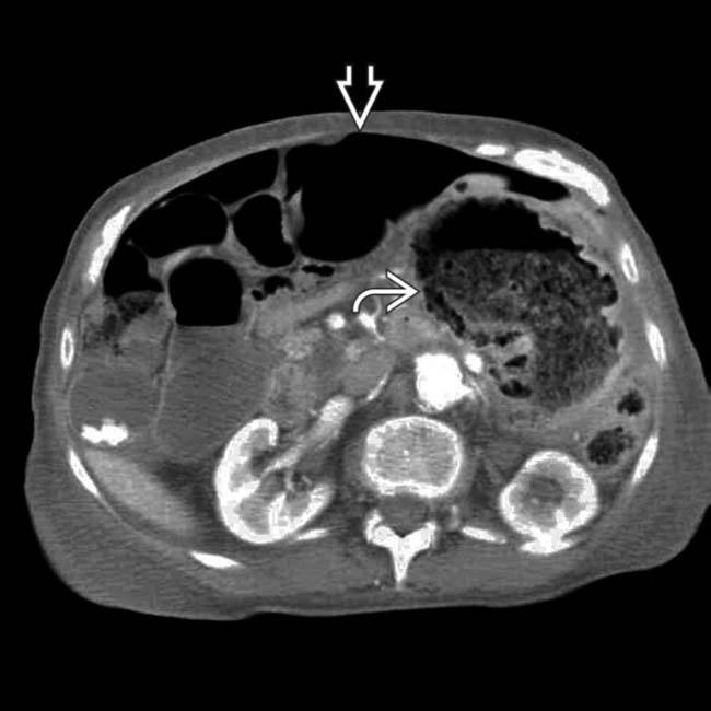
 and massive distention of the rectum and left colon with gas and impacted feces
  .
.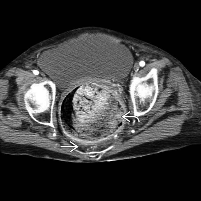
 . Infiltration of the perirectal fat
  suggests stercoral ulceration, confirmed at surgery. The rectal perforation by stercoral colitis was fatal in this case.
