[level-membership-for-opthalmology-category]
CHAPTER 50 Eyelid reconstruction

Anatomy
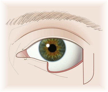
The basic structure of the eyelids is a composite of two layers, or lamellae – an anterior lamella of skin and orbicularis muscle and a posterior lamella of tarsal plate and conjunctiva. The gray line marks the junction of the lamellae at the lid margin (Figs 50.1, 50.2).
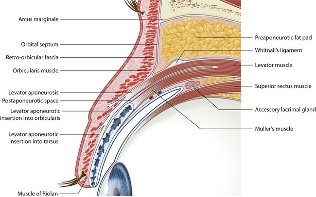
Fig. 50.1 Section through the upper eyelid.
From Tyers & Collin. Color Atlas of Ophthalmic Plastic Surgery. 2008, Elsevier, Butterworth-Heinemann.
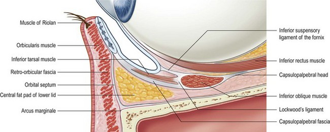
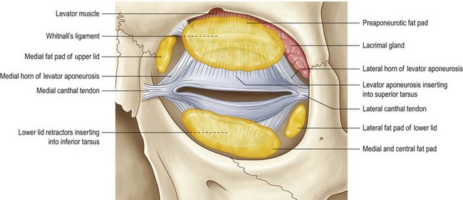
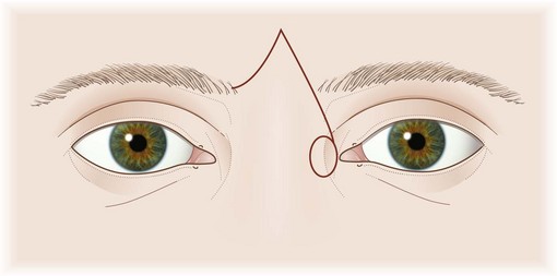
Fig. 50.2 The canthal tendons.
From Tyers & Collin. Color Atlas of Ophthalmic Plastic Surgery. 2008, Elsevier, Butterworth-Heinemann.
The lids are supported at each end by canthal tendons which are attached to the tarsal plates and the orbicularis muscle. The medial canthal tendon is a complex structure, but its essential feature, for the purposes of eyelid reconstruction, is a firm attachment to the posterior lacrimal crest. The lateral canthal tendon attaches to Whitnall’s tubercle just within the lateral orbital rim (Fig. 50.3).
Fundamental principles
Incisions
The ‘relaxed skin tension lines’ (Langer’s lines) mark the preferred direction of skin incisions anywhere on the body surface. In the periocular region they are generally parallel to the lid margins (Fig. 50.4).
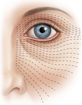
Fig. 50.4 Relaxed skin tension lines around the eye.
From Tyers & Collin. Color Atlas of Ophthalmic Plastic Surgery. 2008, Elsevier, Butterworth-Heinemann.
Correctly placed incisions result in good healing with the least scarring.
Posterior lamellar flaps
If a skin graft is to be used for reconstruction of the anterior lamella of a full-thickness eyelid reconstruction, the posterior lamella must be a flap with an intact blood supply. Few are available. The most commonly used is a tarso-conjunctival flap from the upper lid for reconstruction of lower lid defects (see Landolt–Hughes tarso-conjunctival flap below). An alternative for lower lid defects is the Hewes flap, which is an upper tarsal flap based laterally (see below).
Composite flaps
An alternative to the reconstruction of each eyelid lamella separately, is to use a composite flap of both lamellae. An upper lid defect may be reconstructed with a flap of full-thickness lower lid (see Cutler Beard procedure below). A lower lid defect cannot be reconstructed in this way with a full-thickness flap from the upper lid.
Operation techniques
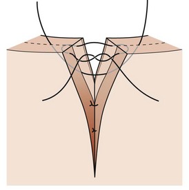
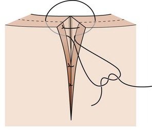
1 Direct closure
Technique
2 Direct closure with extra tissue laterally
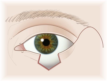
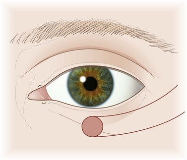
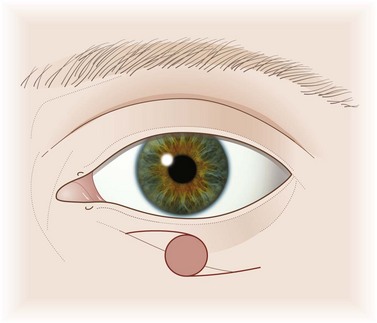
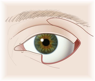
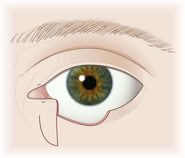
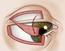
a Lateral cantholysis (Fig. 50.7) also included on the video clip
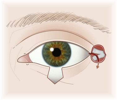
Fig. 50.7 Lateral cantholysis.
Redrawn from Collin. A Manual of Systematic Eyelid Surgery, 3rd edn. 2006, Elsevier.
Technique
b Semicircular flap (Tenzel)
Technique
3 Skin grafts
a Full-thickness skin grafts
b Split-thickness grafts
4 Skin flaps
a Advancement flap
Technique
b O to Z rotation flaps
Technique
c Upper to lower lid transposition flap
Technique
d Nasojugal transposition flap
Technique
e Transposed cheek flap
Technique
f Glabellar flap
Technique
5 Posterior lamellar grafts
a Oral mucosa
Oral mucosa is used not only for reconstruction of the posterior lid lamella but also in socket reconstruction. See Chapter 56 for operative details.
b Tarsal plate
Technique
6 Posterior lamellar flap
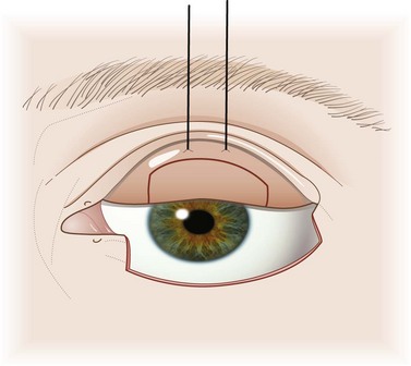
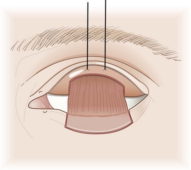
a Landolt–Hughes tarso-conjunctival flap
Technique
b Hewes tarso-conjunctival flap
Technique
7 Combined anterior and posterior flap
a Cutler Beard flap
Technique
8 Canthal support
a Fixation to the posterior lacrimal crest
b Periosteal flap
Borges AF. Relaxed skin tension lines versus other skin lines. Plast Reconstr Surg. 1984;73:144.
Codner MA, McCord CD, Mejia JD, et al. Upper and lower eyelid reconstruction. Plast Reconstr Surg. 2010;126:231e-245e.
Collin JRO. A Manual of Systematic Eyelid Surgery, 3rd edn. Edinburgh: Elsevier; 2005.
Lemke BN, Della Rocca RC. Surgery of the eyelids and orbit, an anatomical approach. Norwalk, CN: Appleton & Lange; 1990.
Levine MR, editor. Manual of Oculoplastic Surgery, 4th edn, NJ, USA: Slack Incorporated, 2010.
Meadows AE, Rhatigan M, Manners RM. Bilobed flap in ophthalmic plastic surgery. Ophthalmic Plast Reconstr Surg. 2005;21:441.
Nerad JA. Techniques in Ophthalmic Plastic Surgery with DVD: A Personal Tutorial. Philadelphia, PA: Saunders; 2010.
Ng SG, Inkster CF, Leatherbarrow B. The rhomboid flap in medial canthal reconstruction. Br J Ophthalmol. 2001;85:556.
Reck AC, Frank H. Tarsal transposition flap to repair inner lamellar defects. Ophthalmic Plast Reconstr Surg. 1995;11:281.
Stephenson CM, Brown BZ. The use of tarsus as a free autogenous graft in eyelid surgery. Ophthalmic Plast Reconstr Surg. 1985;1:43.
Teske SA, Kersten RC, Devoto MH, et al. The modified rhomboid transposition flap in periocular reconstruction. Ophthalmic Plast Reconstr Surg. 1998;14:360.
Tyers AG, Collin JRO. Color Atlas of Ophthalmic Plastic Surgery, 3rd edn. London: Butterworth Heinemann; 2008.
[/level-membership-for-opthalmology-category][not-level-membership-for-opthalmology-category]
CHAPTER 50 Eyelid reconstruction
Anatomy
The basic structure of the eyelids is a composite of two layers, or lamellae – an anterior lamella of skin and orbicularis muscle and a posterior lamella of tarsal plate and conjunctiva. The gray line marks the junction of the lamellae at the lid margin (Figs 50.1, 50.2).
Fig. 50.1 Section through the upper eyelid.
From Tyers & Collin. Color Atlas of Ophthalmic Plastic Surgery. 2008, Elsevier, Butterworth-Heinemann.
Fig. 50.2 The canthal tendons.
From Tyers & Collin. Color Atlas of Ophthalmic Plastic Surgery. 2008, Elsevier, Butterworth-Heinemann.
The lids are supported at each end by canthal tendons which are attached to the tarsal plates and the orbicularis muscle. The medial canthal tendon is a complex structure, but its essential feature, for the purposes of eyelid reconstruction, is a firm attachment to the posterior lacrimal crest. The lateral canthal tendon attaches to Whitnall’s tubercle just within the lateral orbital rim (Fig. 50.3).
Fundamental principles
Incisions
The ‘relaxed skin tension lines’ (Langer’s lines) mark the preferred direction of skin incisions anywhere on the body surface. In the periocular region they are generally parallel to the lid margins (Fig. 50.4).
Fig. 50.4 Relaxed skin tension lines around the eye.
From Tyers & Collin. Color Atlas of Ophthalmic Plastic Surgery. 2008, Elsevier, Butterworth-Heinemann.
Correctly placed incisions result in good healing with the least scarring.
Posterior lamellar flaps
If a skin graft is to be used for reconstruction of the anterior lamella of a full-thickness eyelid reconstruction, the posterior lamella must be a flap with an intact blood supply. Few are available. The most commonly used is a tarso-conjunctival flap from the upper lid for reconstruction of lower lid defects (see Landolt–Hughes tarso-conjunctival flap below). An alternative for lower lid defects is the Hewes flap, which is an upper tarsal flap based laterally (see below).
Composite flaps
An alternative to the reconstruction of each eyelid lamella separately, is to use a composite flap of both lamellae. An upper lid defect may be reconstructed with a flap of full-thickness lower lid (see Cutler Beard procedure below). A lower lid defect cannot be reconstructed in this way with a full-thickness flap from the upper lid.
